. 2020 Nov 16;12(3):1054-1061.
doi: 10.1039/d0sc04889e.

A photo-sensitizable phage for multidrug-resistant Acinetobacter baumannii therapy and biofilm ablation

Affiliations

A photo-sensitizable phage for multidrug-resistant Acinetobacter baumannii therapy and biofilm ablation

Bei Ran et al. Chem Sci. .

Abstract

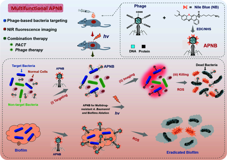
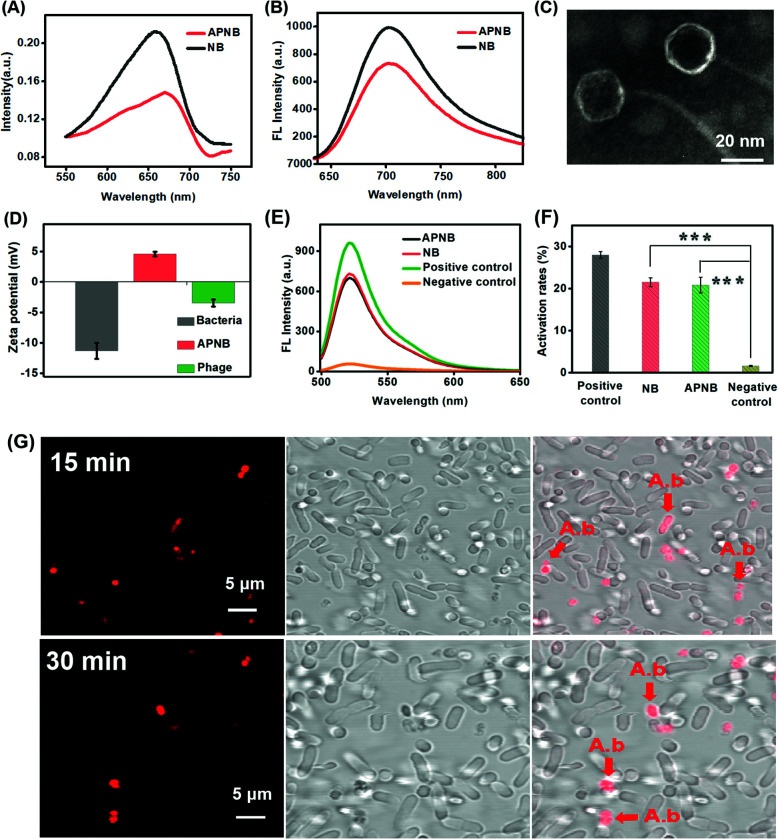
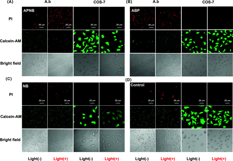
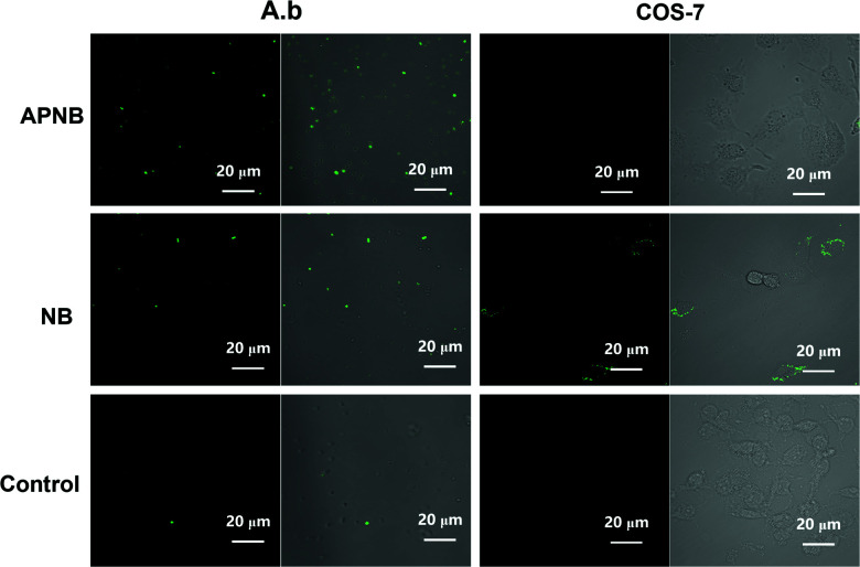
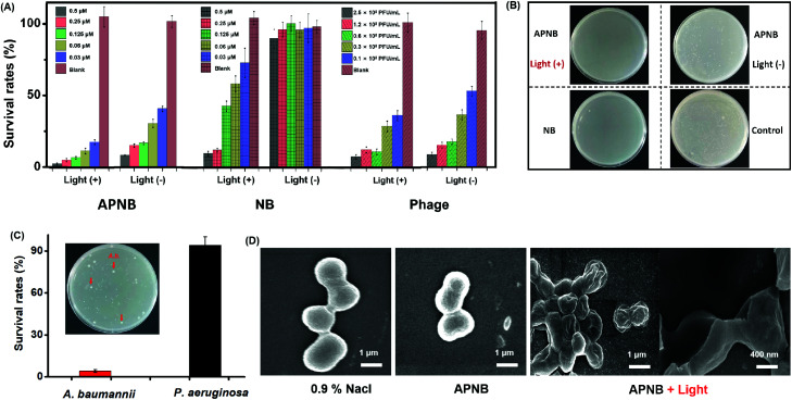
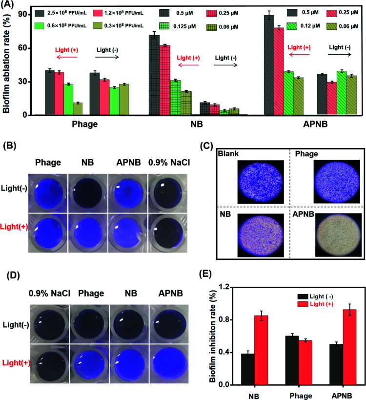
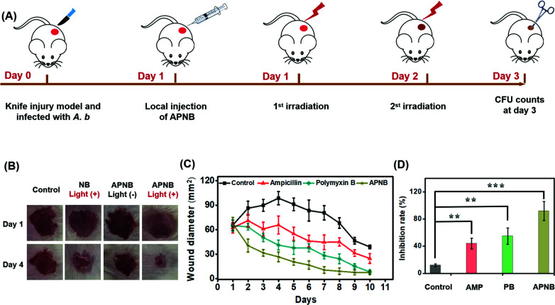
Antibiotic abuse causes the emergence of bacterial resistance. Photodynamic antibacterial chemotherapy (PACT) has great potential to solve serious bacterial resistance, but it suffers from the inefficient generation of ROS and the lack of bacterial targeting ability. Herein, a unique cationic photosensitizer (NB) and bacteriophage (ABP)-based photodynamic antimicrobial agent (APNB) is developed for precise bacterial eradication and efficient biofilm ablation. Thanks to the structural modification of the NB photosensitizer with a sulfur atom, it displays excellent reactive oxygen species (ROS)-production ability. Moreover, specific binding to pathogenic microorganisms can be provided by bacteriophages. The developed APNB has multiple functions, including bacteria targeting, near-infrared fluorescence imaging and combination therapy (PACT and phage therapy). Both in vitro and in vivo experiments prove that APNB can efficiently treat A. baumannii infection. Particularly, the recovery from A. baumannii infection after APNB treatment is faster than that with ampicillin and polymyxin B in vivo. Furthermore, the strategy of combining bacteriophages and photosensitizers is employed to eradicate bacterial biofilms for the first time, and it shows the excellent biofilm ablation effect as expected. Thus, APNB has huge potential in fighting against multidrug-resistant bacteria and biofilm ablation in practice.


Conflict of interest statement

There are no conflicts to declare.

Figures

Scheme 1
Scheme 1. Schematic of the multi-functional antibacterial system (APNB) based on the ABP phage and Nile blue photosensitizer (NB) for the treatment of multi-drug resistant Acinetobacter baumannii and its biofilms. Structural modification of NB with a sulfur atom contributes the high ROS generation ability, and the bacteriophages provide specific binding to pathogenic microorganisms. The developed APNB has multiple functions, including bacteria targeting, near-infrared fluorescence imaging and combination therapy (PACT and phage therapy).
Fig. 1
Fig. 1. (A) UV-vis spectra and (B) fluorescence spectra of NB and APNB. (C) TEM image of the ABP phage (scale bar = 20 nm). (D) Zeta potentials of A. baumanni, the ABP phage and APNB in water. (E) Relative fluorescence intensity and (F) activation rates of 2′,7′-dichlorofluorescein (DCFH) with addition of APNB, NB, the positive control and the negative control upon exposure of 660 nm light (20 mW cm−2). ***P < 0.001. (G) Targeted bacterial imaging of APNB by fluorescence imaging of A. baumanni and P. aeruginosa co-incubated with APNB for 15 min and 30 min. Arrows indicate A. baumannii with staining by APNB (scale bar = 5 μm). [APNB] = 0.5 μM. λex = 660 nm for APNB.
Fig. 2
Fig. 2. Live/dead bacterial viability test by confocal laser scanning microscope (CLSM) imaging. (A) Confocal images of APNB-treated bacteria and COS-7 cells post live/dead staining (scale bar = 20 μm). (B) Confocal images of ABP-treated bacteria and COS-7 cells post live/dead staining (scale bar = 20 μm). (C) Confocal images of NB-treated bacteria and COS-7 cells post live/dead staining (scale bar = 20 μm). (D) Confocal images of control group post live/dead staining (scale bar = 20 μm). The PACT treatment is conducted under 660 nm light irradiation (20 mW cm−2) for 15 min. [APNB] = [NB] = 0.5 μM. [Calcein-AM] = 5 μM. [PI] = 5 μM. λex = 490 nm for Calcein-AM, and λex = 530 nm for PI.
Fig. 3
Fig. 3. Intracellular detection of ROS production in APNB-treated A. baumannii and COS-7 cells using confocal laser scanning microscope (CLSM) imaging (scale bar = 20 μm). [APNB] = [NB] = 0.5 μM. [DCFH] = 10 μM. λex = 488 nm for DCH.
Fig. 4
Fig. 4. Antibacterial evaluation of APNB based PACT in vitro. (A) Survival rates of bacteria and (B) viable bacteria remained in the culture after different treatments using the CFU counting method. (C) A. baumannii and P. aeruginosa are incubated together with APNB. The arrows indicate A. baumannii identified via the colony morphology. (D) SEM images of A. baumannii before and after being treated with APNB (scale bar = 1 μm).
Fig. 5
Fig. 5. (A) In vitro A. baumannii biofilm eradication by APNB without or with light irradiation (15 min at 20 mW cm−2); phage and NB served as the controls. (B) The Photographs and (C) optical microscope photographs of the remaining biofilms after the different treatments. (D) Photographs of in vitro inhibition of biofilm formation by the phage, NB and APNB without or with NIR light irradiation (15 min at 20 mW cm−2); the phage and NB served as the controls. (E) Biofilm inhibition rate of APNB by CFU counting.
Fig. 6
Fig. 6. The antibacterial ability in vivo. (A) Experimental procedure of the in vivo antibacterial evaluation; the different treatments are conducted at day 1 and day 2 post injection of A. baumannii. (B) Photographs and (C) the sizes of the infected areas. (D) the number of bacterial colony-forming units counting from the infected wounds at day 3 post injection of A. baumannii. **P < 0.01 and ***P < 0.001.

References

    1. Baym M. Stone L. K. Kishony R. Science. 2016;351:aad3292. doi: 10.1126/science.aad3292. - DOI - PMC - PubMed
    1. Besser J. Carleton H. A. Gerner-Smidt P. Lindsey R. L. Trees E. Clin. Microbiol. Infect. 2018;24:335–341. doi: 10.1016/j.cmi.2017.10.013. - DOI - PMC - PubMed
    1. Levin B. R. Baquero F. Ankomah P. McCall I. C. Trends Microbiol. 2017;25:878–892. doi: 10.1016/j.tim.2017.07.005. - DOI - PubMed
    1. Chin W. Zhong G. Pu Q. Yang C. Lou W. De Sessions P. F. Periaswamy B. Lee A. Liang Z. C. Ding X. Gao S. Chu C. W. Bianco S. Bao C. Tong Y. W. Fan W. Wu M. Hedrick J. L. Yang Y. Y. Nat. Commun. 2018;9:914–917. doi: 10.1038/s41467-018-03307-8. - DOI - PMC - PubMed
    1. Wong D. Nielsen T. B. Bonomo R. A. Pantapalangkoor P. Luna B. Spellberg B. Clin. Microbiol. Infect. 2017;30:409–447. - PMC - PubMed