Review
. 2021 Jan 26;12(8):2716-2734.
doi: 10.1039/d0sc05419d.

Advances in anion binding and sensing using luminescent lanthanide complexes

Affiliations
Review

Advances in anion binding and sensing using luminescent lanthanide complexes

Samantha E Bodman et al. Chem Sci. .

Abstract

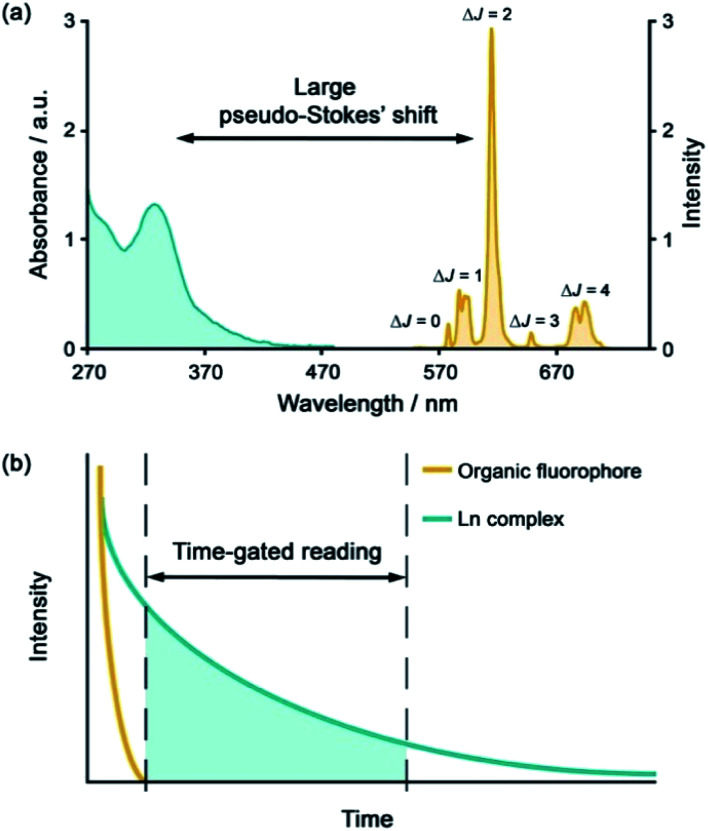
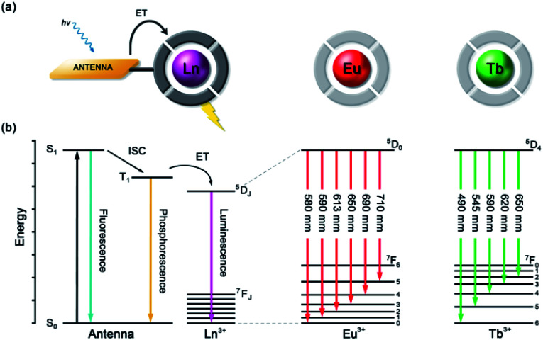
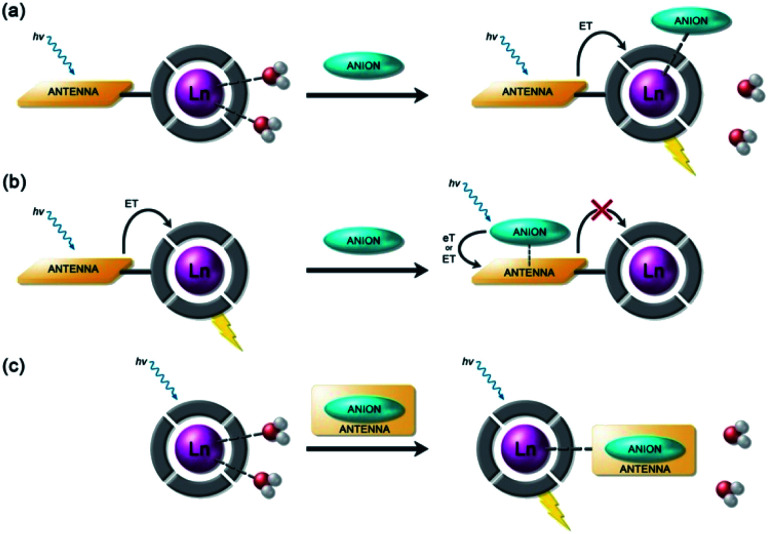
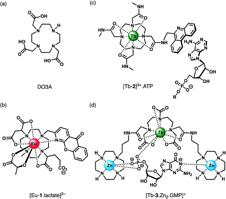
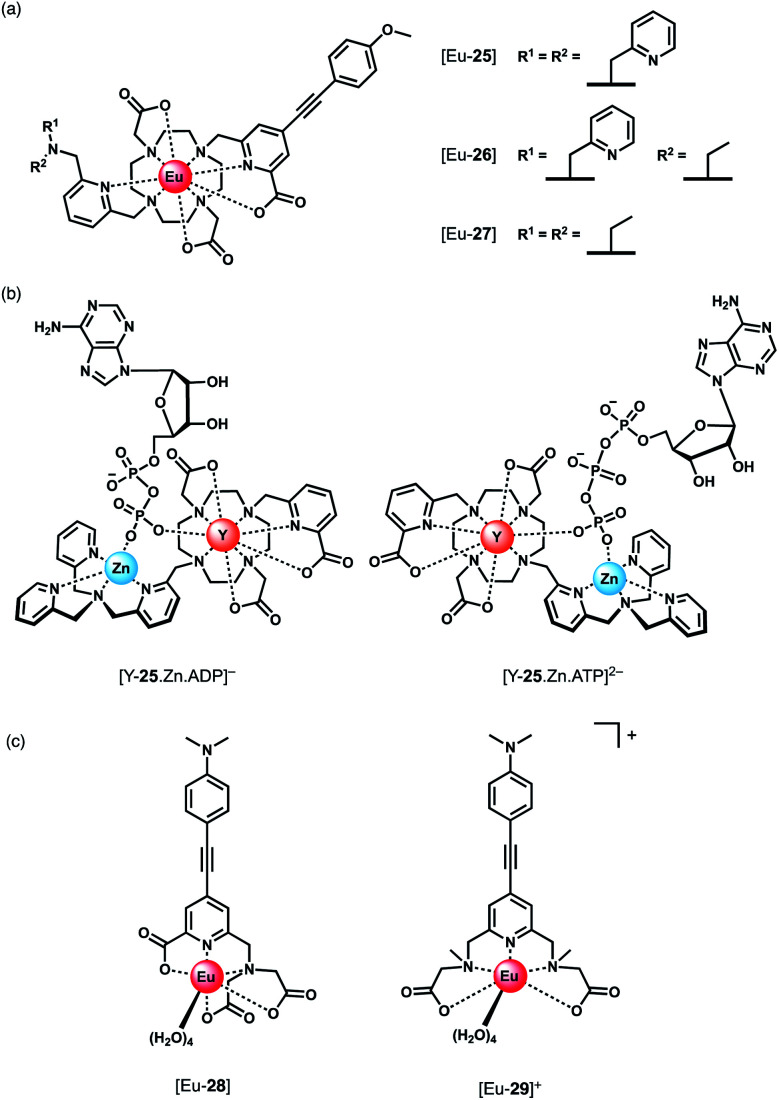
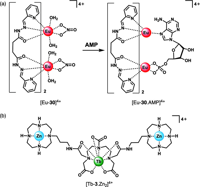
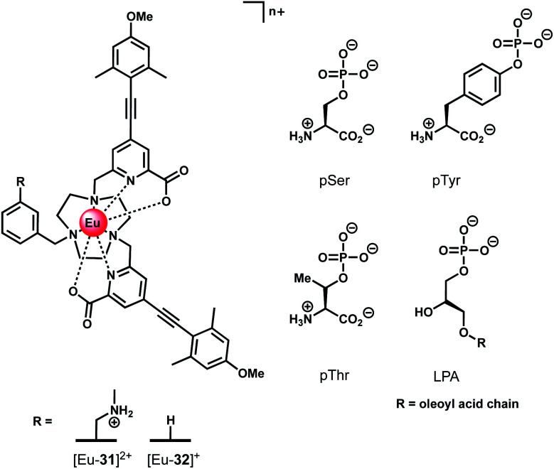
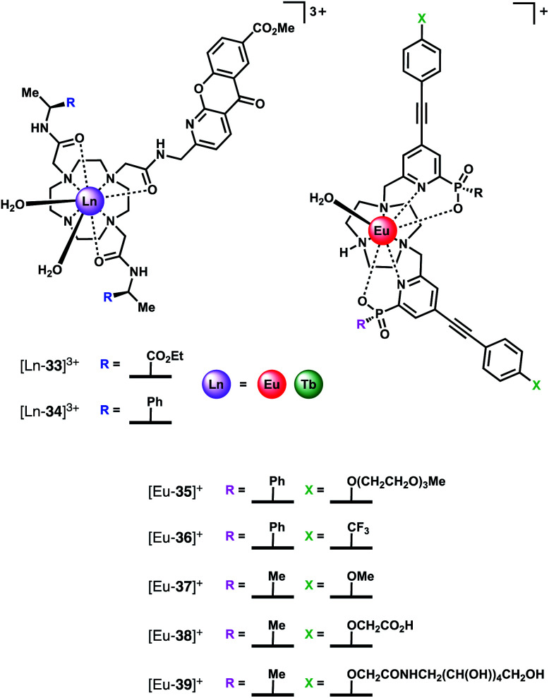
Luminescent lanthanide complexes have been actively studied as selective anion receptors for the past two decades. Ln(iii) complexes, particularly of europium(iii) and terbium(iii), offer unique photophysical properties that are very valuable for anion sensing in biological media, including long luminescence lifetimes (milliseconds) that enable time-gating methods to eliminate background autofluorescence from biomolecules, and line-like emission spectra that allow ratiometric measurements. By careful design of the organic ligand, stable Ln(iii) complexes can be devised for rapid and reversible anion binding, providing a luminescence response that is fast and sensitive, offering the high spatial resolution required for biological imaging applications. This review focuses on recent progress in the development of Ln(iii) receptors that exhibit sufficiently high anion selectivity to be utilised in biological or environmental sensing applications. We evaluate the mechanisms of anion binding and sensing, and the strategies employed to tune anion affinity and selectivity, through variations in the structure and geometry of the ligand. We highlight examples of luminescent Ln(iii) receptors that have been utilised to detect and quantify specific anions in biological media (e.g. human serum), monitor enzyme reactions in real-time, and visualise target anions with high sensitivity in living cells.


Conflict of interest statement

There are no conflicts to declare.

Figures

Fig. 1
Fig. 1. (a) Representative absorption and emission spectra of Eu(iii) complexes highlighting the large pseudo-Stokes'shift; (b) illustration of time-gated detection to eliminate short-lived autofluorescence arising from biomolecules in the sample.
Fig. 2
Fig. 2. (a) Schematic depiction of the antenna effect; (b) simplified Jablonski diagram showing the common pathway leading to lanthanide sensitization, and the 4f–4f transitions of Eu(iii) and Tb(iii) complexes.
Fig. 3
Fig. 3. Schematic representation of different sensing mechanisms for lanthanide(iii)-based anion receptors: (a) coordination of the anion to the metal centre with displacement of inner-sphere water molecules; (b) interaction of the anion with the antenna causing electron or energy transfer, which modulates the sensitisation process, usually quenching luminescence; (c) binding of an anion that possesses an appropriate sensitiser, which ‘switches on’ luminescence.
Fig. 4
Fig. 4. (a) Common macrocyclic ligand DO3A; (b)–(d) examples of Ln(iii) receptors that utilise different mechanisms of anion binding.
Fig. 5
Fig. 5. Series of tripodal Eu(iii) complexes evaluated for the selective detection of HPO42−.
Fig. 6
Fig. 6. (a) Increase in time-delayed (0.1 ms) luminescence of [Eu-6] upon titration with HPO42− (H2O, pH 7.4); (b) luminescence titration data of [Eu-5], [Eu-6], and [Eu-7]+. Inset: Photo of [Eu-6]+ solution in the absence and presence of 10 equiv. of HPO42−. Reproduced from ref. with permission from the American Chemical Society, copyright 2019.
Fig. 7
Fig. 7. (a) Tb(iii)-Based displacement assay for HPO42− and NO3; (b) alkyne-functionalised DO3A complex for assessing bulk phosphate species (PCr, HPO42−); (c) schematic depiction of the detection of HPO42− (Pi) in water samples, using a Eu(iii)–CIP mixture in the presence of the surfactant SDBS.
Fig. 8
Fig. 8. (a) Family of cationic Eu(ii) and Tb(iii) complexes capable of discriminating between nucleoside tri-, di- and monophosphate anions (e.g. ATP, ADP and AMP); (b) Real-time monitoring of elevated ATP levels in the mitochondria of living cells. Time-lapsed LSCM images of NIH-3T3 cells stained with [Eu-21]+ (50 μM, λexc 355 nm, λem 605–720 nm) before (0 min) and after treatment with staurosporine (10 nM), a potent inhibitor of protein kinases; (c) time-dependent increase in emission intensity (605–720 nm) of cells stained with [Eu-21]+ following treatment with staurosporine (10 nM). Reproduced from ref. with permission from Wiley-VCH, copyright 2018.
Fig. 9
Fig. 9. Microplate-based assay for real-time monitoring of kinase activity using [Eu-18]+. (a) Kinase simulation in standard assay conditions (total ATP + ADP = 1 mM, 5 mM MgCl2, 8 μM [Eu-18]+, 10 mM HEPES, pH 7.0), measuring the time-resolved luminescence intensity of differing ratios of ADP/ATP; (b) real-time monitoring of Aurora A kinase reactions at different concentrations of enzyme; (c) inhibition of Aurora A (1 μM) by a range of known inhibitors measured in real-time; (d) IC50 derived from the luminescent titration of staurosporine into Aurora A (50 nM). Reproduced (adapted) from ref. with permission from the Royal Society of Chemistry, copyright 2019.
Fig. 10
Fig. 10. Principle component analysis (PCA) score plots of % change in emission intensity of [Eu-18]+ (8 μM), [Eu-21]+ (13 μM), [Eu-19]+ (10 μM) and [Tb-18]+ (15 μM) with the adenosine and guanosine series of nucleoside polyphosphate anions (1 mM), in 10 mM HEPES, pH 7.0. Reproduced (adapted) from ref. with permission from the Royal Society of Chemistry, copyright 2020.
Fig. 11
Fig. 11. Eu(iii) and Tb(iii) complexes capable of selective binding ATP (and GTP) through π–π stacking and electrostatic interactions, inducing quenching of luminescence.
Fig. 12
Fig. 12. Ratiometric determination of GTP/GDP ratio using time-delayed luminescence of a mixture of [Tb-2]3+ and [Eu-23]. Reproduced from ref. with permission from the Royal Society of Chemistry, copyright 2013.
Fig. 13
Fig. 13. (a) Parker's CPL probes for the discrimination of ATP and ADP. (b) Structures showing differing chirality of the ATP and ADP adducts of complex [Y-25·Zn]2+. Optimised DFT geometries indicate the Δ diastereomer is preferred for ATP and the Λ isomer for ADP. (c) Pentadentate Eu(iii) complexes developed by Schäferling for ATP detection.
Fig. 14
Fig. 14. Ln(iii) receptors for the selective recognition of nucleoside monophosphate anions. (a) Proposed binding mode of AMP to Eu(iii) helicate [Eu-30]4+; (b) trinuclear complex [Tb-3·Zn2]4+ capable of signalling GMP.
Fig. 15
Fig. 15. Structures of emissive Eu(iii) complexes based on TACN, which bind and signal chiral phosphorylated amino acids and LPA, using CPL spectroscopy.
Fig. 16
Fig. 16. Structures of HCO3 selective Ln(iii) receptors, utilised for reporting HCO3 levels in human serum and in living cells.
Fig. 17
Fig. 17. (a) Confocal microscopy images of HeLa cells, showing the mitochondrial region stained by [Eu-33]3+ under 3, 4 and 5% CO2 (1 h incubation, 20 μM complex, 30 min equilibration period between images); (b) variation of Ln(iii) emission intensity from hyper-spectral analysis of microscopy images for HeLa cells stained with [Eu-33]3+; (c) plot of Eu(iii)/Tb(iii) emission intensity ratio for [Ln-34]3+ (600–720 nm vs. 450–570 nm) as a function of pCO2, showing fit of experimental data for an effective Kd = 23.3 (±0.8) mM [HCO3], observed in the mitochondria of NIH-3T3 cells. Reproduced (adapted) from ref. with permission from Wiley-VCH, copyright 2012.
Fig. 18
Fig. 18. Series of Ln(iii) complexes of hexadentate ligands investigated as CPL probes for the detection of HCO3.
Fig. 19
Fig. 19. Structures of Ln(iii) complexes that bind and sense fluoride in aqueous solution.
Fig. 20
Fig. 20. (a) Family of Eu(iii) complexes examined for their ability to signal α-hydroxy acids (e.g. lactate) by CPL spectroscopy; (b) mirror image CPL spectra of [Eu-56]+ following addition of R-(red) and S-(blue) lactate (λexc = 348 nm, 10 μM complex, 50 μM lactate, 295 K, MeOH). Reproduced from ref. with permission from the Royal Society of Chemistry, copyright 2015.
Fig. 21
Fig. 21. Di- and trinuclear Ln(iii) receptors capable of binding dicarboxylate anions (e.g. isophthalate) via a bridging mode.
None
Samantha E. Bodman
None
Stephen Butler

Similar articles

Cited by

References

    1. Bünzli J.-C. G. J. Lumin. 2016;170:866–878. doi: 10.1016/j.jlumin.2015.07.033. - DOI
    1. Butler S. J. Parker D. Chem. Soc. Rev. 2013;42:1652–1666. doi: 10.1039/C2CS35144G. - DOI - PubMed
    1. Mathieu E. Sipos A. Demeyere E. Phipps D. Sakaveli D. Borbas K. E. Chem. Commun. 2018;54:10021–10035. doi: 10.1039/C8CC05271A. - DOI - PubMed
    1. Smith D. G. Pal R. Parker D. Chem.–Eur. J. 2012;18:11604–11613. doi: 10.1002/chem.201201738. - DOI - PubMed
    1. Cordat E. Casey J. R. Biochem. J. 2008;417:423–439. doi: 10.1042/BJ20081634. - DOI - PubMed