

Gastric Mucosa-Associated Lymphoid Tissue Lymphomas Diagnosed by Jumbo Biopsy Using Endoscopic Submucosal Dissection: A Case Report
- PMID: 34164414
- PMCID: PMC8215156
- DOI: 10.3389/fmed.2021.668531
Gastric Mucosa-Associated Lymphoid Tissue Lymphomas Diagnosed by Jumbo Biopsy Using Endoscopic Submucosal Dissection: A Case Report
Abstract
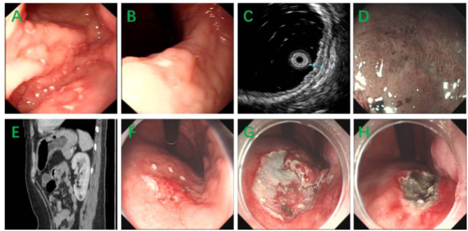
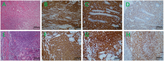
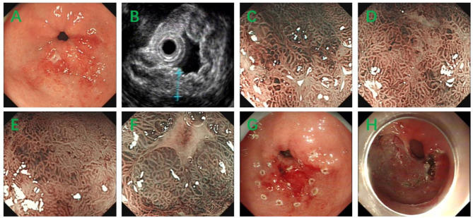
The stomach is the most common primary site of mucosa-associated lymphoid tissue (MALT) lymphoma, and sometimes the histopathological diagnosis is particularly difficult. An endoscopic forceps biopsy is the primary diagnostic test, but false negative results are very common. Therefore, a jumbo biopsy is essential for accurate diagnosis of clinically suspected cases. Here we diagnosed two cases of gastric MALT lymphomas using endoscopic submucosal dissection (ESD). The first patient was suspected of gastric lymphoma at the first endoscopic forceps biopsy, but the second endoscopic forceps biopsy showed chronic inflammation. The second patient was also firstly diagnosed with chronic inflammation by endoscopic forceps biopsy. Both cases were finally confirmed with the diagnosis of gastric MALT lymphoma by jumbo biopsy using ESD. The application of ESD can provide a new diagnostic strategy for clinically suspicious cases of gastric MALT lymphoma with negative endoscopic forceps biopsy.
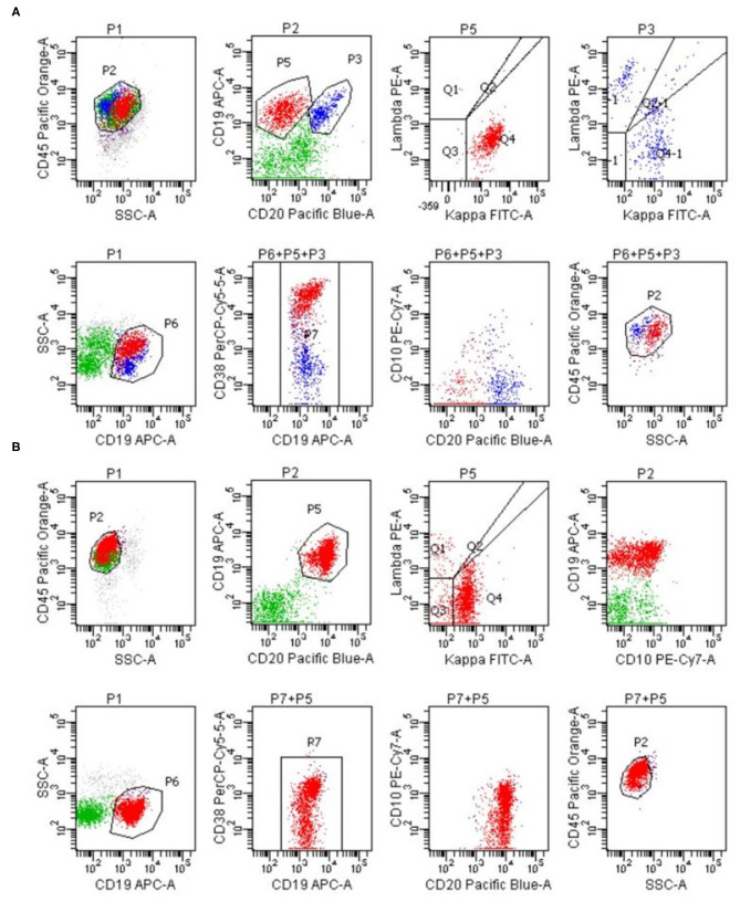
Keywords: endoscopic submucosal dissection; flow cytometry; jumbo biopsy; mucosa-associated lymphoid tissue lymphoma; stomach.
Copyright © 2021 Han, Wang and Xie.
Conflict of interest statement
The authors declare that the research was conducted in the absence of any commercial or financial relationships that could be construed as a potential conflict of interest.
Figures
Similar articles
-
Successful Endoscopic Resection of Primary Rectal Mucosa-Associated Lymphoid Tissue Lymphoma by Endoscopic Submucosal Dissection: A Case Report.Front Med (Lausanne). 2021 Sep 10;8:715256. doi: 10.3389/fmed.2021.715256. eCollection 2021. Front Med (Lausanne). 2021. PMID: 34568373 Free PMC article.
-
A randomized trial to determine the diagnostic accuracy of conventional vs. jumbo forceps biopsy of gastric epithelial neoplasias before endoscopic submucosal dissection; open-label study.Gastric Cancer. 2014 Oct;17(4):661-8. doi: 10.1007/s10120-013-0322-2. Epub 2013 Dec 13. Gastric Cancer. 2014. PMID: 24337434 Clinical Trial.
-
Gastric Mucosa-associated Lymphoid Tissue Lymphoma Mimicking Signet Ring Cell Carcinoma.Korean J Helicobacter Up Gastrointest Res. 2023 Jun;23(2):132-136. doi: 10.7704/kjhugr.2023.0018. Epub 2023 May 30. Korean J Helicobacter Up Gastrointest Res. 2023. PMID: 40502284 Free PMC article.
-
Primary esophageal mucosa-associated lymphoid tissue lymphoma diagnosed by using stacked forceps biopsy.Dis Esophagus. 2016 Oct;29(7):887-890. doi: 10.1111/dote.12309. Epub 2015 Jan 27. Dis Esophagus. 2016. PMID: 25626120 Review.
-
Endoscopic features aiding the diagnosis of gastric mucosa-associated lymphoid tissue lymphoma.Yeungnam Univ J Med. 2019 May;36(2):85-91. doi: 10.12701/yujm.2019.00136. Epub 2019 Feb 26. Yeungnam Univ J Med. 2019. PMID: 31620618 Free PMC article. Review.
Cited by
-
Clinical Management of Patients with Gastric MALT Lymphoma: A Gastroenterologist's Point of View.Cancers (Basel). 2023 Jul 27;15(15):3811. doi: 10.3390/cancers15153811. Cancers (Basel). 2023. PMID: 37568627 Free PMC article. Review.
-
Rare primary colonic T cell lymphoma with curative resection by endoscopic submucosal dissection: A case report.World J Clin Cases. 2024 Aug 6;12(22):5229-5235. doi: 10.12998/wjcc.v12.i22.5229. World J Clin Cases. 2024. PMID: 39109008 Free PMC article.
-
Uncommon presentation of gastric mucosa-associated lymphoid tissue lymphoma in a 13-year-old girl: acute vomiting of blood as the initial symptom.Ann Med Surg (Lond). 2024 Jan 15;86(5):3001-3004. doi: 10.1097/MS9.0000000000001705. eCollection 2024 May. Ann Med Surg (Lond). 2024. PMID: 38694317 Free PMC article.
References
Publication types
LinkOut - more resources
Full Text Sources
Miscellaneous