. 2021 Jun 7:8:629313.
doi: 10.3389/fcvm.2021.629313. eCollection 2021.

Blood Flow Within Bioengineered 3D Printed Vascular Constructs Using the Porcine Model

Affiliations

Blood Flow Within Bioengineered 3D Printed Vascular Constructs Using the Porcine Model

Nhu Thao N Galván et al. Front Cardiovasc Med. .

Abstract

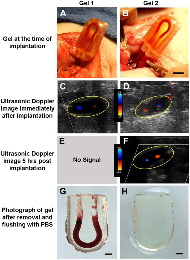
Recently developed biofabrication technologies are enabling the production of three-dimensional engineered tissues containing vascular networks which can deliver oxygen and nutrients across large tissue volumes. Tissues at this scale show promise for eventual regenerative medicine applications; however, the implantation and integration of these constructs in vivo remains poorly studied. Here, we introduce a surgical model for implantation and direct in-line vascular connection of 3D printed hydrogels in a porcine arteriovenous shunt configuration. Utilizing perfusable poly(ethylene glycol) diacrylate (PEGDA) hydrogels fabricated through projection stereolithography, we first optimized the implantation procedure in deceased piglets. Subsequently, we utilized the arteriovenous shunt model to evaluate blood flow through implanted PEGDA hydrogels in non-survivable studies. Connections between the host femoral artery and vein were robust and the patterned vascular channels withstood arterial pressure, permitting blood flow for 6 h. Our study demonstrates rapid prototyping of a biocompatible and perfusable hydrogel that can be implanted in vivo as a porcine arteriovenous shunt, suggesting a viable surgical approach for in-line implantation of bioprinted tissues, along with design considerations for future in vivo studies. We further envision that this surgical model may be broadly applicable for assessing whether biomaterials optimized for 3D printing and cell function can also withstand vascular cannulation and arterial blood pressure. This provides a crucial step toward generated transplantable engineered organs, demonstrating successful implantation of engineered tissues within host vasculature.

Keywords: 3D printed; bioengineered alternative tissue; porcine (pig) model; sterolithography; vascular constructs.


Conflict of interest statement

JSM and BG are cofounders of and hold an equity stake in the startup company Volumetric, Inc. The remaining authors declare that the research was conducted in the absence of any commercial or financial relationships that could be construed as a potential conflict of interest.

Figures

Figure 1
Figure 1
Surgical diagram for inserting constructs into pig models. (A) First, the target artery and vein are isolated and controlled using two silk suture loops. (B) The sutures are pulled to restrict blood flow and a small incision is made partially through the vessel. (C) The tubing (with the vascular construct attached) is inserted into the vessel and clamps are removed, allowing blood to flow through the implanted construct. Tubing is secured within the vessels using silk sutures.
Figure 2
Figure 2
Schematic of PEGDA AV vascular shunt used for implantation in porcine model. (A) Schematic of the modified hydrogel use for implantation in the neck of 5–10 kg piglets. (B) Schematic of 3D printed PC case for hydrogel geometry in (A). (C) Image of a gel implanted as an AV shunt linking the carotid artery to the jugular vein. Scale bar = 5 mm.
Figure 3
Figure 3
Assessment of PEGDA hydrogels implanted in a porcine model. (A,B) Image of gel at time of implantation. Scale bar = 5 mm. (C,D) Ultrasonic Doppler image of gel immediately after wound closure, displaying flow through both channels. (E,F) Ultrasonic Doppler image 5 h after implantation, where no signal was detected for Gel 1. (G,H) Images of gel after explant and flushing with saline. Saline flush dislodges clots which form postmortem but preserve more stable clots seen in a few areas of (H). Scale bar = 2 mm. Gels 1 and 2 in this figure correspond to conditions 4.3 and 4.4 in Table 1, respectively.
Figure 4
Figure 4
Five hundred micrometer thick vibratome sections of gels implanted for 6 h in vivo. (A) Reflected light color photo of a 500 μm thick vibratome section, where the red dotted line indicates the edges of the gel. Scale bar = 1 mm. (B) Phase contrast/Hoechst overlay of Hoechst-stained channel, outlined in white in (A). Scale bar = 250 μm. (C) Zoomed in view of (B) showing individual nuclei. Scale bar = 100 μm.
Figure 5
Figure 5
Resin-based histology of gels implanted in porcine model. (A) Phase contrast image of gel channel, where section is 6 μm thick. (B) Zoomed image of the channel showing the presence of cells adhering to the channel wall. (C) Hoechst stain of the gel demonstrated nucleated cells along the wall. (D) Toluidine blue stain highlighting cell material in dark purple. Scale bars = 100 μm.

References

    1. Reis LA, Chiu LLY, Feric N, Fu L, Radisic M. Biomaterials in myocardial tissue engineering. J Tissue Eng Regen Med. (2016) 10:11–28. 10.1002/term.1944 - DOI - PMC - PubMed
    1. Miller JS. The billion cell construct: will three-dimensional printing get us there? PLoS Biol. (2014) 12:e1001882. 10.1371/journal.pbio.1001882 - DOI - PMC - PubMed
    1. Nugent HM, Edelman ER. Tissue engineering therapy for cardiovascular disease. Circ Res. (2003) 92:1068–78. 10.1161/01.RES.0000073844.41372.38 - DOI - PubMed
    1. Kang Y, Mochizuki N, Khademhosseini A, Fukuda J, Yang Y. Engineering a vascularized collagen-β-tricalcium phosphate graft using an electrochemical approach. Acta Biomater. (2015) 11:449–58. 10.1016/j.actbio.2014.09.035 - DOI - PMC - PubMed
    1. McFadden TM, Duffy GP, Allen AB, Stevens HY, Schwarzmaier SM, Plesnila N, et al. . The delayed addition of human mesenchymal stem cells to pre-formed endothelial cell networks results in functional vascularization of a collagen-glycosaminoglycan scaffold in vivo. Acta Biomater. (2013) 9:9303–16. 10.1016/j.actbio.2013.08.014 - DOI - PubMed

LinkOut - more resources