Review
. 2021 Jun 25;129(1):195-215.
doi: 10.1161/CIRCRESAHA.121.318174. Epub 2021 Jun 24.

Cerebral Cavernous Malformation: From Mechanism to Therapy

Affiliations
Review

Cerebral Cavernous Malformation: From Mechanism to Therapy

Daniel A Snellings et al. Circ Res. .

Erratum in

Abstract

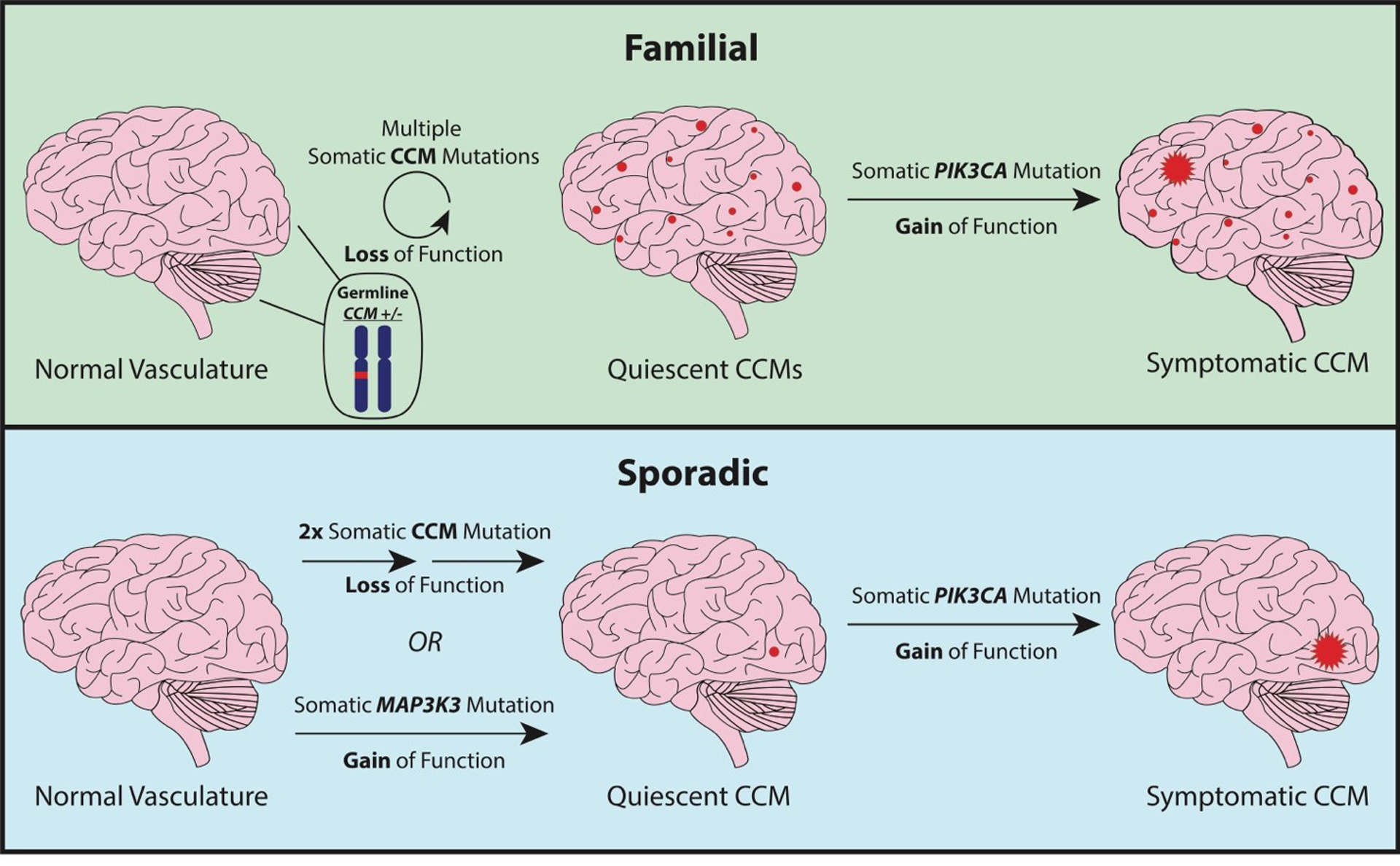
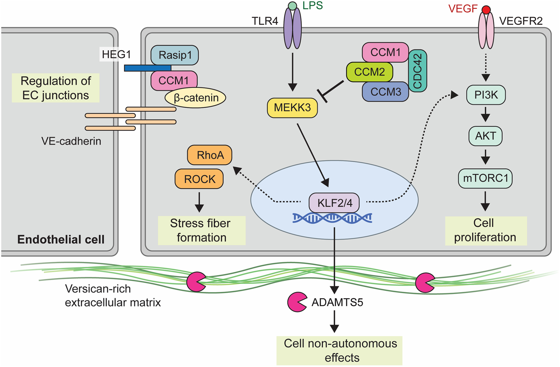
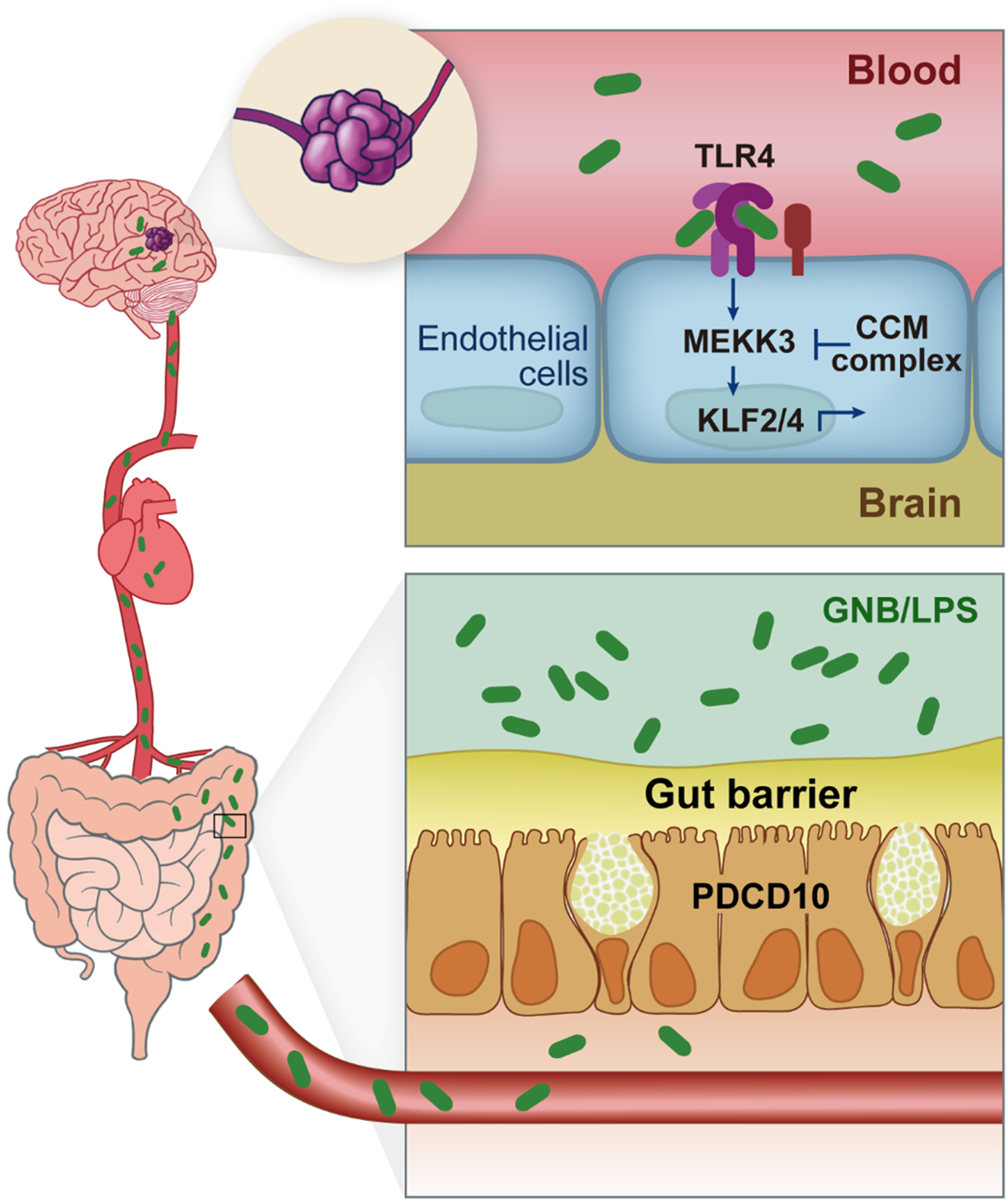
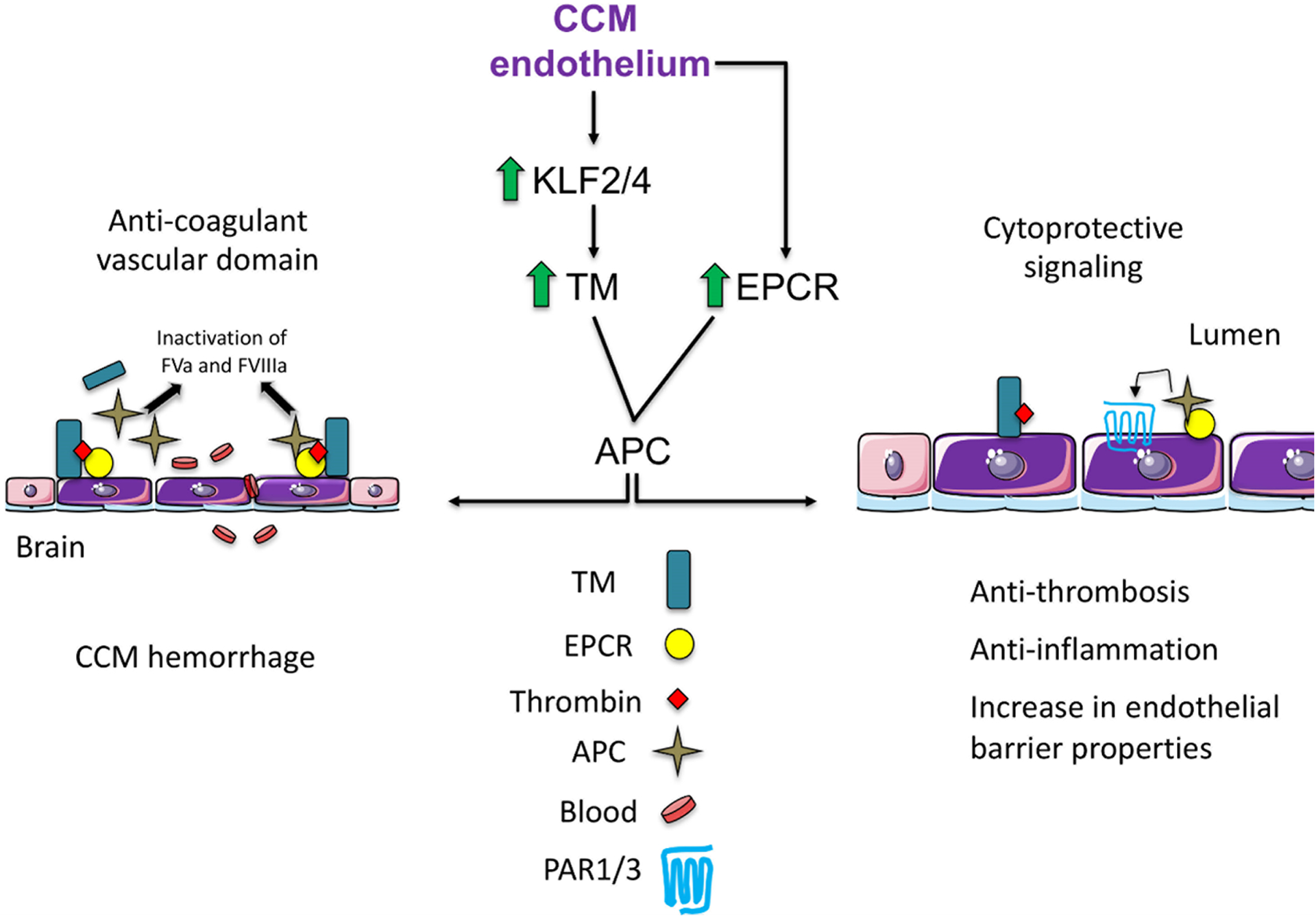
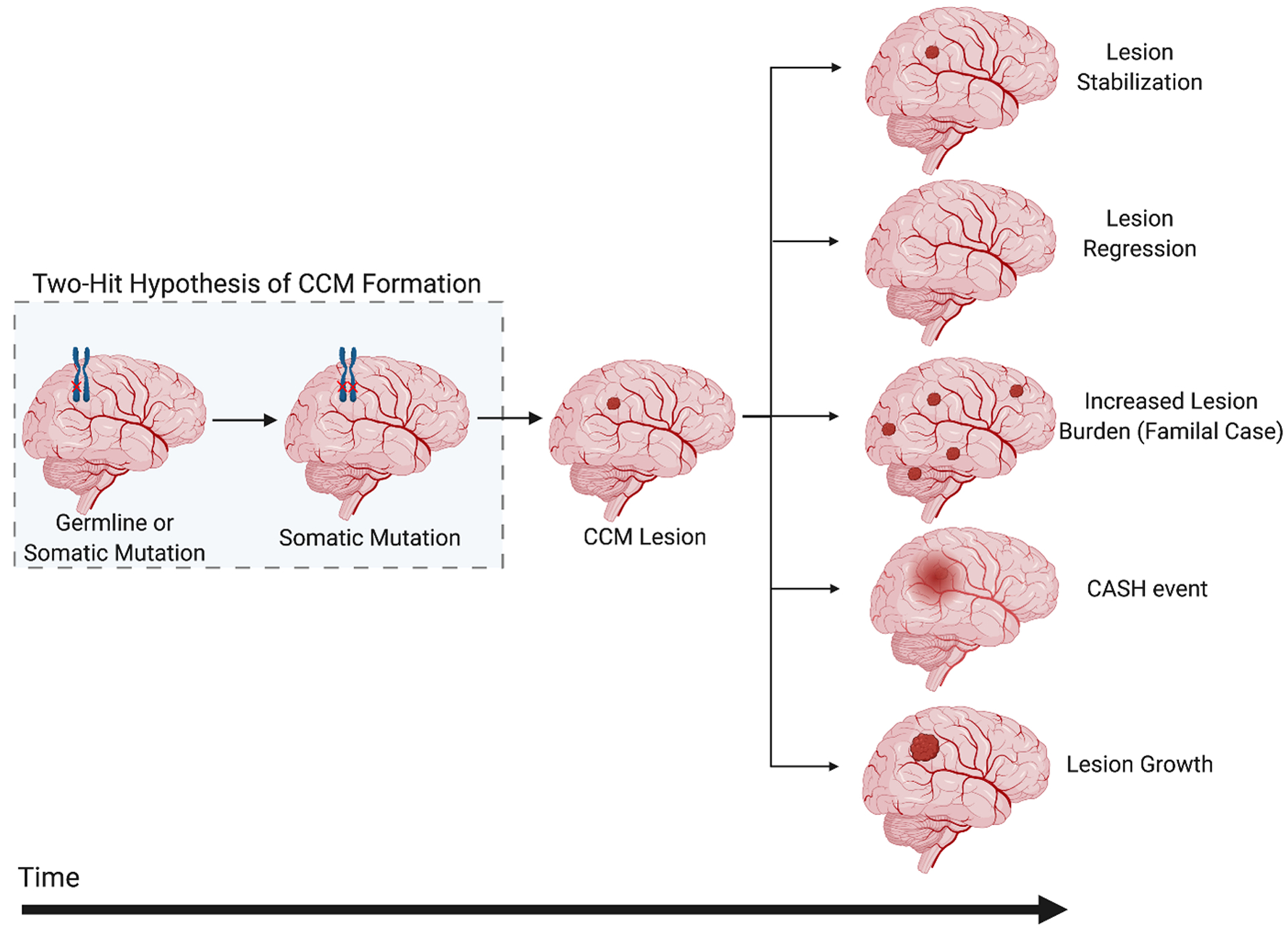
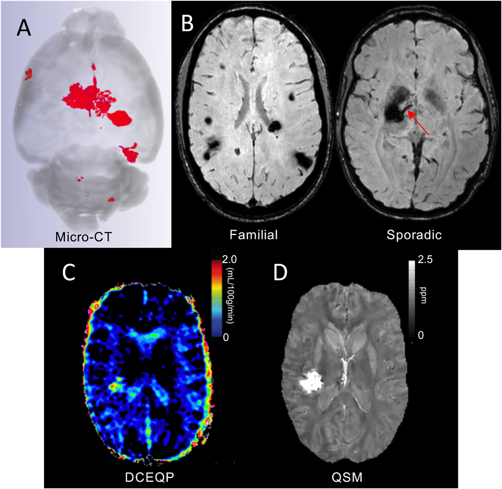
Cerebral cavernous malformations are acquired vascular anomalies that constitute a common cause of central nervous system hemorrhage and stroke. The past 2 decades have seen a remarkable increase in our understanding of the pathogenesis of this vascular disease. This new knowledge spans genetic causes of sporadic and familial forms of the disease, molecular signaling changes in vascular endothelial cells that underlie the disease, unexpectedly strong environmental effects on disease pathogenesis, and drivers of disease end points such as hemorrhage. These novel insights are the integrated product of human clinical studies, human genetic studies, studies in mouse and zebrafish genetic models, and basic molecular and cellular studies. This review addresses the genetic and molecular underpinnings of cerebral cavernous malformation disease, the mechanisms that lead to lesion hemorrhage, and emerging biomarkers and therapies for clinical treatment of cerebral cavernous malformation disease. It may also serve as an example for how focused basic and clinical investigation and emerging technologies can rapidly unravel a complex disease mechanism.

Keywords: central nervous system; endothelial cells; hemorrhage; stroke; vascular malformations.


Figures

Figure 1.
Figure 1.. Somatic mutations driving CCM progression.
In familial CCM (top), lesion formation is initiated by a somatic mutation in a CCM gene resulting in biallelic loss of function which may occur multiple times resulting in the formation of multiple quiescent CCMs. A subset of these CCMs acquire a somatic gain of function mutation in PIK3CA which fuels lesion growth. Sporadic CCM formation (bottom) requires either two somatic CCM mutations to occur in the same cell resulting in biallelic loss of function or a single gain of function somatic mutation in MAP3K3. A CCM at this stage may or may not acquire a somatic gain of function in PIK3CA which would fuel lesion growth. The order in which the mutations occur is not yet known. This figure shows the CCM/MAP3K3 mutations as occurring first, however the PIK3CA mutation may occur first in some or all cases.
Figure 2.
Figure 2.. Molecular signaling pathways involved in CCM lesion formation.
Shown is a partial summary of the various upstream inputs and downstream effectors implicated in CCM signaling. The heterotrimeric CCM complex, consisting of CCM1 (KRIT1), CCM2, and CCM3 (PDCD10), is an inhibitor of MAP3K3 Kinase, MEKK3. Inflammatory signals via TLR4 serve as the upstream input into MEKK3 signaling, as may shear forces associated with blood flow (not shown). Loss of CCM and subsequent activation of MEKK3 leads to upregulation of transcription factors KLF2 and KLF4. Downstream KLF2/4 transcriptional targets include both cell autonomous pathways through PI3K signaling and RHO/ROCK signaling, as well as cell non-autonomous effects via metalloprotease ADAMTS5 and extracellular matrix cleavage. VEGF is shown as a possible distinct input for PI3K-mTOR signaling while HEG1 has been associated with regulation of endothelial cell junctions. Of note, numerous other pathways have been found to be affected by loss of CCM function. This diagram focuses on those that are presently best understood and those that have been investigated using mouse and human genetic studies and/or are presently targeted therapeutically.
Figure 3.
Figure 3.. Gut-brain axis of CCM disease.
The gut microbiome and gut barrier are drivers of cavernoma formation. Gram-negative bacteria (GNB) and GNB-derived lipopolysaccharide (LPS) translocate across the mucosal and epithelial gut barrier into blood stream. LPS activates Toll-like Receptor 4 (TLR4) on brain endothelial cells and serves as an inflammatory upstream input into the MEKK3-KLF2/4 signaling pathway. PDCD10, unlike KRIT1 and CCM2, is required for the secretion of mucus by intestinal goblet cells. Germline loss of one PDCD10 allele reduces the gut barrier, thereby increasing translocation of LPS to the blood and accelerating the growth of CCM lesions in the brain. Adapted from Tang et al.,
Figure 4.
Figure 4.. Mechanism of increase in TM and EPCR in CCM.
CCM endothelium is associated with locally elevated expression of anticoagulant endothelial receptors TM and EPCR. TM upregulation is due to upregulation of KLF2 and KLF4 transcription factors. Increased levels of vascular TM and EPCR result in enhanced APC and the anticoagulation cascade by inactivation of FVa and FVIIIa, thus contributing to an increase in lesion bleeding (Anti-coagulant vascular domain). Proposed cytoprotective signaling in CCMs, retention of APC to endothelial receptor EPCR allows activation of PAR1/3 and subsequent cytoprotective signaling in CCM endothelium. TM-thrombin complex reduces fibrin generation, and TM exerts anti-inflammatory properties. TM=thrombomodulin, EPCR= endothelial protein C, APC=activated protein C. Adapted from Lopez-Ramirez et al. and Mosnier et al.
Figure 5.
Figure 5.. Progression of CCM Lesion Pathogenesis.
The pathogenesis of CCMs begins with an inherited or somatic mutation, followed by somatic mutations resulting in lesion genesis and growth. The natural history of CCM disease is thought to lead to five clinically relevant outcomes (1) lesion stabilization, (2) lesion regression, (3) increased lesion burden, (4) symptomatic hemorrhage (SH), and/or (5) lesional growth (shown from top to bottom). CASH, Cavernous Angioma with Symptomatic Hemorrhage.
Figure 6.
Figure 6.. Imaging Biomarkers of CCM.
(A). Micro-CT is used to assess lesion burden, volume, and stage ex-vivo in murine brains. (B). Susceptibility weighted MRI assesses lesion burden in familial cases, and the presence of an associated developmental venous anomaly (red arrow) in sporadic cases. (C). Dynamic contrast enhanced quantitative perfusion (DCEQP) MRI assesses the vascular permeability of CCM lesions and background brain. (D). Quantitative susceptibility mapping (QSM) MRI assesses iron content in CCM lesions.

References

    1. Bicknell JM, Carlow TJ, Kornfeld M, Stovring J and Turner P. Familial cavernous angiomas. Arch Neurol. 1978;35:746–9. - PubMed
    1. Clark JV. Familial occurrence of cavernous angiomata of the brain. J Neurol Neurosurg Psychiatry. 1970;33:871–6. - PMC - PubMed
    1. Kidd HA and Cumings JN. Cerebral angiomata in an Icelandic family. Lancet. 1947;1:747. - PubMed
    1. Laberge-le Couteulx S, Jung HH, Labauge P, Houtteville JP, Lescoat C, Cecillon M, Marechal E, Joutel A, Bach JF and Tournier-Lasserve E. Truncating mutations in CCM1, encoding KRIT1, cause hereditary cavernous angiomas. Nat Genet. 1999;23:189–93. - PubMed
    1. Sahoo T, Johnson EW, Thomas JW, Kuehl PM, Jones TL, Dokken CG, Touchman JW, Gallione CJ, Lee-Lin SQ, Kosofsky B, Kurth JH, Louis DN, Mettler G, Morrison L, Gil-Nagel A, Rich SS, Zabramski JM, Boguski MS, Green ED and Marchuk DA. Mutations in the gene encoding KRIT1, a Krev-1/rap1a binding protein, cause cerebral cavernous malformations (CCM1). Hum Mol Genet. 1999;8:2325–33. - PubMed

Publication types

MeSH terms