
Recurrent emergence of SARS-CoV-2 spike deletion H69/V70 and its role in the Alpha variant B.1.1.7

Bo Meng et al. Cell Rep. .

Abstract

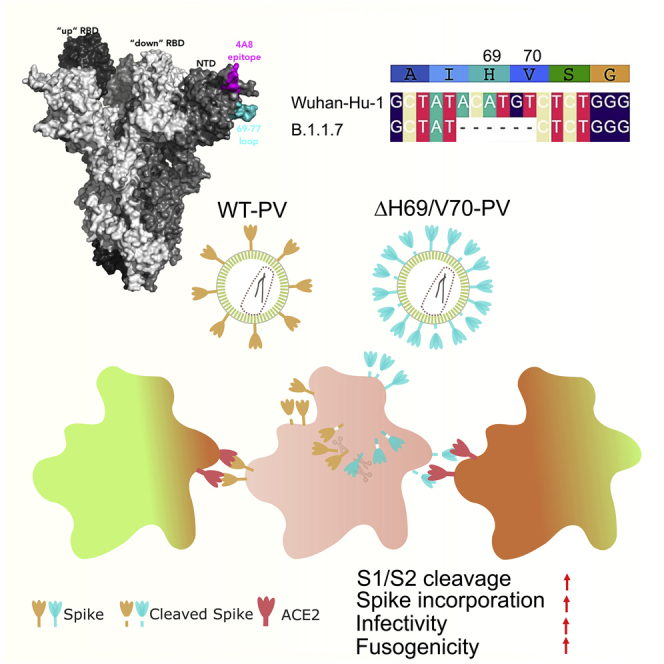
We report severe acute respiratory syndrome coronavirus 2 (SARS-CoV-2) spike ΔH69/V70 in multiple independent lineages, often occurring after acquisition of receptor binding motif replacements such as N439K and Y453F, known to increase binding affinity to the ACE2 receptor and confer antibody escape. In vitro, we show that, although ΔH69/V70 itself is not an antibody evasion mechanism, it increases infectivity associated with enhanced incorporation of cleaved spike into virions. ΔH69/V70 is able to partially rescue infectivity of spike proteins that have acquired N439K and Y453F escape mutations by increased spike incorporation. In addition, replacement of the H69 and V70 residues in the Alpha variant B.1.1.7 spike (where ΔH69/V70 occurs naturally) impairs spike incorporation and entry efficiency of the B.1.1.7 spike pseudotyped virus. Alpha variant B.1.1.7 spike mediates faster kinetics of cell-cell fusion than wild-type Wuhan-1 D614G, dependent on ΔH69/V70. Therefore, as ΔH69/V70 compensates for immune escape mutations that impair infectivity, continued surveillance for deletions with functional effects is warranted.

Keywords: Alpha variant; B.1.1.7; COVID-19; SARS-CoV-2; antibody escape; deletion; infectivity; neutralizing antibodies; resistance; spike mutation.


Conflict of interest statement

Declaration of interests A.D.M., C.S., K.C., E.C., L.P., and D.A.C. are employees of Vir Biotechnology and may hold shares in Vir Biotechnology. R.K.G. has received consulting fees from UMOVIS lab, Gilead Sciences, and ViiV Healthcare and a research grant from InvisiSmart Technologies.

Figures

None
Graphical abstract
Figure 1
Figure 1
Global snapshot of SARS-CoV-2 lineages associated with ΔH69/V70 and RBM mutations (A) Maximum-likelihood phylogeny of global SARS-CoV-2 whole-genome sequences, highlighting those with specific mutations in spike: ΔH69/V70, N439K, Y453F, and N501Y. The tree is subsampled, and tips are colored by geographic region (see key). Grey bars on the right show the presence or absence of the ΔH69/V70 and amino acid variants N439K, Y453F, and N501Y. Pangolin lineages are shown. (B and C) Cumulative occurrences of SARS-CoV-2 sequences with ΔH69/V70 by month for (B) ΔH69/V70 with or without N439K/Y453F and (C) ΔH69/V70 with N501Y. Indicated frequencies are by month, and all data were collected from the GISAID database (http://gisaid.org, accessed May 21, 2021) and sorted by reporting country and sampling date.
Figure 2
Figure 2
Spike ΔH69/V70 does not reduce sensitivity to neutralizing antibodies (A) Surface representation of the spike homotrimer in the open conformation (PDB: 7C2L), with each monomer shown in different shades of gray. On the monomer shown positioned to the right, the exposed loop consisting of residues 69–77 is shown in cyan and the neutralizing antibody (4A8)-binding NTD epitope in magenta. (B) Prediction of conformational change in the spike NTD because of deletion of residues His69 and Val70. The pre-deletion structure is shown in cyan, except for residues 69 and 70, which are shown in red. The predicted post-deletion structure is shown in green. Residues 66–77 of the pre-deletion structure are shown in stick representation and colored by atom (nitrogen in blue, oxygen in coral). Yellow lines connect aligned residues 66–77 of the pre- and post-deletion structures, and the distance of 6 Å between aligned alpha carbons of Thr73 in the pre- and post-deletion conformation is labeled. (C) Neutralization of spike ΔH69/V70 PV and WT (D614G background) by convalescent sera from 15 donors. GMT (geometric mean titer) with SD presented is representative of two independent experiments, each with two technical repeats. Wilcoxon matched-pairs signed-rank test; ns, not significant. (D) Ten example neutralization curves. Indicated is serum log10 inverse dilution against percent neutralization. Data points represent means of technical replicates, and error bars represent SD. Curves are representative of two independent experiments. (E–G) Kinetics of binding to WT and ΔH69/V70 NTD of 12 NTD-specific mAbs. (E) Biolayer interferometry analysis of binding to wild-type (WT; black) and WT ΔH69/V70 (red) NTDs by 12 NTD-targeting mAbs. Dotted lines separate the association phase from the dissociation phase. Shown is 1 of 2 independent experiments. (F) Side-by-side comparison of binding to WT (black) and ΔH69/V70 (red) NTDs by 11 NTD-targeting mAbs. Binding is shown as area under the curve (AUC). The S2L28 mAb is not shown because of too little response measured (<0.10 nm). (G) Binding to NTD of the 11 mAbs shown in (B), expressed as fold change of the AUC of the WT compared with ΔH69/V70. Data are representative of two independent experiments.
Figure 3
Figure 3
Spike ΔH69/V70 enhances entry and is accompanied by increased spike S2 incorporation into virions (A) Single-round infectivity on different cell targets by spike ΔH69/V70 versus the WT PV produced in HEK293T cells. Data are representative of at least three independent experiments. Data are shown with mean and SEM, and the statistics were performed using unpaired Student’s t test. (B) Infectivity of ΔH69/V70 PV on target HeLa cells transduced with ACE2, expressed as fold change relative to the WT. Mean and SEM are shown; one-sample t test, ∗∗p < 0.01 (C–E) Western blots and quantification of virions with infectivity shown in (B) and of cell lysates of HEK293T producer cells following transfection with plasmids expressing lentiviral vectors and SARS-CoV-2 S ΔH69/V70 versus the WT (all with D614G), probed with antibodies for HIV-1 p24 and SARS-Cov-2 S2 (C). (D) Quantification of spike:p24 ratio in supernatants for WT virus with ΔH69/V70 versus the WT alone across multiple replicate experiments. Mean and SEM are shown; one-sample t test, ∗∗∗p < 0.001. (E). Quantification of cleaved S2 spike: FL spike for WT virus with ΔH69/V70 versus the WT alone in virions and cell lysates. Each data point represents a single experiment. (F) Infectivity of ΔH69/V70 PV produced in H1299 lung epithelial cells on target HEK293T cells transiently expressing ACE2 and TMPRSS2. The statistical analysis was performed using unpaired Student’s t test. (G) Western blots of virions and cell lysates of H1299 lung epithelial producer cells following transfection with plasmids expressing lentiviral vectors and SARS-CoV-2 S ΔH69/V70 versus the WT (all with D614G). (H) Quantification of the S2:FL ratio in purified virions from H1299 lung epithelial producer cells. Data from at least two independent experiments are shown. RLU: relative light units; RT: reverse transcriptase.
Figure 4
Figure 4
The route of SARS-CoV-2 S-mediated virus entry in cell lines is not altered by ΔH69/V70 spike (A) Schematic illustrating spike in producer cells with CMK targeting and blocking furin cleavage (left panel). In target cells, camostat inhibits TMPRSS2 and, therefore, cell fusion at the plasma membrane, and E64D blocks cathepsins and targets endocytic viral entry (right panel). (B) Western blots show that CMK inhibits spike S1/S2 cleavage in producer cells transfected with the S ΔH69/V70 plasmid, and spikes with altered S1/S2 cleavage are incorporated into the virions. Antibodies against HIV-1 p24 and spike S2 were used with anti-GAPDH as a loading control. (C) The viruses produced from transfected HEK293T cells in the presence of CMK were used to transduce target cells. The luciferase reading is used as a surrogate for the spike infectivity bearing with various S2/FL ratios. The data shown are technical triplicates or quadruplicates, and statical analysis was done using an unpaired t test. (D) Comparison of the infectivity of spike with the PBCS deleted (ΔPBCS) with and without ΔH69/V70. The effect of ΔH69/V70 is independent of the PBCS. (E) ΔH69/V70 does not alter the virus entry route. S pseudotyped lentiviruses bearing WT S, ΔH69/V70 S, or VSV-G were used to transduce 293T-ACE2 or 293T-ACE2/TMPRSS2 cells in the presence of E64D or camostat at different drug concentrations. The cells were then harvested after 2 days and assayed for luciferase expression, which was then normalized against the non-drug control (set as 100%). The data shown are technical duplicates. The data are representative of at least two independent experiments.
Figure 5
Figure 5
ΔH69/V70 appears after spike N439K and Y453F and compensates for their reduced infectivity (A and B) Maximum-likelihood phylogeny of global sequences carrying Spike mutant (A) N439K and (B) Y453F. All sequences in the GISAID database containing S:439K or S:Y453F (February 18, 2021) were downloaded, realigned to Wuhan-Hu-1 using MAFFT, and deduplicated. (C) Representation of the Spike RBM:ACE2 interface (PDB: 6M0J) with residues N439, Y453, and N501, highlighted as spheres colored by element. (D–F) Spike mutant ΔH69/V70 compensates for the infectivity defect of spike RBD mutations and is associated with increased spike incorporation into virions. (D) Infectivity of spike (D614G) ΔH69/V70 in the absence and presence of spike RBD mutations. Shown is single-round infection by luciferase-expressing lentiviruses pseudotyped with SARS-CoV-2 spike protein on target HeLa cells stably transduced with ACE2. Mean and SEM are shown. (E) Representative western blot of purified virions and cell lysates probed with antibodies against HIV-1 p24, SARS-CoV-2 spike S2, and GAPDH. (F and G) Densitometric quantification of the (F) spike:p24 and (G) cleaved S2 spike:FL spike ratios for spike (D614G) ΔH69/V70 in the absence and presence of spike RBD mutations across multiple experiments in pelleted viruses. U, unit of reverse transcriptase (RT) activity. Data are representative of at least two independent experiments. Student’s t test, ∗∗∗p < 0.001.
Figure 6
Figure 6
Spike ΔH69/V70 in B.1.1.7 enhances spike infectivity (A) Surface representation of the spike homotrimer in open conformation with one upright RBD overlaid with ribbon representation (PDB: 6ZGG; Wrobel et al., 2020), with different monomers shown in black, pale blue, and gold. The deleted residues H69 and V70 and the residues involved in amino acid substitutions (501, 570, 716, 982, and 1118) and the deletion at position 144 are colored red on each monomer and labeled on the monomer with an upright RBD shown in black. Scissors mark the approximate location of an exposed loop (residues 677–688) containing the furin cleavage site and including residue 681, which is absent from the structure. (B) Representative infectivity of B.1.1.7 with replacement of H69 and V70 versus B.1.1.7 containing spike ΔH69/V70 and WT (D614G) spike; single-round infection by luciferase-expressing lentiviruses pseudotyped with SARS-CoV-2 spike protein on HeLa cells transduced with ACE2. The data represent technical triplicates. (C) Fold change of luciferase expression over the replacement of H69V70 in ACE2-transfected and ACE2- and TMPRSS2-transfected HEK293T cells, A459-ACE2/TMPRSS2 cells, and H1299 cells. The data shown are from three independent experiments, each in technical triplicates (one-sample t test). (D) Representative western blot analysis following transfection of HEK293T cells with spike and lentiviral plasmids. Virion loading was normalized for input virus using RT activity. Antibodies against HIV-1 p24 and spike S2 were used with anti-GAPDH as a loading control. (E and F) S2 to FL spike was analyzed by densitometry, and the S2:FL cleavage ratio was calculated for virions (E) and cell lysates (F). (G) Quantification of the spike:p24 ratio for B.1.1.7 and B.1.1.7 with H69/V70 replacement across three independent experiments. ∗∗p < 0.01, ∗∗∗∗p < 0.0001.
Figure 7
Figure 7
ΔH69/V70 significantly accelerates cell-cell fusion activity of Alpha variant B.1.1.7 spike protein (A) Schematic of the cell-cell fusion assay (created with BioRender). (B) Reconstructed images at 6 h of HEK293T cells co-transfected with the indicated spike mutants and mCherry-expressing plasmid mixed with green dye-labeled Vero acceptor cells. Scale bars represent 100 mm. Green identifies acceptor cells, and red marks donor cells. Merged green-red indicates syncytia. (C) Quantification of cell-cell fusion kinetics, showing percentage of green and red overlap area over time. Mean is plotted, with error bars representing SEM. (D) Quantification of cell-cell fusion of the indicated spike mutants 6 h after transfection. Mean is plotted, with error bars representing SEM. (E) Representative western blot of cells transfected with the indicated spike mutants (detected with anti-S2 antibody). The S2 subunit is indicated by an arrowhead. β-Actin is shown as a loading control. Data are representative of at least three independent experiments. p < 0.05, unpaired t test.

References

    1. Baum A., Fulton B.O., Wloga E., Copin R., Pascal K.E., Russo V., Giordano S., Lanza K., Negron N., Ni M. Antibody cocktail to SARS-CoV-2 spike protein prevents rapid mutational escape seen with individual antibodies. Science. 2020;369:1014–1018. - PMC - PubMed
    1. Bazykin G., Stanevich O., Danilenko D., Fadeev A., Komissarova K., Ivanova A., Sergeeva M., Safina K., Nabieva E., Klink G. 2021. Emergence of Y453F and δ69-70HV mutations in a lymphoma patient with long-term COVID-19.https://virological.org/t/emergence-of-y453f-and-69-70hv-mutations-in-a-...
    1. Bernhart S.H., Hofacker I.L., Will S., Gruber A.R., Stadler P.F. RNAalifold: improved consensus structure prediction for RNA alignments. BMC Bioinformatics. 2008;9:474. - PMC - PubMed
    1. Bhullar K., Waglechner N., Pawlowski A., Koteva K., Banks E.D., Johnston M.D., Barton H.A., Wright G.D. Antibiotic resistance is prevalent in an isolated cave microbiome. PLoS ONE. 2012;7:e34953. - PMC - PubMed
    1. Braga L., Ali H., Secco I., Chiavacci E., Neves G., Goldhill D., Penn R., Jimenez-Guardeno J.M., Ortega-Prieto A.M., Bussani R. Drugs that inhibit TMEM16 proteins block SARS-CoV-2 Spike-induced syncytia. Nature. 2021;594:88–93. - PMC - PubMed

Publication types

Substances