

Case Reports
doi: 10.1016/j.clinimag.2021.06.019.
Epub 2021 Jun 18.
Acute myocarditis after a second dose of the mRNA COVID-19 vaccine: a report of two cases
Affiliations
- PMID: 34166884
- PMCID: PMC8216670
- DOI: 10.1016/j.clinimag.2021.06.019
Item in Clipboard
Case Reports
Acute myocarditis after a second dose of the mRNA COVID-19 vaccine: a report of two cases
Clin Imaging.
2021 Oct.
Abstract
We report two cases of myocarditis, in two young and previously healthy individuals, temporally related to the second dose of the mRNA-COVID-19 vaccine. Both patients developed acute chest pain, changes on electrocardiogram (ECG), and elevated serum troponin within two days of receiving their second dose. Cardiac magnetic resonance (CMR) findings were consistent with acute myocarditis.
Keywords: Acute myocarditis; COVID-19 vaccination; Cardiac magnetic resonance; Myocarditis; mRNA-COVID-19 vaccine.
Copyright © 2021 Elsevier Inc. All rights reserved.
Figures

Magnetic Resonance Imaging of Case 1. Post contrast magnitude inversion recovery (MAG-IR) images in short axis (A) and four-chamber long axis (B) views show subepicardial enhancement in the anterolateral wall of the mid ventricle and apex (arrowheads). Native T1 map shows corresponding abnormality (arrowheads in C) with elevated values (D) in the anterolateral wall as compared to the interventricular septum. T2 mapping also showed abnormality in this region (arrows in E) with elevated values (F) when compared to the interventricular septum.
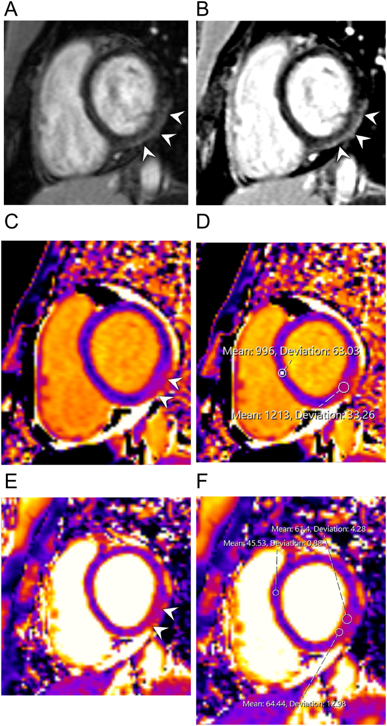
Magnetic Resonance Imaging of Case 2. Post-contrast magnitude inversion recovery (MAG-IR) (A) and phase sensitive inversion recovery (PSIR) (B) images in short axis views show subepicardial enhancement in the inferolateral wall at the base (arrowheads). Native T1 map shows corresponding abnormality (arrowheads in C) with elevated values (D) in the inferolateral wall as compared to the interventricular septum. T2 mapping also showed abnormality in this region (arrows in E) with elevated values (F) when compared to the interventricular septum.
References
-
- Yamamoto H., Hashimoto T., Ohta-Ogo K., et al. A case of biopsy-proven eosinophilic myocarditis related to tetanus toxoid immunization. Cardiovasc Pathol. 2018;37:54–57. - PubMed
Publication types
MeSH terms
Substances
LinkOut - more resources
Full Text Sources
Medical
