

Mechanisms of Photosensitivity in Autoimmunity
- PMID: 34167786
- PMCID: PMC8688579
- DOI: 10.1016/j.jid.2021.05.007
Mechanisms of Photosensitivity in Autoimmunity
Abstract
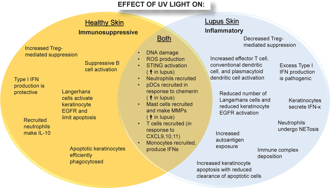
Aberrant responses to UV light frequently lead to the formation of skin lesions and the activation of systemic inflammation in some autoimmune diseases, especially systemic lupus erythematosus. Whereas the effects of UV light on the skin have been studied for decades, only recently have some of the mechanisms that contribute to abnormal responses to UV light in patients with autoimmune diseases been uncovered. This review will discuss the biology of UV in the epidermis and discuss the abnormal epidermal and inflammatory mechanisms that contribute to photosensitivity. Further research is required to fully understand how to normalize UV-mediated inflammation in patients with autoimmune diseases.
Copyright © 2021 The Authors. Published by Elsevier Inc. All rights reserved.
Conflict of interest statement
Figures
References
-
- Apel F, Andreeva L, Knackstedt LS, Streeck R, Frese CK, Goosmann C, et al. The cytosolic DNA sensor cGAS recognizes neutrophil extracellular traps. Sci Signal 2021;14(673). - PubMed
-
- Avalos-Díaz E, Alvarado-Flores E, Herrera-Esparza R. UV-A irradiation induces transcription of IL-6 and TNF alpha genes in human keratinocytes and dermal fibroblasts. Revue du rhumatisme (English ed) 1999;66(1):13–9. - PubMed
Publication types
MeSH terms
Grants and funding
LinkOut - more resources
Full Text Sources
Medical
