. 2021 Jul 1;35(13-14):1005-1019.
doi: 10.1101/gad.348320.121. Epub 2021 Jun 24.

Targeting the m6A RNA modification pathway blocks SARS-CoV-2 and HCoV-OC43 replication

Affiliations

Targeting the m6A RNA modification pathway blocks SARS-CoV-2 and HCoV-OC43 replication

Hannah M Burgess et al. Genes Dev. .

Abstract

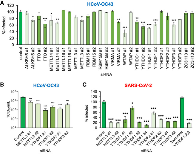
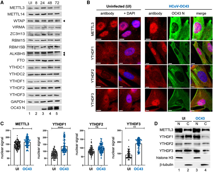
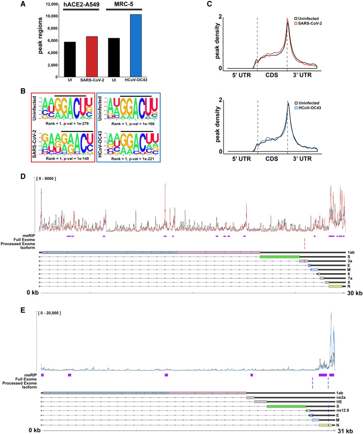
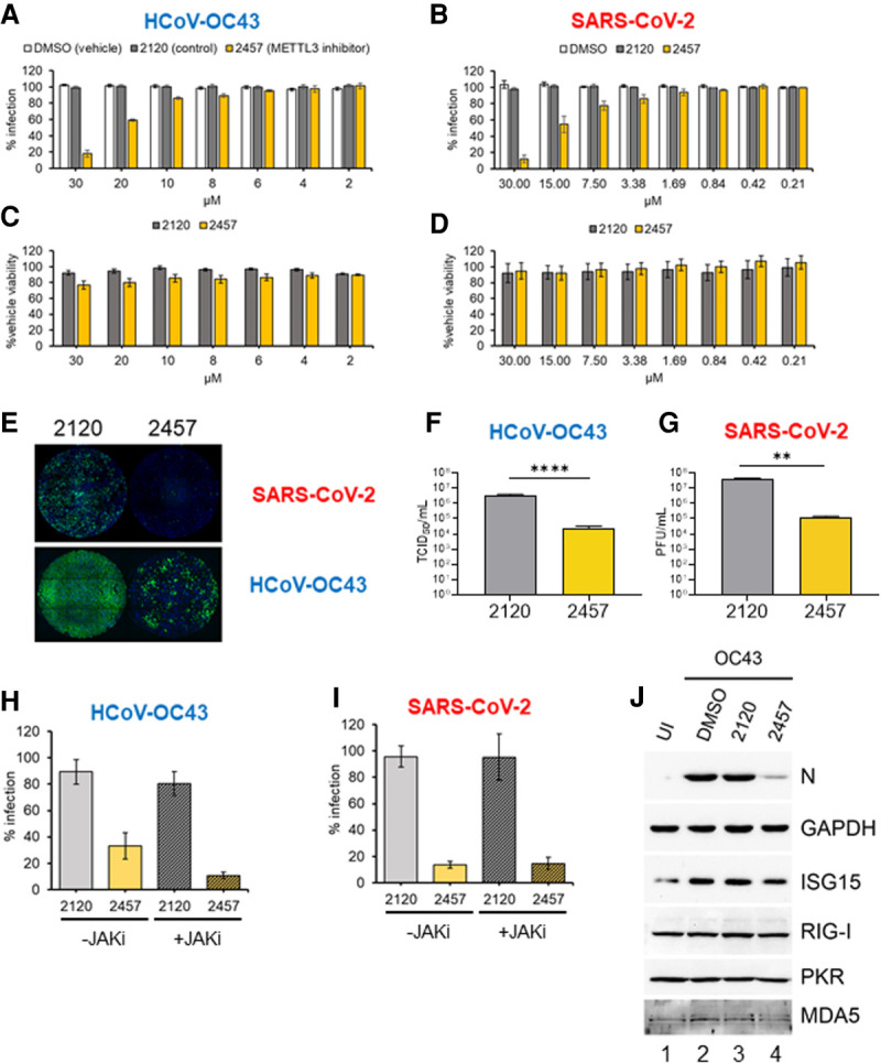
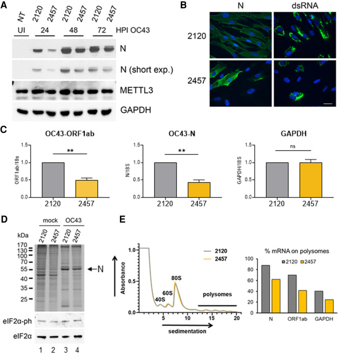
N6-methyladenosine (m6A) is an abundant internal RNA modification, influencing transcript fate and function in uninfected and virus-infected cells. Installation of m6A by the nuclear RNA methyltransferase METTL3 occurs cotranscriptionally; however, the genomes of some cytoplasmic RNA viruses are also m6A-modified. How the cellular m6A modification machinery impacts coronavirus replication, which occurs exclusively in the cytoplasm, is unknown. Here we show that replication of SARS-CoV-2, the agent responsible for the COVID-19 pandemic, and a seasonal human β-coronavirus HCoV-OC43, can be suppressed by depletion of METTL3 or cytoplasmic m6A reader proteins YTHDF1 and YTHDF3 and by a highly specific small molecule METTL3 inhibitor. Reduction of infectious titer correlates with decreased synthesis of viral RNAs and the essential nucleocapsid (N) protein. Sites of m6A modification on genomic and subgenomic RNAs of both viruses were mapped by methylated RNA immunoprecipitation sequencing (meRIP-seq). Levels of host factors involved in m6A installation, removal, and recognition were unchanged by HCoV-OC43 infection; however, nuclear localization of METTL3 and cytoplasmic m6A readers YTHDF1 and YTHDF2 increased. This establishes that coronavirus RNAs are m6A-modified and host m6A pathway components control β-coronavirus replication. Moreover, it illustrates the therapeutic potential of targeting the m6A pathway to restrict coronavirus reproduction.

Keywords: HCoV-OC43; N6-methyladenosine; RNA modification; SARS-CoV-2; coronavirus; direct RNA sequencing; nanopore sequencing; virus–host interactions.


Figures

Figure 1.
Figure 1.
Focused RNA interference screen implicates METTL3 and the YTHDF readers in control of β-coronavirus replication. (A) Human MRC-5 normal lung fibroblasts were transfected with a set of validated siRNAs (two per factor) targeting 14 host components of the m6A pathway and cultured for 72 h before infection with HCoV-OC43 at MOI = 0.001. After 48 h, cells were fixed and the infection assessed by indirect immunofluorescence using an antibody to the viral nucleocapsid protein (N). The percentage of N-positive cells per well was determined using a CellInsight CX7 LZR high-content screening platform. Each assay was performed three times with technical duplicates and normalized to control siRNA treated cells. (B) The impact of siRNA depletion on infectious viral titer was determined by collecting supernatant culture media from A and establishing TCID50 on MRC-5 cells. (C) A549+ACE2 cells were transfected with validated siRNAs targeting METTL3, YTHDF1, YTHDF2, and YTHDF3, either individually or as a mix of single siRNAs to all three YTHDF proteins using siRNA#1 in each case. After 72 h, the cells were infected with icSARS-CoV-2-mNG at MOI = 0.1 for 48 h and then fixed and scored for green fluorescence. The extent of spread was normalized to cells transfected with control siRNA. In each case, an ANOVA test with Dunnett multiple comparison correction was used to establish statistical significance compared with control siRNA. (*) P < 0.033, (**) P < 0.002, (***) P < 0.001.
Figure 2.
Figure 2.
HCoV-OC43 infection results in the nuclear accumulation of METTL3, YTHDF1, and YTHDF2. (A) Lysates from uninfected (UI) or HCoV-OC43-infected MRC-5 cells were prepared at 8, 24, 48, and 72 h postinfection at MOI = 3 and probed by immunoblotting using antibodies to methyltransferase subunits (METTL3, METTL14, WTAP, RBM15, and RBM15B), demethylases (ALKBH5, FTO) and m6A binding proteins (YTHDC1, YTHDF1, YTHDF2, and YTHDF3). Arrowheads denote target bands. (B) Indirect immunofluorescence images of representative uninfected or HCoV-OC43-infected MRC5 cells (24 hpi, MOI = 3) with primary antibodies to METTL3, YTHDF1, YTHDF2, and YTHDF3. Scale bar, 10 µm. (C) Quantitation of nuclear signal for each primary antibody used in panel B after normalization to background fluorescence. Analysis was performed on ≥100 cells/condition and statistical significance determined using an F-test. (****) P ≤ 0.0001, (**) P ≤ 0.0047, (ns) not significant. (D) Uninfected and HCoV-OC43-infected MRC-5 cells (as in B) were rapidly lysed to produce insoluble particulate (nuclear [N]) and soluble (cytosolic [C]) fractions and probed by immunoblotting using antibodies to METTL3, YTHDF1, YTHDF2, and YTHDF3. Lysis and subcellular fractionation efficiency were assessed using antibodies to cytoplasmic β-tubulin and nuclear histone H3.
Figure 3.
Figure 3.
SARS-CoV-2 and HCoV-OC43 RNAs are m6A-modified. (A) meRIP-seq data sets profiling of SARS-CoV-2-infected A549+ACE2 cells and HCOV-OC43-infected MRC-5 cells were analyzed to determine numbers of significant (Padj < 0.05) peak ranges, enriched over input, in each comparison. (B) Sequence motif analysis of peak regions present in the cellular meRIP-seq data sets show enrichment for the classical RRACH/DRACH motif associated with installation of m6A. (C) Metagene analysis of m6A-peak region distribution across cellular RNAs with annotated 5′ and 3′ untranslated regions (UTR) and coding sequences (CDS). (D) Integration of putative m6A peak ranges (purple) identified by meRIP-seq of SARS-CoV-2-infected A549+ACE2 cells with candidate m6A sites (red) identified through comparative profiling of nanopore direct RNA sequencing (DRS) data sets (SARS-CoV-2-infected A549+ACE2 cells with or without STM2457). The top track shows the normalized coverage for a representative biological replicate of the meRIP-seq paired INPUT (black) and IP (red) data sets (see Supplemental Fig. S4 for additional replicates). To maximize sensitivity and accuracy, comparative DRS analyses were performed using DRUMMER at three different levels: full exome (i.e., all reads), processed exome (i.e., only reads containing the leader sequence), and isoform level (i.e., data sets aligned to the transcriptome rather than genome). The canonical SARS-CoV-2 transcriptome structure is shown below. (E) As in D but for HCOV-OC43-infected MRC-5 cells with or without STM2457.
Figure 4.
Figure 4.
Inhibition of METTL3 activity suppresses β-coronavirus replication. (A) MRC-5 cells were infected with HCoV-OC43 at MOI = 0.001 for 48 h in the presence of METTL3 inhibitor (STM2457, yellow), inactive control compound (STM2120, gray), or vehicle (DMSO, white) at the indicated concentrations. Identification of infected cells by indirect immunofluorescence for nucleocapsid protein was as described in Figure 1A. (B) A549+ACE2 cells were infected with icSARS-CoV-2-mNG at MOI = 0.1 for 48 h, and the percentage of cells infected was established by green fluorescence and normalized to infection of nontreated cells. (C, D) The viability of MRC-5 cells (C) and A549+ACE2 cells (D) in the presence of concentrations of STM2120 or STM2457 used in the infection assays shown in A and B was assessed using a commercial ATP quantitation assay. Cells were maintained at either 33°C or 37°C, respectively, in culture medium containing diluted compound for 48 h prior to lysis. Each experiment was conducted three times with internal duplicates, normalized to DMSO-treated cells processed in parallel and plotted as the mean ± SEM. (E) Representative montages showing wells from the infections quantified in A and B that were treated with 30 µM STM2120 or STM2457 and infected with either icSARS-CoV-2-mNG or HCoV-OC43 as indicated. The signal for the OC43-N antibody and Alexa Fluor 647 secondary antibody is represented in green. (F) Infectious viral titers from MRC-5 cells infected with HCoV-OC43 at MOI = 0.001 treated with 30 µM either STM2120 or STM2457 was determined by TCID50 assay. (G) Infectious virus titers from A549+ACE2 cells infected with icSARS-CoV-2-mNG at MOI = 0.1 and treated with 30 µM either STM2120 or STM2457 was determined by plaque assay. (H,I) MRC-5 and A549+ACE2 cells were infected with OC43 or icSARS-CoV-2-mNG, respectively, as in (A) and (B) in the presence of 30 µM STM2120 or STM2457 and 10 µM JAK inhibitor (pyridone-6) or vehicle control (DMSO) and the percent infected cells quantified. Each experiment was conducted three times with internal duplicates, normalized to DMSO-treated cells processed in parallel, and plotted as the mean ± SEM. (J) Immunoblot analysis of lysates from MRC-5 cells infected with HCoV-OC43 at MOI = 0.001 in the presence of 30 µM STM2120 or STM2457 as in A and collected at 48 hpi and probed for viral N protein, or host ISGs (ISG15, RIG-I, PKR, and MDA5) and GAPDH.
Figure 5.
Figure 5.
Initial HCoV-OC43 RNA synthesis and protein expression is reduced by inhibition of METTL3 catalysis. (A) Immunoblot analysis of lysates from MRC-5 cells infected with HCoV-OC43 at MOI = 3 in the presence of 30 µM STM2120 or STM2457 collected at 24, 48, or 72 hpi and probed for viral N protein, or host METTL3 and GAPDH. (B) Representative images showing detection of viral N protein or viral dsRNA by indirect immunofluorescence assay (green) in HCoV-OC43-infected MRC-5 cells at 24 hpi in the presence of 30 µM STM2120 or STM2457. Cell nuclei were stained with DAPI. Scale bar, 20 µm. (C). The relative abundance of HCoV-OC43 gRNA (ORF1ab) or sgRNA (N) or host GAPDH at 24 hpi MOI = 3 in the presence of 30 µM STM2120 or STM2457 was determined by RT-qPCR using transcript-specific primers and normalizing to 18S rRNA and plotted as the mean ± SEM (n = 3). (D) MRC-5 cells infected with HCoV-OC43 at MOI = 3 in the presence of 30 µM STM2120 or STM2457 were metabolically pulse-labeled with 35S amino acids for 1 h. Lysates were separated by SDS-PAGE and the fixed, dried gel exposed to film. Migration of molecular weight standards is shown at the left. The arrow at the right indicates migration of virus-encoded N protein. Additionally, the relative levels of total eIF2α and phospho-eIF2α in the same lysates was assessed by immunoblotting. Note that an ultrasensitive enhanced chemiluminescent substrate (SuperSignal West Femto) was required to visualize the very low levels of phospho-eIF2α. (E, left) Cytoplasmic lysates from HCoV-OC43-infected MRC-5 cells treated with STM2120 (gray) or STM2457 (yellow) for 24 h prior to harvest were fractionated over a 10%–50% sucrose gradient, and the absorbance at 254 nm is shown with ribosomal peaks indicated. (Right) The relative abundance of viral N and ORF1a/b RNAs and host GAPDH mRNA in each gradient fraction was determined by RT-qPCR, and the sum of polysomal mRNA (fractions 13–20) under each drug treatment is presented. A representative experiment of two biological replicates in which similar results were obtained is shown.

Similar articles

Cited by

References

    1. Alarcón CR, Lee H, Goodarzi H, Halberg N, Tavazoie SF. 2015. N6-methyladenosine marks primary microRNAs for processing. Nature 519: 482–485. 10.1038/nature14281 - DOI - PMC - PubMed
    1. Barbieri I, Tzelepis K, Pandolfini L, Shi J, Millán-Zambrano G, Robson SC, Aspris D, Migliori V, Bannister AJ, Han N, et al. 2017. Promoter-bound METTL3 maintains myeloid leukaemia by m6A-dependent translation control. Nature 552: 126–131. 10.1038/nature24678 - DOI - PMC - PubMed
    1. Bokar JA, Shambaugh ME, Polayes D, Matera AG, Rottman FM. 1997. Purification and cDNA cloning of the AdoMet-binding subunit of the human mRNA (N6-adenosine)-methyltransferase. RNA 3: 1233–1247. - PMC - PubMed
    1. Bray NL, Pimentel H, Melsted P, Pachter L. 2016. Near-optimal probabilistic RNA-seq quantification. Nat Biotechnol 34: 525–527. 10.1038/nbt.3519 - DOI - PubMed
    1. Burgess HM, Mohr I. 2015. Cellular 5′′-3′′ mRNA exonuclease Xrn1 controls double-stranded RNA accumulation and anti-viral responses. Cell Host Microbe 17: 332–344. 10.1016/j.chom.2015.02.003 - DOI - PMC - PubMed

Publication types

MeSH terms

LinkOut - more resources