. 2021 Aug 6;373(6555):642-648.
doi: 10.1126/science.abi9745. Epub 2021 Jun 24.

Structural basis for enhanced infectivity and immune evasion of SARS-CoV-2 variants

Affiliations

Structural basis for enhanced infectivity and immune evasion of SARS-CoV-2 variants

Yongfei Cai et al. Science. .

Abstract

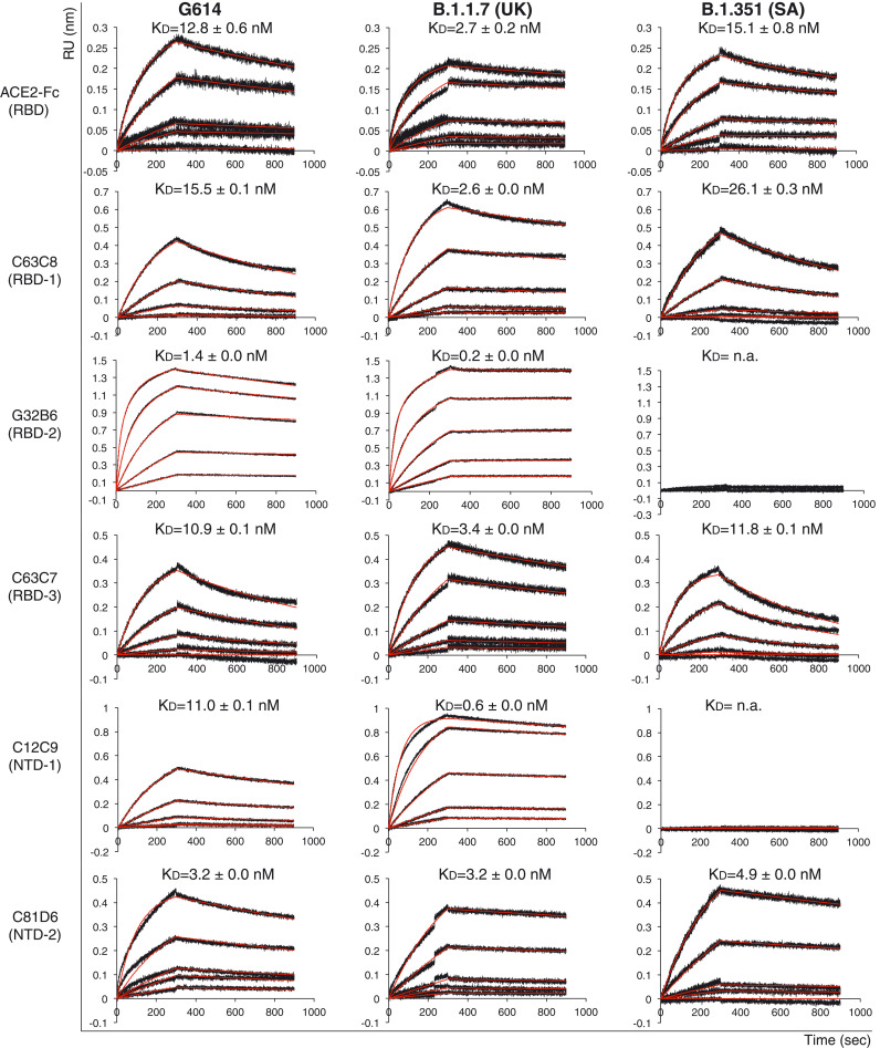
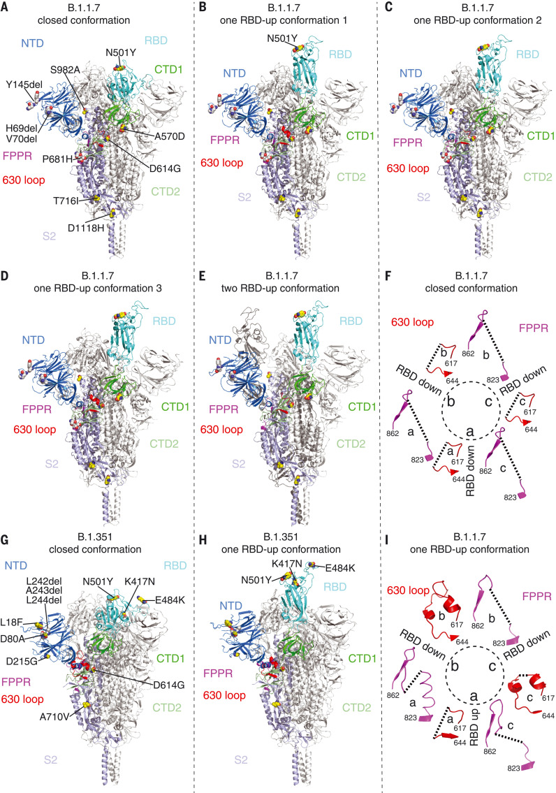
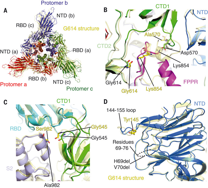
Several fast-spreading variants of severe acute respiratory syndrome coronavirus 2 (SARS-CoV-2) have become the dominant circulating strains in the COVID-19 pandemic. We report here cryo-electron microscopy structures of the full-length spike (S) trimers of the B.1.1.7 and B.1.351 variants, as well as their biochemical and antigenic properties. Amino acid substitutions in the B.1.1.7 protein increase both the accessibility of its receptor binding domain and the binding affinity for receptor angiotensin-converting enzyme 2 (ACE2). The enhanced receptor engagement may account for the increased transmissibility. The B.1.351 variant has evolved to reshape antigenic surfaces of the major neutralizing sites on the S protein, making it resistant to some potent neutralizing antibodies. These findings provide structural details on how SARS-CoV-2 has evolved to enhance viral fitness and immune evasion.


Figures

Fig. 1.
Fig. 1.
Antigenic properties of the purified full-length SARS-CoV-2 S proteins. BLI analysis of the association of prefusion S trimers from the G614 “parent” strain and the B.1.1.7 and B.1.351 variants derived from it with soluble ACE2 constructs and with a panel of antibodies representing five epitopic regions on the RBD and NTD [see fig. S4A and (35)]. For ACE2 binding, purified S proteins were immobilized to AR2G (Amine Reactive 2nd Generation) biosensors and dipped into the wells containing ACE2 at different concentrations. For antibody binding, various antibodies were immobilized to AHC (Anti-human IgG Fc Capture) biosensors and dipped into the wells containing each purified S protein at different concentrations. Binding kinetics were evaluated using a 1:1 Langmuir model except for dimeric ACE2 and antibody G32B6 targeting the RBD-2, which were analyzed by a bivalent binding model. The sensorgrams are in black and the fits in red. Binding constants are also summarized here and in table S1. All experiments were repeated at least twice with essentially identical results. KD, dissociation constant; n.a., not available; RU, response unit.
Fig. 2.
Fig. 2.. Cryo-EM structures of the full-length SARS-CoV-2 S proteins from the B.1.1.7 and B.1.351 variants.
(A to E) The structures of the closed prefusion conformation (A), three one-RBD-up conformations [(B) to (D)], and a two-RBD-up conformation (E) of the B.1.1.7 S trimer are shown in ribbon diagram, with one protomer colored as NTD in blue, RBD in cyan, CTD1 in green, CTD2 in light green, S2 in light blue, the 630 loop in red, and the FPPR in magenta. (G and H) The structures of the closed prefusion conformation (G) and one-RBD-up conformation (H) of the B.1.351 S trimer are shown in ribbon diagram with the same color scheme as in (A) to (E). All mutations in the new variants, as compared with the original virus (D614), are highlighted as sphere models. (F and I) Structures, in the B.1.1.7 trimer, of segments (residues 617 to 644) containing the 630 loop (red) and segments (residues 823 to 862) containing the FPPR (magenta) from each of the three protomers (a, b, and c). The position of each RBD is indicated. Dashed lines indicate gaps in the chain trace (disordered loops).
Fig. 3.
Fig. 3.
Structural impact of the mutations in the B.1.1.7 S. (A) Top views of superposition of the structure of the B.1.1.7 S trimer in ribbon representation, with the structure of the prefusion trimer of the G614 S [Protein Data Bank (PDB) ID: 7KRQ] shown in yellow. The NTD and RBD of each protomer are indicated. (B) A close-up view of the region near the A570D mutation with superposition of the B.1.1.7 trimer structure (one-RBD-up) in green (CTD1) and magenta (FPPR) and the G614 trimer (closed) in yellow. Residues Ala570, Asp570, two Gly614, and two Lys854 from both structures are shown in stick model. (C) A view of the region near the S982A mutation with superposition of the B.1.1.7 trimer structure (closed) in green (CTD1) and magenta (FPPR) and the G614 trimer (closed) in yellow. (D) Superposition of the NTD structure of the B.1.1.7 S trimer in blue with the NTD of the G614 S trimer in yellow. Locations of Tyr145 and the disordered loop containing residues 69 to 76 are indicated.
Fig. 4.
Fig. 4.
Structural impact of the mutations in the B.1.351 S. (A) Top views of superposition of the structure of the B.1.351 S trimer in ribbon representation, with the structure of the prefusion trimer of the G614 S (PDB ID: 7KRQ) shown in yellow. The NTD and RBD of each protomer are indicated. (B) Superposition of the RBD structure of the B.1.351 S trimer in blue with the RBD of the G614 S trimer in yellow. Locations of mutations K417N, E484K, and N501Y are indicated, and these residues are shown in stick model. (C) A view of the NTDs from superposition of the structure of the B.1.351 S trimer in blue and the G614 S in yellow. Locations of mutations L18F, D80A, and D215G; the disulfide bond between Cys15 and Cys136; and replacement of Leu242-Ala243-Leu244 by His245-Arg246-Ser247 are indicated, and the residues are shown in stick model. (D) Superposition of the NTD structure of the B.1.351 S trimer in blue with the NTD of the G614 S trimer in yellow. Displacement of the segment 169 to 188 and the location of Arg246 in the B.1.351 structure are indicated.

Update of

References

    1. Zhou P., Yang X.-L., Wang X.-G., Hu B., Zhang L., Zhang W., Si H.-R., Zhu Y., Li B., Huang C.-L., Chen H.-D., Chen J., Luo Y., Guo H., Jiang R.-D., Liu M.-Q., Chen Y., Shen X.-R., Wang X., Zheng X.-S., Zhao K., Chen Q.-J., Deng F., Liu L.-L., Yan B., Zhan F.-X., Wang Y.-Y., Xiao G.-F., Shi Z.-L., A pneumonia outbreak associated with a new coronavirus of probable bat origin. Nature 579, 270–273 (2020). 10.1038/s41586-020-2012-7 - DOI - PMC - PubMed
    1. Robson F., Khan K. S., Le T. K., Paris C., Demirbag S., Barfuss P., Rocchi P., Ng W.-L., Coronavirus RNA proofreading: Molecular basis and therapeutic targeting. Mol. Cell 79, 710–727 (2020). 10.1016/j.molcel.2020.07.027 - DOI - PMC - PubMed
    1. H. Tegally, E. Wilkinson, M. Giovanetti, A. Iranzadeh, V. Fonseca, J. Giandhari, D. Doolabh, S. Pillay, E. J. San, N. Msomi, K. Mlisana, A. von Gottberg, S. Walaza, M. Allam, A. Ismail, T. Mohale, A. J. Glass, S. Engelbrecht, G. Van Zyl, W. Preiser, F. Petruccione, A. Sigal, D. Hardie, G. Marais, M. Hsiao, S. Korsman, M.-A. Davies, L. Tyers, I. Mudau, D. York, C. Maslo, D. Goedhals, S. Abrahams, O. Laguda-Akingba, A. Alisoltani-Dehkordi, A. Godzik, C. K. Wibmer, B. T. Sewell, J. Lourenço, L. C. J. Alcantara, S. L. Kosakovsky Pond, S. Weaver, D. Martin, R. J. Lessells, J. N. Bhiman, C. Williamson, T. de Oliveira, Emergence and rapid spread of a new severe acute respiratory syndrome-related coronavirus 2 (SARS-CoV-2) lineage with multiple spike mutations in South Africa. medRxiv 2020.12.21.20248640 [Preprint] (2020). 10.1101/2020.12.21.20248640. - DOI
    1. Grabowski F., Preibisch G., Kochańczyk M., Lipniacki T., SARS-CoV-2 variant of concern 202012/01 has about twofold replicative advantage and acquires concerning mutations. Viruses 13, 392 (2021). 10.3390/v13030392 - DOI - PMC - PubMed
    1. Voloch C. M., da Silva Francisco R. Jr., de Almeida L. G. P., Cardoso C. C., Brustolini O. J., Gerber A. L., Guimarães A. P. C., Mariani D., da Costa R. M., Ferreira O. C. Jr., Cavalcanti A. C., Frauches T. S., de Mello C. M. B., Leitão I. C., Galliez R. M., Faffe D. S., Castiñeiras T. M. P. P., Tanuri A., de Vasconcelos A. T. R.; Covid19-UFRJ Workgroup; LNCC Workgroup , Genomic characterization of a novel SARS-CoV-2 lineage from Rio de Janeiro, Brazil. J. Virol. 95, e00119–e00121 (2021). 10.1128/JVI.00119-21 - DOI - PMC - PubMed

Publication types

MeSH terms