. 2021 Aug 6;373(6555):eabi6226.
doi: 10.1126/science.abi6226. Epub 2021 Jun 24.

Effect of natural mutations of SARS-CoV-2 on spike structure, conformation, and antigenicity

Affiliations

Effect of natural mutations of SARS-CoV-2 on spike structure, conformation, and antigenicity

Sophie M-C Gobeil et al. Science. .

Abstract

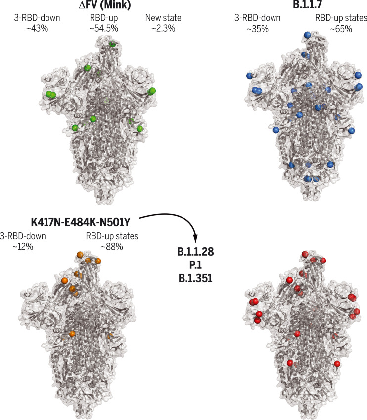
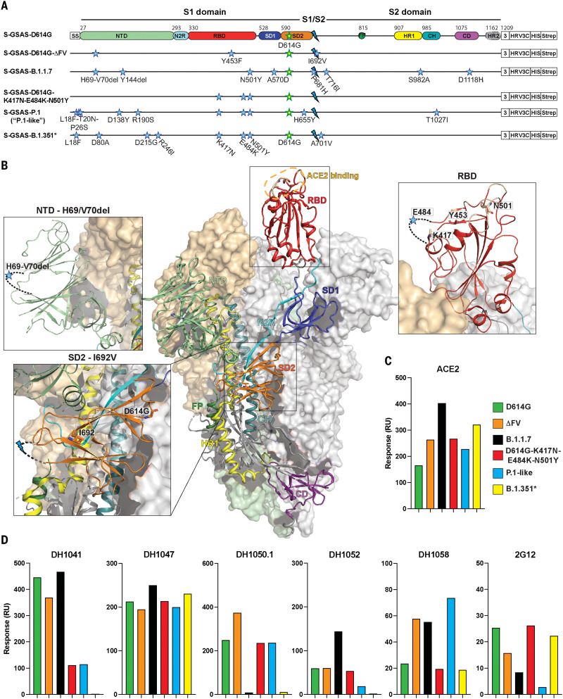
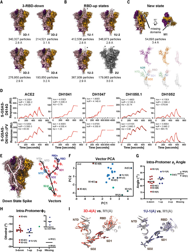
Severe acute respiratory syndrome coronavirus 2 (SARS-CoV-2) variants with multiple spike mutations enable increased transmission and antibody resistance. We combined cryo-electron microscopy (cryo-EM), binding, and computational analyses to study variant spikes, including one that was involved in transmission between minks and humans, and others that originated and spread in human populations. All variants showed increased angiotensin-converting enzyme 2 (ACE2) receptor binding and increased propensity for receptor binding domain (RBD)-up states. While adaptation to mink resulted in spike destabilization, the B.1.1.7 (UK) spike balanced stabilizing and destabilizing mutations. A local destabilizing effect of the RBD E484K mutation was implicated in resistance of the B.1.1.28/P.1 (Brazil) and B.1.351 (South Africa) variants to neutralizing antibodies. Our studies revealed allosteric effects of mutations and mechanistic differences that drive either interspecies transmission or escape from antibody neutralization.


Figures

None
Cryo-EM structures of SARS-CoV-2 spike ectodomains.
Naturally occurring amino acid variations are represented by colored spheres. Spike mutations from a mink-associated (ΔFV) (top left), B.1.1.7 (top right), B.1.351 (bottom right), and a spike with three RBD mutations (bottom left) are shown. Relative proportions of the RBD down and up populations are indicated for each. The three amino acid substitutions in the RBD—K417N/T, E484K, and N501Y—were found in the B.1.1.28 variant and are shared with the P.1 and B.1.351 lineages.
Fig. 1.
Fig. 1.. SARS-CoV-2 spike (S) protein ectodomains for characterizing structures and antigenicity of S protein variants.
(A) Domain architecture of the SARS-CoV-2 spike protomer. The S1 subunit contains a signal sequence (SS), the NTD (N-terminal domain, pale green), N2R (NTD-to-RBD linker, cyan), RBD (receptor binding domain, red), and SD1 and SD2 (subdomains 1 and 2, dark blue and orange). The S2 subunit contains the FP (fusion peptide, dark green), HR1 (heptad repeat 1, yellow), CH (central helix, teal), CD (connector domain, purple), and HR2 (heptad repeat 2, gray) regions. The transmembrane domain (TM) and cytoplasmic tail (CT) have been truncated and replaced by a foldon trimerization sequence (3), an HRV3C cleavage site (HRV3C), a His-tag (HIS), and a strep-tag (Strep). The D614G mutation (yellow star with green outline) is in SD2. The S1/S2 furin cleavage site (RRAR) has been mutated to GSAS (blue lightning). The substitutions in each variant are indicated by blue stars. *A few ectodomain constructs were prepared on the B.1.351 spike backbone; these differed in their NTD mutations (see table S1). Binding data for the other constructs, including the one representing the dominant circulating form (L18F, D80A, D215G, Δ242-244, K417N, E484K, N501Y, D614G, A701V), are shown in figs. S2 and S3. The construct shown here was used for determining the cryo-EM structure (Fig. 6). The “P.1-like” spike was prepared in the P.1 backbone but retained the K417N RBD substitution (instead of the K417T in the P.1 spike; see table S1). (B) Representation of the trimeric SARS-CoV-2 spike ectodomain in a prefusion conformation with one RBD up (PDB ID 7KDL). The S1 subunit on an RBD-down protomer is shown as a pale orange molecular surface; the S2 subunit is shown in pale green. The subdomains on an RBD-up protomer are colored according to (A) on a ribbon diagram. Each inset corresponds to the spike regions harboring mutations included in this study. (C and D) Binding of ACE2 (C) and of RBD-directed antibodies DH1041 and DH1047, NTD-directed antibodies DH1050.1 and DH1052, and S2-directed antibodies DH1058 and 2G12 (D) to spike variants measured by SPR. Data are representative of two independent experiments.
Fig. 2.
Fig. 2.. Structures and antigenicity of the mink-associated ΔFV spike ectodomain.
(A to C) Cryo-EM reconstructions of the ΔFV ectodomain colored by protomer chains. (A) 3-RBD-down states: 3D-1 (EMDB 23549, PDB 7LWL), 3D-3 (EMDB 23548, PDB 7LWK), 3D-2 (EMDB 23546, PDB 7LWI), 3D-4 (EMDB 23547, PDB 7LWJ). (B) RBD-up states, including 3 1-RBD-up states: 1U-1 (EMDB 23550, PDB 7LWM), 1U-2 (EMDB 23551, PDB 7LWN), 1U-3 (EMDB 23552, PDB 7LWO), and a 2-RBD-up state (EMDB 23553, PDB 7LWP). The asterisks are placed next to the RBD in the up position. (C) M1 (EMDB 23554, PDB 7LWQ), a state lacking the S1 subunit and SD2 subdomain of one of the three protomers. Top: Two views of the cryo-EM reconstruction rotated by 90°; middle, the individual protomers colored to match the colors in the top panel; bottom, the protomers with RBDs colored salmon, NTDs green, SD1 blue, SD2 orange, and the S2 subunit gray. (D) Binding of ACE2 receptor ectodomain (RBD-directed) and antibodies DH1041 and DH1047 (RBD-directed, neutralizing), DH1050.1 (NTD-directed, neutralizing), and DH1052 (NTD-directed, non-neutralizing) to D614G (top row) and B.1.1.7 (bottom row) spikes, measured by SPR using single-cycle kinetics. The red lines are the binding sensorgrams; the black lines show fits of the data to a 1:1 Langmuir binding model. The on-rate (kon, M–1 s–1), off-rate (koff, s–1), and binding affinity (KD, nM) for each interaction are indicated. RU, response units. The binding of DH1047 to spike was too tight to allow accurate affinity measurement. (E to I) Vector analysis defining changes in intraprotomer domain dispositions. (E) Left: Map of the 3-RBD-down spike highlighting vector positions. Right: Schematic showing angles and dihedrals between different structural elements in the SARS-CoV-2 S ectodomain. (F) Principal components analysis of the intraprotomer vector magnitudes, angles, and dihedrals. Dot color indicates K-means cluster assignment. (G) Intraprotomer θ3 angles formed by NTD′, SD2, and SD1. (H) Intraprotomer ϕ3 dihedral angle describing rotation of the NTD′ relative to the RBD about an SD2-to-SD1 axis. (I) Chain A of the M1 protomer aligned to the chain A of 3D-4 (left) and chain A of 1U-1 (right). The protomers were aligned on SD2; for clarity, only secondary structural elements are shown.
Fig. 3.
Fig. 3.. Antigenicity and structures of the B.1.1.7 spike.
(A) Binding of ACE2 receptor ectodomain (RBD-directed) and antibodies DH1041 and DH1047 (RBD-directed, neutralizing), DH1050.1 (NTD-directed, neutralizing), and DH1052 (NTD-directed, non-neutralizing) to B.1.1.7 (top) and N501Y (bottom) measured by SPR using single-cycle kinetics. The red lines are the binding sensorgrams; the black lines show fits of the data to a 1:1 Langmuir binding model. The on-rate (kon, M–1 s–1), off-rate (koff, s–1), and binding affinity (KD, nM) for each interaction are indicated. (B to D) Cryo-EM reconstructions of 3-RBD-down states (B), 1-RBD-up states (C), and 1-RBD-up states with disordered RBD (D). The asterisks are placed next to the RBD in the up position. (E) Residue His1118 in the B.1.1.7 spike (PDB 7LWS) and Asp1118 in the D614G spike (PDB 7DKH). (F) Ile716 in the B.1.1.7 spike and Thr716 in the D614G spike. Dashed line shows H-bond with backbone carbonyl of Gln1071.
Fig. 4.
Fig. 4.. Details of the B.1.1.7 spike modulation of the Ser982-Ala570 latch.
(A) Zoomed-in view of the region of the A570D (red spheres) and S982A (orange spheres) substitutions in the B.1.1.7 spike; S protomers are colored pale cyan and salmon. (B) Overlay of 3-RBD-down structures of the D614G spike (PDB 7KDK; orange and slate blue) and the B.1.1.7 spike (PDB 7LWS; pale cyan and salmon). (C) Zoomed-in view of region around the B.1.1.7 spike S982A substitution (PDB 7LWS). Residues Ala982 and Thr547 are shown in sticks. (D) Overlay of 3-RBD-down (PDB 7KDK, orange and slate blue) and 1-RBD-up (PDB 7KDL, teal) structures of S-GSAS-D614G, showing movement of the Thr547 and Ala570 loops and loss in H-bond between Thr547 and Ser982 upon transition from the down to the up state. (E) Overlay of 3-RBD-down structures of S-GSAS-D614G (PDB 7KDK, orange and slate blue) and S-GSAS-B.1.1.7 (PDB 7LWS, pale cyan and salmon), and 1-RBD-up structure of S-GSAS-B.1.1.7 (PDB 7LWV, green). Relative to the S-GSAS-D614G down state, the Thr547 loop in the B.1.1.7 spike down state protomer is shifted toward the loop position in the up protomer. Residues 908 to 1035 were used for the overlays. Hydrogen bonds are shown as dashed lines. (F) Top left: Zoomed-in view of the S1 interaction network spanning ProtomerA and ProtomerB highlighting the locations of the NTD′s, SD2s, SD1s, and the interprotomer contact point between SD1 and the NTD′. Top right: S ectodomain trimer indicating the zoomed-in location. Bottom: Vector network connecting the protomer NTD′, SD2, and SD1 domains. The SD2 anchor point (SD2a) is indicated by the asterisk. Interactive, interprotomer contact units involving SD1/RBD to NTD/NTD′ pairs are identified with RBD-to-RBD communication (Com) points highlighted. Dashed box indicates the visible region in the structure at upper left. (G) Angular measures for the interprotomer network. Top left: Angle formed by SD2, SD2a, and SD1s. Top right: Angle formed by NTD′, SD2, and SD2a. Bottom left: Interprotomer dihedral rotation of SD2a relative to SD2 about an SD1-to-NTD′ axis. Bottom right: Interprotomer dihedral rotation between SD1 and SD2 about an NTD′-to-SD2 axis.
Fig. 5.
Fig. 5.. Antigenic and conformational analysis of the RBD E484K substitution.
(A) Binding of RBD-directed antibodies DH1041, DH1043, and DH1047 and NTD-directed antibodies DH1050.1 and DH1052 to WT RBD, RBD-K417N, RBD-N501Y, and RBD-E484K, measured by SPR. (B) Binding of ACE2; RBD-directed antibodies DH1041, DH1043, and DH1047; and NTD-directed antibodies DH1050.1 and DH1052 to spike variants, measured by SPR. The black dotted lines represent D614G spike binding levels. (C) Binding of ACE2 receptor ectodomain (RBD-directed) and antibodies DH1041 and DH1047 (RBD-directed, neutralizing), DH1050.1 (NTD-directed, neutralizing), and DH1052 (NTD-directed, non-neutralizing) to S-GSAS-D614G-E484K (top row) and S-GSAS-D614G-K417N-E484K-N501Y (“triple mutant spike”) (bottom row), measured by SPR using single-cycle kinetics. The red lines are the binding sensorgrams; the black lines show fits of the data to a 1:1 Langmuir binding model. The on-rate (kon, M–1 s–1), off-rate (koff, s–1), and affinity (KD, nM) for each interaction are indicated. The binding of DH1047 to spike was too tight to allow accurate affinity measurement. (D and E) State probabilities from the WT RBD [(D), left] and the K417N-E484K-N501Y variant RBD [(E), left] Markov model stationary distribution. Error bars indicate the 95% confidence interval. The Hook and Disordered states of the WT RBD with 25 configurations are shown in translucent gray [(D), right)]. The K417N-E484K-N501Y variant RBD Hook and Disordered states with 25 configurations are shown in translucent gray [(E), right)]. Residue 484 is depicted in stick representation.
Fig. 6.
Fig. 6.. Analysis of S-GSAS-D614G-K417N-E484K-N501Y (“triple mutant spike”) and S-GSAS-B.1.351 (B.1.351 spike).
(A and B) Cryo-EM reconstructions of (A) triple mutant spike and (B) B.1.351 spike, in rainbow colors. (C) Binding ACE2 receptor ectodomain (RBD-directed) and antibodies DH1041 and DH1047 (RBD-directed, neutralizing), DH1050.1 (NTD-directed, neutralizing), and DH1052 (NTD-directed, non-neutralizing) to the B.1.351 spike measured by SPR using single-cycle kinetics. The red lines are the binding sensorgrams; the black lines show fits of the data to a 1:1 Langmuir binding model. The on-rate (kon, M–1 s–1), off-rate (koff, s–1), and affinity (KD, nM) for each interaction are indicated. The binding of DH1047 to spike was too tight to allow accurate affinity measurement. (D) Cartoon helix and sheet secondary structure elements of the triple mutant spike variant SD2 aligned S1 protomers (left) and B.1.351 variant SD2 aligned S1 protomers (right). (E) Angle and dihedral measures for the interprotomer SD2-SD1-NTD′ network. From left to right: RBD to adjacent NTD distance, NTD′-to-SD2 angle, SD1-to-NTD′ dihedral, and NTD′-to-SD2 dihedral.
Fig. 7.
Fig. 7.. Comparison of interprotomer network and RBD-to-RBD quaternary structure.
(A) Left: RBD and NTD vectors, angles, and dihedrals. Anchor points are identified with asterisks. Right: Simplified schematic of the SD2, SD2a, SD1, and NTD′ interprotomer contact network. (B) Principal components analysis of the interprotomer network and RBD-to-RBD vector measures. Dot color indicates K-means cluster assignment. Clusters correspond to a GSAS-D614G (D614G)–like cluster (red), a u1S2q-like cluster (blue), and outlier ΔFV (ΔFV) 3D-4 (green). (C) Top three contributors to PC1 for ProtomerA″. (D) RBD-to-NTD distance for the variants including the previously determined D614G structures and the asymmetric u1S2q structure. (E) Significant correlations between the interprotomer angle measures (N = 12, P < 0.05). Pink outlines identify relationships plotted in (F). Square outline identifies nonsignificant correlation in the full structure set that was significant in the D614G cluster–only correlations. (F) Selected vector relationship plots. Dot color indicates K-means cluster assignment from the PCA analysis in (B).

Update of

References

    1. Wang Z., Schmidt F., Weisblum Y., Muecksch F., Barnes C. O., Finkin S., Schaefer-Babajew D., Cipolla M., Gaebler C., Lieberman J. A., Oliveira T. Y., Yang Z., Abernathy M. E., Huey-Tubman K. E., Hurley A., Turroja M., West K. A., Gordon K., Millard K. G., Ramos V., Da Silva J., Xu J., Colbert R. A., Patel R., Dizon J., Unson-O’Brien C., Shimeliovich I., Gazumyan A., Caskey M., Bjorkman P. J., Casellas R., Hatziioannou T., Bieniasz P. D., Nussenzweig M. C., mRNA vaccine-elicited antibodies to SARS-CoV-2 and circulating variants. Nature 592, 616–622 (2021). - PMC - PubMed
    1. Wibmer C. K., Ayres F., Hermanus T., Madzivhandila M., Kgagudi P., Oosthuysen B., Lambson B. E., de Oliveira T., Vermeulen M., van der Berg K., Rossouw T., Boswell M., Ueckermann V., Meiring S., von Gottberg A., Cohen C., Morris L., Bhiman J. N., Moore P. L., SARS-CoV-2 501Y.V2 escapes neutralization by South African COVID-19 donor plasma. Nat. Med. 27, 622–625 (2021). - PubMed
    1. Oude Munnink B. B., Sikkema R. S., Nieuwenhuijse D. F., Molenaar R. J., Munger E., Molenkamp R., van der Spek A., Tolsma P., Rietveld A., Brouwer M., Bouwmeester-Vincken N., Harders F., Hakze-van der Honing R., Wegdam-Blans M. C. A., Bouwstra R. J., GeurtsvanKessel C., van der Eijk A. A., Velkers F. C., Smit L. A. M., Stegeman A., van der Poel W. H. M., Koopmans M. P. G., Transmission of SARS-CoV-2 on mink farms between humans and mink and back to humans. Science 371, 172–177 (2021). 10.1126/science.abe5901 - DOI - PMC - PubMed
    1. Turoňová B., Sikora M., Schürmann C., Hagen W. J. H., Welsch S., Blanc F. E. C., von Bülow S., Gecht M., Bagola K., Hörner C., van Zandbergen G., Landry J., de Azevedo N. T. D., Mosalaganti S., Schwarz A., Covino R., Mühlebach M. D., Hummer G., Krijnse Locker J., Beck M., In situ structural analysis of SARS-CoV-2 spike reveals flexibility mediated by three hinges. Science 370, 203–208 (2020). - PMC - PubMed
    1. Ke Z., Oton J., Qu K., Cortese M., Zila V., McKeane L., Nakane T., Zivanov J., Neufeldt C. J., Cerikan B., Lu J. M., Peukes J., Xiong X., Kräusslich H.-G., Scheres S. H. W., Bartenschlager R., Briggs J. A. G., Structures and distributions of SARS-CoV-2 spike proteins on intact virions. Nature 588, 498–502 (2020). 10.1038/s41586-020-2665-2 - DOI - PMC - PubMed

Publication types

MeSH terms