. 2021 Nov;92(11):1197-1205.
doi: 10.1136/jnnp-2021-326854. Epub 2021 Jun 24.

Genotype-associated cerebellar profiles in ALS: focal cerebellar pathology and cerebro-cerebellar connectivity alterations

Affiliations

Genotype-associated cerebellar profiles in ALS: focal cerebellar pathology and cerebro-cerebellar connectivity alterations

Peter Bede et al. J Neurol Neurosurg Psychiatry. 2021 Nov.

Abstract

Objective: Cerebellar disease burden and cerebro-cerebellar connectivity alterations are poorly characterised in amyotrophic lateral sclerosis (ALS) despite the likely contribution of cerebellar pathology to the clinical heterogeneity of the condition.

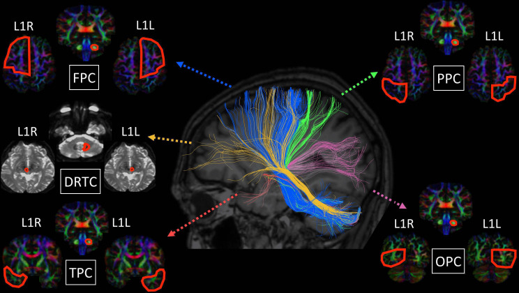
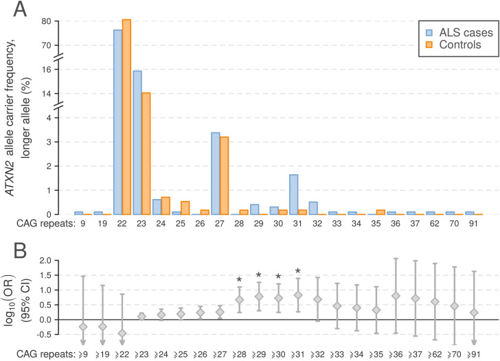
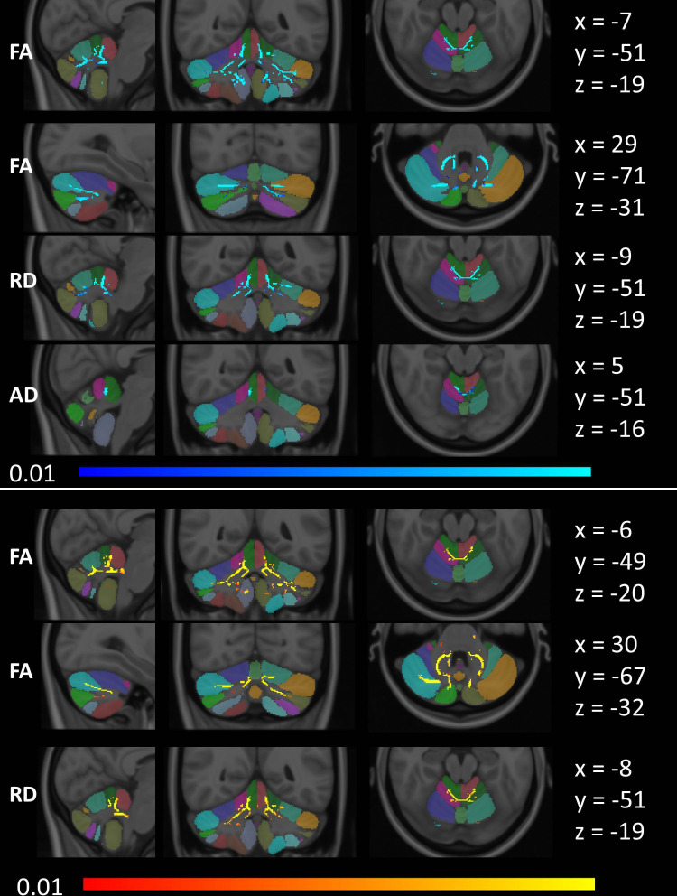
Methods: A prospective imaging study has been undertaken with 271 participants to systematically evaluate cerebellar grey and white matter alterations, cerebellar peduncle integrity and cerebro-cerebellar connectivity in ALS. Participants were stratified into four groups: (1) patients testing positive for GGGGCC repeat expansions in C9orf72, (2) patients carrying an intermediate-length repeat expansion in ATXN2, (3) patients without established ALS-associated mutations and (4) healthy controls. Additionally, the cerebellar profile of a single patient with ALS who had an ATXN2 allele length of 62 was evaluated. Cortical thickness, grey matter and white matter volumes were calculated in each cerebellar lobule complemented by morphometric analyses to characterise genotype-associated atrophy patterns. A Bayesian segmentation algorithm was used for superior cerebellar peduncle volumetry. White matter diffusivity parameters were appraised both within the cerebellum and in the cerebellar peduncles. Cerebro-cerebellar connectivity was assessed using deterministic tractography.

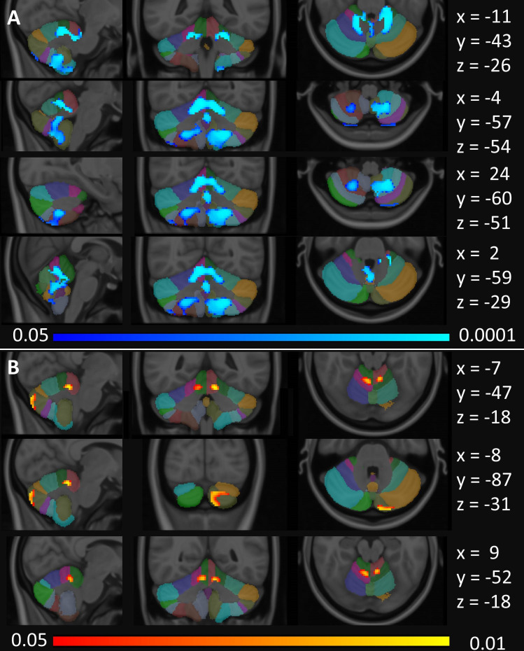
Results: Cerebellar pathology was confined to lobules I-V of the anterior lobe in patients with sporadic ALS in contrast to the considerable posterior lobe and vermis disease burden identified in C9orf72 mutation carriers. Patients with intermediate ATXN2 expansions did not exhibit significant cerebellar pathology.

Conclusions: Focal rather than global cerebellar degeneration characterises ALS. Pathognomonic ALS symptoms which are typically attributed to other anatomical regions, such as dysarthria, dysphagia, pseudobulbar affect, eye movement abnormalities and cognitive deficits, may be modulated, exacerbated or partially driven by cerebellar changes in ALS.


Conflict of interest statement

Competing interests: None declared.

Figures

Figure 1
Figure 1
Tractography methods and anatomical targets for the fronto-ponto-cerebellar (FPC), parieto-ponto-cerebellar (PPC), occipito-ponto-cerebellar (OPC), temporo-ponto-cerebellar (TPC) and dentate-rubro-thalamo-cortical (DRTC) tracts.
Figure 2
Figure 2
Profile of ATXN2 repeat expansions in 977 Irish patients with ALS and 562 population-matched healthy controls. (A) Allele frequencies for ALS cases and controls for the larger ATXN2 allele observed per individual. (B) OR ±95% CI for ATXN2 repeat expansions in ALS, thresholded by each observed allele as a lower limit. Asterisks indicate repeat expansion size categories significantly associated with ALS (Fisher’s exact test p<2.4×10⁻³). ALS, amyotrophic lateral sclerosis.
Figure 3
Figure 3
Cerebellar grey matter alterations as indicated by focal partial volume reductions at p<0.05 FWE TFCE corrected for age, gender and total intracranial volumes. (A) Focal changes in ALS-C9 are indicated in blue and (B) changes in ALS-NEG patients in red-yellow. The Diedrichsen probabilistic cerebellar atlas is presented as underlay to aid localisation. MNI coordinates are provided on the right side of the figure for sagittal (x), coronal (y) and axial (z) views. ALS, amyotrophic lateral sclerosis; ALS-C9, patients who tested positive for GGGGCC repeat expansions in C9orf72; ALS-NEG, patients who tested negative for both ATXN2 and C9orf72; FWE, family-wise error; MNI, Montreal Neurological Institute; TFCE, threshold-free cluster enhancement.
Figure 4
Figure 4
Tract-based white matter changes in ALS as identified by FA, AD and RD alterations at p<0.01 TFCE adjusted for age and gender. Changes in ALS-C9 are indicated in blue (top), and changes in ALS-NEG are shown in red-yellow (bottom). The Diedrichsen probabilistic cerebellar atlas is presented as underlay to aid localisation. MNI coordinates are provided on the right side of the figure for sagittal (x), coronal (y) and axial (z) views. AD, axial diffusivity; ALS, amyotrophic lateral sclerosis; ALS-C9, patients who tested positive for GGGGCC repeat expansions in C9orf72; ALS-NEG, patients who tested negative for both ATXN2 and C9orf72; FA, fractional anisotropy; MNI, Montreal Neurological Institute; RD, radial diffusivity; TFCE, threshold-free cluster enhancement.

Comment in

  • The cerebellum in ALS: friend or foe?
    Pradat PF. Pradat PF. J Neurol Neurosurg Psychiatry. 2021 Nov;92(11):1137. doi: 10.1136/jnnp-2021-327216. Epub 2021 Aug 2. J Neurol Neurosurg Psychiatry. 2021. PMID: 34341140 No abstract available.

References

    1. Elden AC, Kim H-J, Hart MP, et al. . Ataxin-2 intermediate-length polyglutamine expansions are associated with increased risk for ALS. Nature 2010;466:1069–75. 10.1038/nature09320 - DOI - PMC - PubMed
    1. Tan RH, Devenney E, Dobson-Stone C, et al. . Cerebellar integrity in the amyotrophic lateral sclerosis-frontotemporal dementia continuum. PLoS One 2014;9:e105632. 10.1371/journal.pone.0105632 - DOI - PMC - PubMed
    1. Tan RH, Kril JJ, McGinley C, et al. . Cerebellar neuronal loss in amyotrophic lateral sclerosis cases with ATXN2 intermediate repeat expansions. Ann Neurol 2016;79:295–305. 10.1002/ana.24565 - DOI - PubMed
    1. Al-Sarraj S, King A, Troakes C, et al. . P62 positive, TDP-43 negative, neuronal cytoplasmic and intranuclear inclusions in the cerebellum and hippocampus define the pathology of C9orf72-linked FTLD and MND/ALS. Acta Neuropathol 2011;122:691–702. 10.1007/s00401-011-0911-2 - DOI - PubMed
    1. Yunusova Y, Plowman EK, Green JR, et al. . Clinical measures of bulbar dysfunction in ALS. Front Neurol 2019;10:106. 10.3389/fneur.2019.00106 - DOI - PMC - PubMed

Publication types

Substances