. 2021 Jun 24;12(7):644.
doi: 10.1038/s41419-021-03925-z.

Inverse and reciprocal regulation of p53/p21 and Bmi-1 modulates vasculogenic differentiation of dental pulp stem cells

Affiliations

Inverse and reciprocal regulation of p53/p21 and Bmi-1 modulates vasculogenic differentiation of dental pulp stem cells

Zhaocheng Zhang et al. Cell Death Dis. .

Abstract

Dental pulp stem cells (DPSC) are capable of differentiating into vascular endothelial cells. Although the capacity of vascular endothelial growth factor (VEGF) to induce endothelial differentiation of stem cells is well established, mechanisms that maintain stemness and prevent vasculogenic differentiation remain unclear. Here, we tested the hypothesis that p53 signaling through p21 and Bmi-1 maintains stemness and inhibits vasculogenic differentiation. To address this hypothesis, we used primary human DPSC from permanent teeth and Stem cells from Human Exfoliated Deciduous (SHED) teeth as models of postnatal mesenchymal stem cells. DPSC seeded in biodegradable scaffolds and transplanted into immunodeficient mice generated mature human blood vessels invested with smooth muscle actin-positive mural cells. Knockdown of p53 was sufficient to induce vasculogenic differentiation of DPSC (without vasculogenic differentiation medium containing VEGF), as shown by increased expression of endothelial markers (VEGFR2, Tie-2, CD31, VE-cadherin), increased capillary sprouting in vitro; and increased DPSC-derived blood vessel density in vivo. Conversely, induction of p53 expression with small molecule inhibitors of the p53-MDM2 binding (MI-773, APG-115) was sufficient to inhibit VEGF-induced vasculogenic differentiation. Considering that p21 is a major downstream effector of p53, we knocked down p21 in DPSC and observed an increase in capillary sprouting that mimicked results observed when p53 was knocked down. Stabilization of ubiquitin activity was sufficient to induce p53 and p21 expression and reduce capillary sprouting. Interestingly, we observed an inverse and reciprocal correlation between p53/p21 and the expression of Bmi-1, a major regulator of stem cell self-renewal. Further, direct inhibition of Bmi-1 with PTC-209 resulted in blockade of capillary-like sprout formation. Collectively, these data demonstrate that p53/p21 functions through Bmi-1 to prevent the vasculogenic differentiation of DPSC.


Conflict of interest statement

The authors declare no competing interests.

Figures

Fig. 1
Fig. 1. Characterization of MSC and endothelial markers in dental pulp stem cells.
Cell surface markers detected by flow cytometry in DPSC, SHED, and HDMEC under standard culture conditions for each cell type. a Flow plots depicting expression of MSC markers (i.e., CD73, CD90, CD105, CD34, CD44) using IgG-APC as isotype-matched control. b Flow plots depicting expression of endothelial cell-related proteins (VEGFR1 and CD146) using IgG-PE and IgG-FITC were used as isotype-matched controls. c RT-PCR for odontogenic/osteogenic markers (DSPP, DMP-1), endothelial cell-related markers (VEGFR1, VEGFR2, CD146, CD31, VE-cadherin) in primary human odontoblasts, dental pulp tissue, DPSC, SHED, or HDMEC. GAPDH was used as a loading control.
Fig. 2
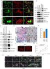
Fig. 2. Silencing p53 enhances vasculogenic differentiation of DPSC in vitro and in vivo.
a Photomicrographs depicting dental pulp stem cells that differentiated into functional blood vessels in vivo. GFP-transduced DPSC were seeded in scaffolds and transplanted in the subcutaneous space of the dorsum of SCID mice. Six weeks later, scaffolds were retrieved and tissue sections were prepared for immunofluorescence staining. Scattered cells showed positive staining for both, GFP (green), and SMA-alpha (red). In mature blood vessels, GFP was detected in the endothelial cells, SMA-alpha was stained the surrounding smooth muscle cells. Endothelial cells in the inner layer were stained for CD31 (green), while surrounding smooth muscle cells stained positive for SMA-alpha (red). Arrows point to DPSC-derived endothelial cells (green cells). Scale bar: 50 µm. b Western blots for VEGFR2, CD31, PDGFR-a, PDGFR-β, SMA-a, p53, and Bmi-1 in DPSC, SHED, HUASMC, and HDMEC. c Endothelial cell differentiation: shRNA-transduced DPSC were cultured with 5%FBS–MEM for 14 days, western blots for p53, p21, VEGFR2, Tie-2, CD31, and VE-cadherin. The density of protein expression was normalized with β-actin. d shRNA-p53-mediated vasculogenic differentiation in vivo: H&E and IHC staining. Blood vessels were revealed by H&E staining and detected with anti-factor VIII antibody, scale bar: 50 µm. e Graph depicting the blood vessel density observed in d stained with Factor VIII. Asterisk indicates p = 0.00035, as determined by t test. f DPSC cells were seeded in matrigel and cultured with EGM2 for 8 days. The Matrigel was fixed, and the sprouts were revealed by IF staining for CD31. Scale bar: 100 µm. g In all, 1 × 104 shRNA-transduced DPSC were seeded in growth factor-reduced matrigel-coated 12-well plate and cultured in endothelial differentiation medium (EGM2) for indicated time points. Sprouts were photographed, scale bar:100 µm. h Graph depicting the numbers of sprout formed in g. Three independent experiments using triplicate wells per condition were performed. Asterisk indicates p < 0.001, as determined by one-way ANOVA followed by a post hoc test (Tukey’s test).
Fig. 3
Fig. 3. Stabilization of p53 by blockade of the p53-MDM2 binding inhibits vasculogenic differentiation of dental pulp stem cells.
a shRNA-p53 transduced DPSC were treated with 0–2.5 µM MI-773 and 0–10 µM APG-115 for 24 h, western blots were performed for P53, MDM2, and p21, β-actin was used as loading control. b DPSC were cultured with endothelial differentiation medium in the presence of 50 ng/ml VEGF and 0–0.25 µM MI-773 for 14 days, western blots for p53, MDM2, p21, Bmi-1, VEGFR2, Tie-2, and CD31. The density of protein expression was normalized with β-actin. c, d 1 × 104 DPSC were seeded in growth factor-reduced matrigel and cultured in endothelial differentiation medium (EGM2) in the presence of 0–0.5 µM MI-773 for 13 days. c photographs of sprout, scale bar:100 µm. d Graph depicting the number of sprouts formed in c. Asterisk indicates p < 0.001, as determined by one-way ANOVA followed by a post hoc test (Tukey’s test).
Fig. 4
Fig. 4. p21 is a downstream effector of p53-mediated regulation of the vasculogenic fate of dental pulp stem cells.
a DPSC were transduced with shRNA scramble and shRNA-p21, western blot was performed for p53, MDM2, and Bmi-1. b, c shRNA scramble and shRNA-p21 transduced DPSC were seeded in growth factor-reduced matrigel and cultured in endothelial differentiation medium for different time points. b photographs of sprouts, scale bar:100 µm. c Graph depicting the number of sprouts formed in b. Asterisk indicates p < 0.001, as determined by one-way ANOVA followed by a post hoc test (Tukey’s test).
Fig. 5
Fig. 5. p53-dependent vasculogenic differentiation requires inverse and reciprocal regulation of p21 and Bmi-1.
ad Western blot for p53, MDM2, p21, and Bmi-1 in dental pulp cells. a Untransduced and shRNA-transduced DPSC. bd Dental pulp cells were treated with 0–10 µM MI-773 (b), 0–10 µM APG-115 (c), and 0–10 µM Bmi-1 inhibitor (PTC-209) (d) for 24 h. e SHED were cultured with 5% FBS–MEM in the presence of 50 ng/ml VEGF with or without 0–2.5 µM PTC-209 for 14 days, western blots for VEGFR2, Tie-2, Bmi-1, and p21, GAPDH was used as a loading control. The density of protein expression was normalized with GAPDH. f, g 1 × 104 SHED were seeded in growth factor-reduced matrigel and cultured with EGM2 in the presence of 0–2.5 µM PTC-209 for different time points. f photographs of sprout in SHED. Scale bar: 100 µm. g Graph depicting the number of sprouts formed in f, Asterisk indicates p < 0.001, as determined by one-way ANOVA followed by a post hoc test (Tukey’s test).
Fig. 6
Fig. 6. Ubiquitin/proteasome activity regulates Bmi-1 and vasculogenic differentiation of pulp stem cells.
a shRNA-transduced DPSC were treated with 1 µM MI-773 for 24 h, RT-PCR for p53, p21, and Bmi-1. b, c shRNA-transduced DPSC were cultured with 0–20 µM proteasome inhibitor MG132 for 24 h (b), or 5 µM MG132 for indicated time points (c), western blots for p53, MDM2, p21, and Bmi-1. d, e SHED (d) and DPSC (e) were exposed to ultraviolet (UV) for 1 min, then cultured for time points, western blots were performed for p53, MDM2, p21, p-JNK, JNK, p-c-Jun, c-Jun, and Bmi-1. f DPSC and SHED were treated with 0–100 nM ubiquitin aldehyde for 24 h, western blots were performed for p53, MDM2, p21, and Bmi-1. g, h 1 × 104 DPSC were seeded in growth factor-reduced matrigel and cultured with EGM2 in the presence of 0–2.5 nM ubiquitin aldehyde for different time points. g photographs of sprout in DPSC. Scale bar: 100 µm. h Graph depicting the number of sprouts formed in g. Asterisk indicates p < 0.001, as determined by one-way ANOVA followed by a post hoc test (Tukey’s test).
Fig. 7
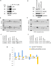
Fig. 7. Expression of downstream targets of p53 in DPSC cells.
a DPSC were treated with 1 µM MI-773 (or vehicle control) for 24 hours. Western blots for p53, MDM2, and p21 were used to verify the effect of MI-773 treatment on p53. b, c Protein arrays for evaluation of MI-773-treated DPSC versus control-treated DPSC. c Selected genes from b (full results are in Suppl Table 1). d DPSC were stably transduced with shRNA-control or shRNA-p53. Westerns blot for p53, MDM2, and p21 to verify the effect of shRNA constructs. e, f Protein array for evaluation of shRNA-p53 transduced DPSC versus shRNA-Control-transduced DPSC. f Selected genes from e (full results are in Suppl Table 2). g Graph depicting the fold change in protein density from MI-773/vehicle from MI-773/vehicle c and from shRNA-p53/shRNA-control f showing inverse correlations in several p53 targets (e.g., p21, SMAC, and TRAILR2).

Similar articles

Cited by

References

    1. Bronner-Fraser M. Segregation of cell lineage in the neural crest. Curr Opin Genet Dev. 1993;3:641–7. doi: 10.1016/0959-437X(93)90101-T. - DOI - PubMed
    1. Chai Y, Jiang X, Ito Y, Bringas P, Han J, Rowitch DH, et al. Fate of the mammalian cranial neural crest during tooth and mandibular morphogenesis. Development. 2000;127:1671–9. doi: 10.1242/dev.127.8.1671. - DOI - PubMed
    1. Lumsden AG. Spatial organization of the epithelium and the role of neural crest cells in the initiation of the mammalian tooth germ. Development. 1988;103:155–69. doi: 10.1242/dev.103.Supplement.155. - DOI - PubMed
    1. Gronthos S, Mankani M, Brahim J, Robey PG, Shi S. Postnatal human dental pulp stem cells (DPSCs) in vitro and in vivo. Proc Natl Acad Sci USA. 2000;97:13625–30. doi: 10.1073/pnas.240309797. - DOI - PMC - PubMed
    1. Miura M, Gronthos S, Zhao M, Lu B, Fisher LW, Robey PG, et al. SHED: Stem cells from human exfoliated deciduous teeth. Proc Natl Acad Sci USA. 2003;100:5807–12. doi: 10.1073/pnas.0937635100. - DOI - PMC - PubMed

Publication types

MeSH terms

Substances