

doi: 10.1038/s41592-021-01183-7.
Epub 2021 Jun 24.
Automatic detection of synaptic partners in a whole-brain Drosophila electron microscopy data set
Affiliations
- PMID: 34168373
- PMCID: PMC7611460
- DOI: 10.1038/s41592-021-01183-7
Item in Clipboard
Automatic detection of synaptic partners in a whole-brain Drosophila electron microscopy data set
Nat Methods.
2021 Jul.
Abstract
We develop an automatic method for synaptic partner identification in insect brains and use it to predict synaptic partners in a whole-brain electron microscopy dataset of the fruit fly. The predictions can be used to infer a connectivity graph with high accuracy, thus allowing fast identification of neural pathways. To facilitate circuit reconstruction using our results, we develop CIRCUITMAP, a user interface add-on for the circuit annotation tool CATMAID.
Conflict of interest statement
Competing Interests Statement
Stephan Gerhard is the founder and CEO of UniDesign Solutions GmbH, which provides IT services.
Figures
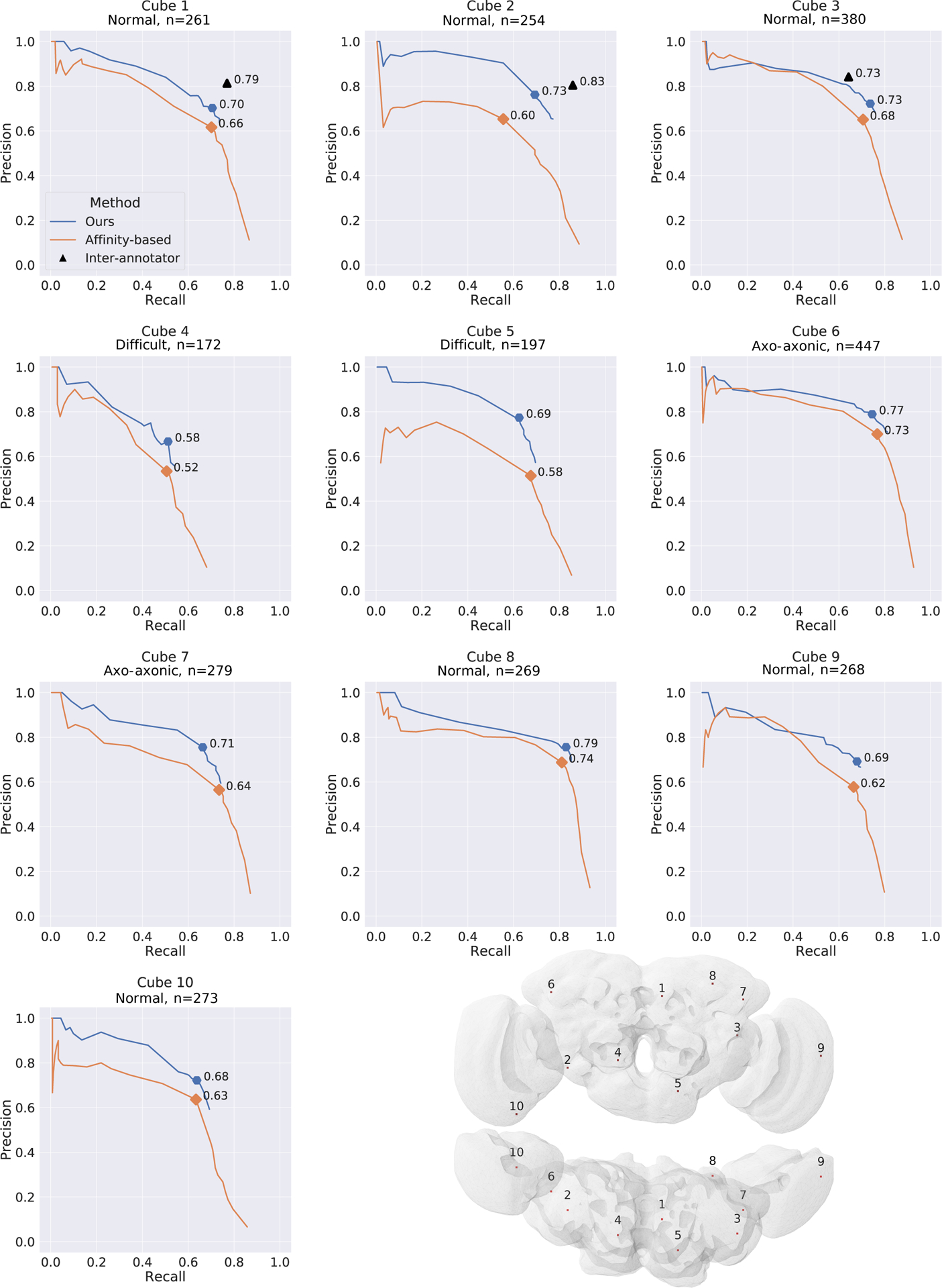
Precision and recall over 10 densely annotated cubes in different brain regions (dataset DenseCubes). Highlights show best f-score over the detection threshold. Top row: Black triangle marks show the inter-human variance between two annotations, with one annotation treated as ground-truth evaluated against the other one. Bottom right: Visualization of the cube locations within the FAFB dataset.
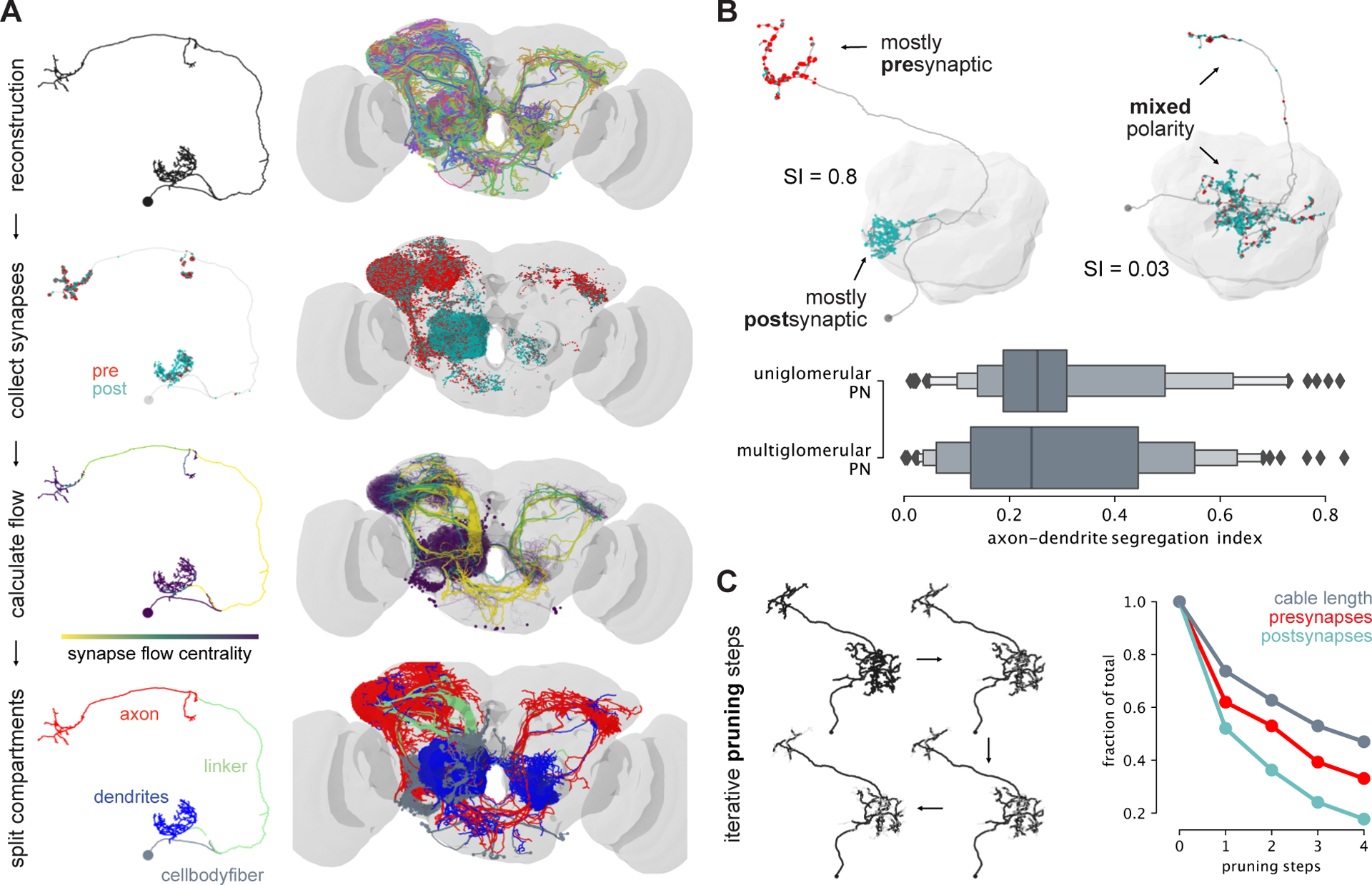
a, Outline of axon-dendrite splitting procedure. b, Exemplary well segregated (left) and unsegregated (right) PN. Boxplot shows segregation index (SI) for all PNs separated into uni- and multiglomerular PNs. c, Impact of completeness of neuronal reconstruction on recovery of synapses.
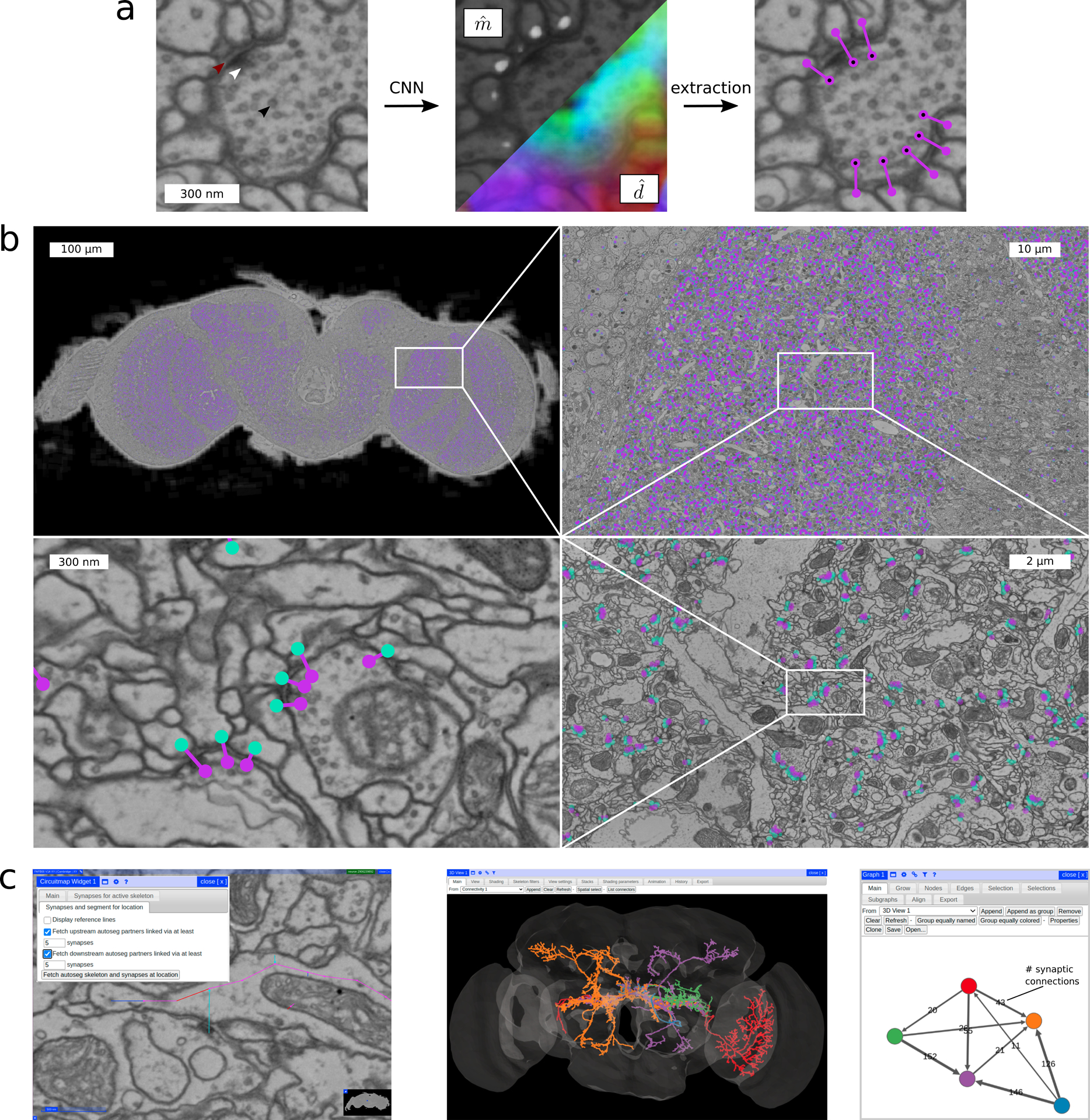
Method overview for synaptic partner prediction, application to the “full adult fly brain” (FAFB) dataset and usage of Circuit Map for circuit reconstruction and analysis in Catmaid . a CNN predictions of post-synaptic sites () and direction vectors pointing to the pre-synaptic site (, 3D vectors shown as RGB-color) together with final detections after post-processing. Arrows show synaptic cleft (red), T-bar (white), and vesicles (black). b Sample section of the FAFB dataset with predicted synaptic partners (pre-synaptic site purple, post-synaptic site turquoise). c Using Circuit Map (left image), predicted synaptic partners are available in Catmaid to allow exploration of automatically reconstructed neural circuits. The example shows five automatically segmented neurons from [3] (middle image) together with their predicted number of synaptic connections (right image, node colors match the segmentation). See also Supplementary Video.
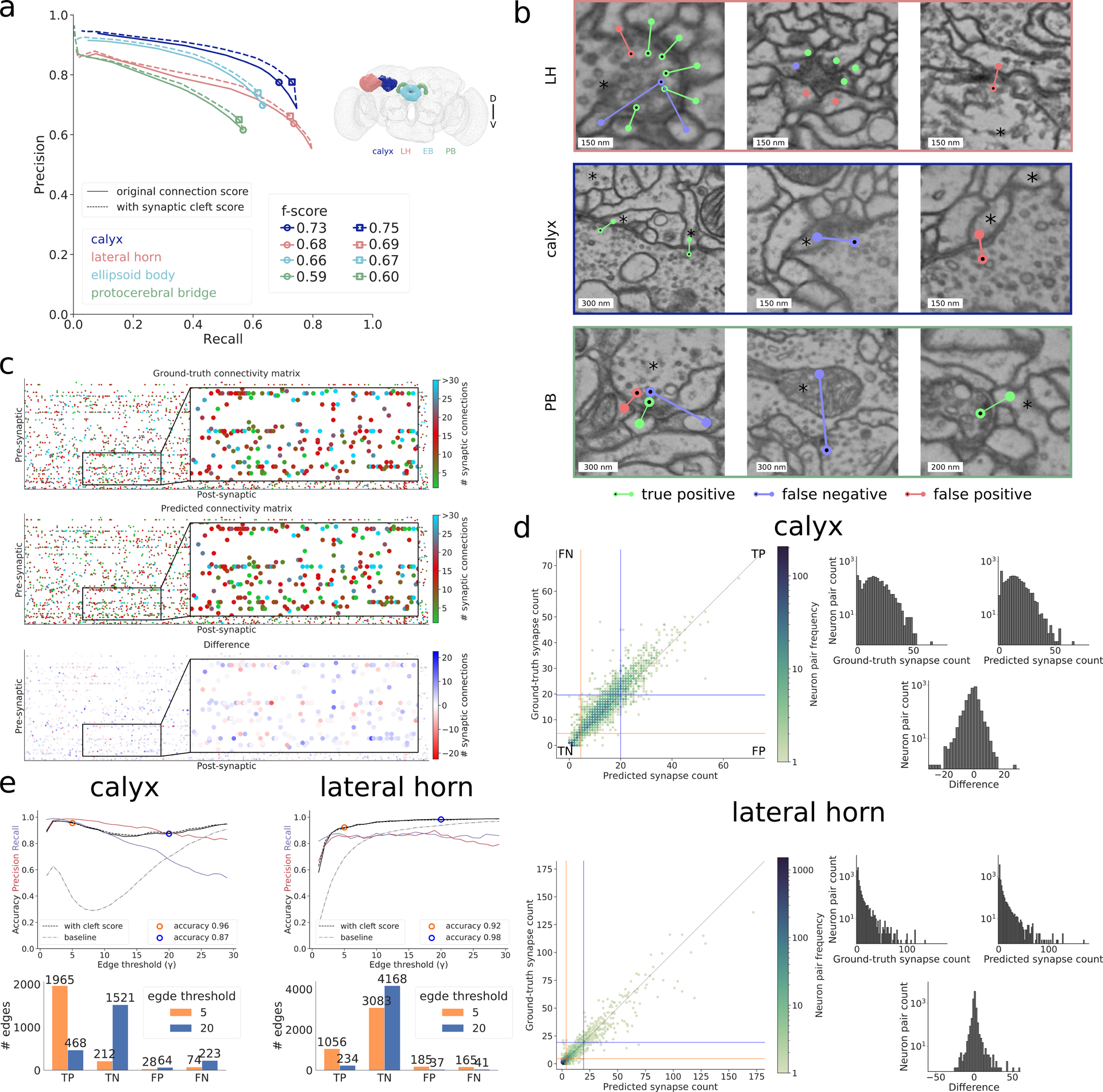
Results in whole-brain FAFB dataset. a Precision-recall curves using the Cremi metric for the four different brain regions calyx (dark blue), lateral horn (peach), ellipsoid body (light blue) and protocerebral bridge (green) over different prediction score thresholds, with (solid) and without (dashed) filtering via synaptic cleft predictions [6] (Supplementary Note 2). Markers highlight validation best score threshold (f-score with cleft predictions: 0.75, 0.69, 0.67, 0.60 for calyx, lateral horn, ellipsoid body, and protocerebral bridge, respectively). b Examples of identified true positives, false positives, false negatives in lateral horn (top row), calyx (middle row), and protocerebral bridge (bottom row) on ground-truth neurons (marked with an asterisk). False positive in top row left, false positives and false negative in top row middle are examples of ambiguous cases, false negative in bottom row middle is an example of a missed axo-axonic connection. c Ground-truth, predicted, and difference of number of synaptic connections between 138 projection neurons (pre-synaptic) and 528 Kenyon cells (post-synaptic) in calyx. d For calyx, same data as in (c), shown as ground-truth versus predicted count of number of synaptic connections between pairs of neurons and equivalent plot for lateral horn. Pairs with both zero connections in ground-truth and prediction are omitted. Orange/blue line quadrants highlight false negative (FN), true positive (TP), false positive (FP), and true negative (TN) edges for two connectivity criteria, i.e., number of synapses equal or greater than γ = 5 (orange) and γ = 20 (blue). Histograms on the right show the respective frequency distributions of number of synaptic connections per edge. e Edge accuracy, precision, and recall over different connectivity thresholds γ for calyx (left) and lateral horn (right). Accuracy results for thresholds γ = 5 and γ = 20 are highlighted and their absolute numbers of TPs, TNs, FPs, FNs are provided in the bar plots below. Connectivity derived from neuron-proximity is shown as “baseline” (Supplementary Note 2).
References
-
- Denk Winfried, Briggman Kevin L, and Helmstaedter Moritz. Structural neurobiology: missing link to a mechanistic understanding of neural computation. Nature Reviews Neuroscience, 13(5):351–358, 2012. - PubMed
-
- Li Peter H., Lindsey Larry F., Januszewski Michal, Zheng Zhihao, Bates Alexander Shakeel, Taisz István, Tyka Mike, Nichols Matthew, Li Feng, Perlman Eric, Maitin-Shepard Jeremy, Blakely Tim, Leavitt Laramie, Jefferis Gregory S. X. E., Bock Davi, and Jain Viren. Automated Reconstruction of a Serial-Section EM Drosophila Brain with Flood-Filling Networks and Local Realignment. bioRxiv, April2019. doi: 10.1101/605634. - DOI
-
- Dorkenwald Sven, Schubert Philipp J, Killinger Marius F, Urban Gregor, Mikula Shawn, Svara Fabian, and Kornfeld Joergen. Automated synaptic connectivity inference for volume electron microscopy. Nature methods, 14 (4):435, 2017. - PubMed
-
- Motta Alessandro, Berning Manuel, Boergens Kevin M, Staffler Benedikt, Beining Marcel, Loomba Sahil, Hennig Philipp, Wissler Heiko, and Helmstaedter Moritz. Dense connectomic reconstruction in layer 4 of the somatosensory cortex. Science, 366(6469), 2019. - PubMed
Publication types
MeSH terms
Grants and funding
LinkOut - more resources
Full Text Sources
Molecular Biology Databases
