Evolution of the Laser-Induced Spallation Technique in Film Adhesion Measurement
- PMID: 34168374
- PMCID: PMC8208493
- DOI: 10.1115/1.4050700
Evolution of the Laser-Induced Spallation Technique in Film Adhesion Measurement
Abstract
Laser-induced spallation is a process in which a stress wave generated from a rapid, high-energy laser pulse initiates the ejection of surface material opposite the surface of laser impingement. Through knowledge of the stress-wave amplitude that causes film separation, the adhesion and interfacial properties of a film-on-substrate system are determined. Some advantages of the laser spallation technique are the noncontact loading, development of large stresses (on the order of GPa), and high strain rates, up to 108/s. The applicability to both relatively thick films, tens of microns, and thin films, tens of nm, make it a unique technique for a wide range of materials and applications. This review combines the available knowledge and experience in laser spallation, as a state-of-the-art measurement tool, in a comprehensive pedagogical publication for the first time. An historical review of adhesion measurement by the laser-induced spallation technique, from its inception in the 1970s through the present day, is provided. An overview of the technique together with the physics governing the laser-induced spallation process, including functions of the absorbing and confining materials, are also discussed. Special attention is given to applications of laser spallation as an adhesion quantification technique in metals, polymers, composites, ceramics, and biological films. A compendium of available experimental parameters is provided that summarizes key laser spallation experiments across these thin-film materials. This review concludes with a future outlook for the laser spallation technique, which approaches its semicentennial anniversary.
Copyright © 2021 by ASME.
Figures
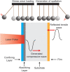
![Schematic of (a) laser impingement for a confined configuration and (b) pressure profiles for direct and confined configurations (Adapted with permission from Meng et al. [55]. Copyright 2017 by Multidisciplinary Digital Publishing Institute).](https://cdn.ncbi.nlm.nih.gov/pmc/blobs/7307/8208493/1d1f0e98c910/amr-20-1087_030802_g001.gif)


![(a) Representative voltage trace obtained from the Michelson interferometer setup during calibration experiments. (b) The x-axis, time, is artificially set to zero at the arrival of the mechanical wave, indicated by dashed red box in (a). (c) Displacement measurements for relatively high (solid), medium (dashed), and low (small dashed) laser fluences. Displacement curve in (c) for relatively high fluence (solid) is produced from the voltage curve in (a) and (b) using Eqs. (7) and (8). (d) Substrate stress profile obtained using Eq. (10) for corresponding laser fluences in (c). The calibration specimen that produced these data is a 1 mm glass slide with a titanium test film, an aluminum absorbing layer, and a waterglass confining layer (Adapted with permission from Boyd et al. [52]. Copyright 2021 by Elsevier).](https://cdn.ncbi.nlm.nih.gov/pmc/blobs/7307/8208493/665b9e39f502/amr-20-1087_030802_g005.gif)
![Schematic of volumetric, cohesive, and spectral elements implemented in FEA of stress-wave propagation through a substrate and thin film (Adapted with permission from Tran et al. [86]. Copyright 2008 by Elsevier).](https://cdn.ncbi.nlm.nih.gov/pmc/blobs/7307/8208493/ac19aab4e2fb/amr-20-1087_030802_g006.gif)
![(a) Example of laser spallation schematic shown by Gupta et al. including some of the optics and lasers employed during testing. (b) Interface strength of polyimide films on silicon nitride decreases as humidity increases. Each figure is adapted with permission from Gupta et al. [87]. Copyright 2000 by Elsevier.](https://cdn.ncbi.nlm.nih.gov/pmc/blobs/7307/8208493/b0ddb5af2519/amr-20-1087_030802_g007.gif)
![(a) Example of setup used for loading with mixed-mode failure as well as the propagation of shear wave. (b) Image of tearing that occurs in thin metallic films after loading with high-amplitude shear stress waves. (c) Typical data (from raw voltage curve to tensile interface stress) for mixed-mode failure, including arrival and turning point for the shear wave. Each figure is adapted with permission from Wang et al. [31]. Copyright 2003 by Springer Nature.](https://cdn.ncbi.nlm.nih.gov/pmc/blobs/7307/8208493/8909a4de0441/amr-20-1087_030802_g008.gif)
![(a) Image of delamination that occurred due to interface failure of the thin aluminum strips used in an experiment to measure interfacial fracture energy; adhered strips can be observed in the back of the image (Adapted with permission from Kandula et al. [103]. Copyright 2008 by AIP Publishing). (b) Evolution of Au film failure with increasing interface stress for two different interfacial chemistries (Adapted with permission from Grady et al. [60]. Copyright 2014 by American Chemical Society). (c) Measured adhesion strength values for various interfacial chemistries (Adapted with permission from Grady et al. [60]. Copyright 2014 by American Chemical Society).](https://cdn.ncbi.nlm.nih.gov/pmc/blobs/7307/8208493/8b2208a961da/amr-20-1087_030802_g009.gif)
![(a) Evolution of film failure with increased loading for PBO film; initial delamination occurs at lower fluence values before ejection of film occurs (Adapted with permission from Kandula et al. [59]. Copyright 2008 by Elsevier). (b) Comparison of substrate stress profiles of multiple measurements at fixed laser fluence values to validate repeatability of stress-wave generation (Adapted with permission from Grady et al. [41]. Copyright 2014 by Elsevier). (c) Failure occurring from loading of polyurea films from a side view; depth of failure is indicated by bracketed line (Adapted with permission from Youssef and Gupta [90]. Copyright 2018 by Springer Nature). (d) Spall strength of polyurea measured by Youssef and Gupta (Adapted with permission from Youssef and Gupta [90]. Copyright 2018 by Springer Nature).](https://cdn.ncbi.nlm.nih.gov/pmc/blobs/7307/8208493/2ff5da899646/amr-20-1087_030802_g010.gif)
![(a) Interfacial mechanophore specimen preparation. (b) Top row depicts optical images of 1-photopatterned epoxy and 2-maleimide-anthracene functionalized silica substrate, while the bottom row depicts the same images under fluorescence where the higher amplitude stress waves have activated the specimens, denoted by the fluorescence present. Each figure is adapted with permission from Sung et al. [91]. Copyright 2018 by American Chemical Society.](https://cdn.ncbi.nlm.nih.gov/pmc/blobs/7307/8208493/fa9a8baaa182/amr-20-1087_030802_g011.gif)
![(a) Representative micrography of damage resulting from shock wave propagation inside a bonded CFRP with a very low adhesion rate due to release agent contamination. (b) Laser spallation applied to composite substrates to determine bond strength. (c) Internal failure of a composite observed using thermal imaging of layers after loading (Adapted with permission from Ecault et al. [92]. Copyright 2014 by Emerald Publishing Limited).](https://cdn.ncbi.nlm.nih.gov/pmc/blobs/7307/8208493/fa6532183202/amr-20-1087_030802_g012.gif)
![(a) Example micrograph of porous ceramic coating (Adapted with permission from Kobayashi et al. [97]. Copyright 2004 by Elsevier). (b) Stress of failure for zirconia films at different spraying distances (Adapted with permission from Kobayashi et al. [97]. Copyright 2004 by Elsevier). (c) Laser spallation and interferometric calibration setup for ceramic films is similar to setups observed for other materials (Adapted with permission from Ikeda et al. [95]. Copyright 2004 by IOP Publishing).](https://cdn.ncbi.nlm.nih.gov/pmc/blobs/7307/8208493/fe2e61ec67fa/amr-20-1087_030802_g013.gif)
![(a) Optical microscopy of diamond film failure after laser spallation loading (Adapted with permission from Ikeda et al. [95]. Copyright 2004 by IOP Publishing). (b) Interfacial stresses computed from compression and expansion surface displacements (Adapted with permission from Ikeda et al. [95]. Copyright 2004 by IOP Publishing). (c) Failure of PZT sol–gel films; at high fluence full ejection occurs similar to previously discussed films (Adapted with permission from Berfield et al. [96]. Copyright 2016 by Elsevier). (d) Impact of different functionalized surfaces on the interfacial strength of PZT sol–gel films; adhesion to SiO2/Si is much higher compared to ODS/Si (Adapted with permission from Berfield et al. [96]. Copyright 2016 by Elsevier).](https://cdn.ncbi.nlm.nih.gov/pmc/blobs/7307/8208493/ce309a00a619/amr-20-1087_030802_g014.gif)
![(a) Laser spallation setup used during loading of biological samples; often liquid is used to maintain functionality of biological films so laser spallation techniques are oriented differently compared to nonbiological materials. (b) Optical images for a region of singular neuron cells before and after loading occur; dashed circle indicates the loaded region within which detachments of cells can be observed. (c) Fluence of failure versus cell growth time indicates that higher energy is needed to detach cells with a longer growth time possibly due to increased adhesion, until this effect reaches saturation. Each figure is adapted with permission from Hu et al. [35]. Copyright 2006 by AIP Publishing.](https://cdn.ncbi.nlm.nih.gov/pmc/blobs/7307/8208493/95990cb736dd/amr-20-1087_030802_g015.gif)
![(a) SEM image of region with detached cells after loading occurs, a circle is used to indicate spalled region; see online version for better image contrast. (b) Adhesion strength of cells on fibronectin-coated and uncoated surfaces; the fibronectin coating results in a larger stress needed for detachment. Each figure is adapted with permission from Hagerman et al. [51]. Copyright 2007 by Wiley.](https://cdn.ncbi.nlm.nih.gov/pmc/blobs/7307/8208493/14fe5f76c838/amr-20-1087_030802_g016.gif)
![(a) Cultured biofilms with loading before and after indicated, multiple regions are loaded on a single biofilm to account for the variability associated with biological films, with Example substrate assembly inlayed (Adapted with permission from Boyd et al. [48]. Copyright 2019 by Springer Nature). (b) Evolution of failure for bacterial biofilms as well as cell monolayers with increased stress values on smooth and rough titanium (Adapted with permission from Boyd et al. [52]. Copyright 2021 by Elsevier).](https://cdn.ncbi.nlm.nih.gov/pmc/blobs/7307/8208493/560597358134/amr-20-1087_030802_g017.gif)
![(a) Weibull analysis performed on failure of cells and bacterial biofilms at increasing interface strength; roughened titanium surfaces have a significant impact on cell adhesion but minimal effect on bacterial adhesion (Adapted with permission from Boyd et al. [52]. Copyright 2021 by Elsevier). (b) Image detecting software is used to measure distance of testing spot from center of biofilm and a graph illustrating this effect on percentage of region spalled (Adapted with permission from Kearns et al. [117]. Copyright 2020 by American Chemical Society). (c) Schematic of reflective panel attached to both smooth and roughened titanium surface in order to measure effect of stress-wave generation (Adapted with permission from Boyd and Grady [118]. Copyright 2020 by AIP Publishing). (d) Average peak substrate stresses generated on both smooth and rough titanium using the reflective panel at two fixed fluences (Adapted with permission from Boyd and Grady [118]. Copyright 2020 by AIP Publishing).](https://cdn.ncbi.nlm.nih.gov/pmc/blobs/7307/8208493/251690852289/amr-20-1087_030802_g018.gif)
References
-
- Freund, L. B. , and Suresh, S. , 2004, Thin Film Materials: Stress, Defect Formation and Surface Evolution, Cambridge University Press, Cambridge, UK.
-
- Mittal, K. L. , and Pizzi, A. , 1999, Adhesion Promotion Techniques: Technological Applications, CRC Press, Boca Raton, FL.
-
- Gent, A. , and Kaang, S. , 1987, “ Effect of Peel Angle Upon Peel Force,” J. Adhes., 24(2–4), pp. 173–181.10.1080/00218468708075425 - DOI
-
- Kaelble, D. H. , 1965, “ Peel Adhesion: Micro‐Fracture Mechanics of Interfacial Unbonding of Polymers,” Trans. Soc. Rheol., 9(2), pp. 135–163.10.1122/1.549022 - DOI
-
- Kim, K.-S. , and Kim, J. , 1988, “ Elasto-Plastic Analysis of the Peel Test for Thin Film Adhesion,” ASME J. Eng. Mater. Technol., 110(3), pp. 266–273.10.1115/1.3226047 - DOI
Publication types
Grants and funding
LinkOut - more resources
Full Text Sources
