Case Reports
. 2021 Jun 9;16(8):2154-2157.
doi: 10.1016/j.radcr.2021.04.070. eCollection 2021 Aug.

Two case reports: Breast schwannoma and a rare case of an axillary schwannoma imitating an axillary lymph node metastasis

Affiliations
Case Reports

Two case reports: Breast schwannoma and a rare case of an axillary schwannoma imitating an axillary lymph node metastasis

Martin Duehrkoop et al. Radiol Case Rep. .

Abstract

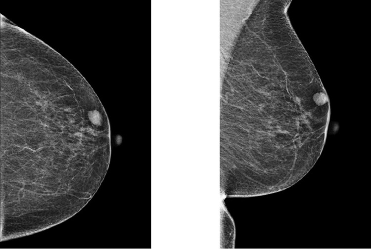
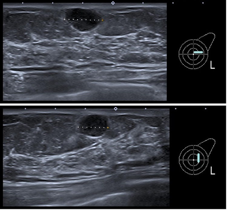
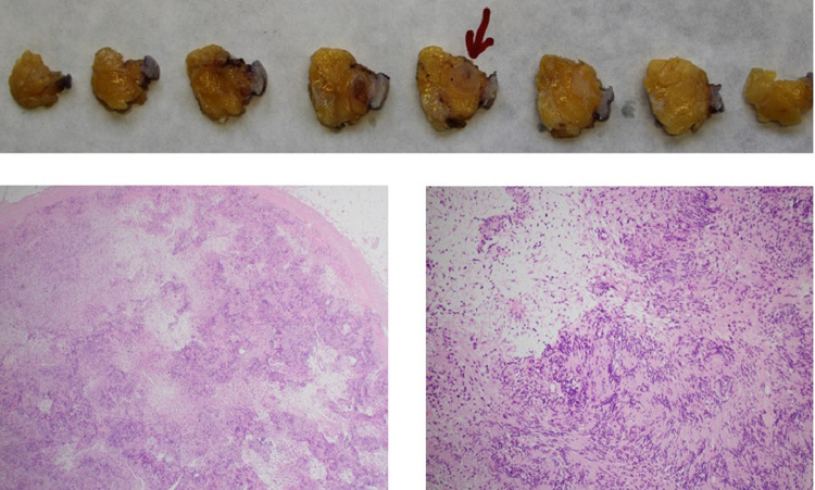
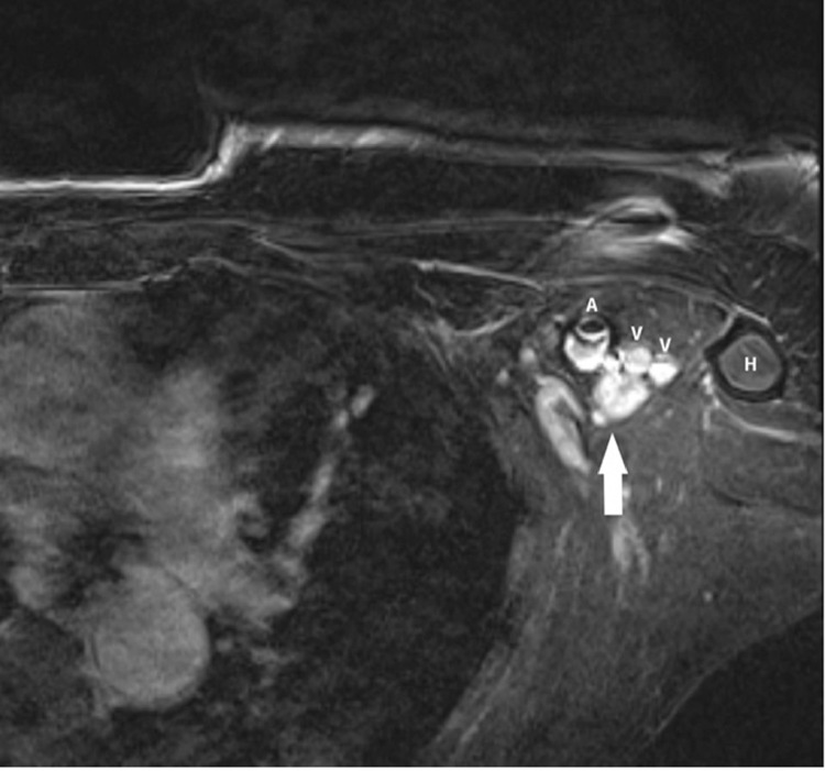
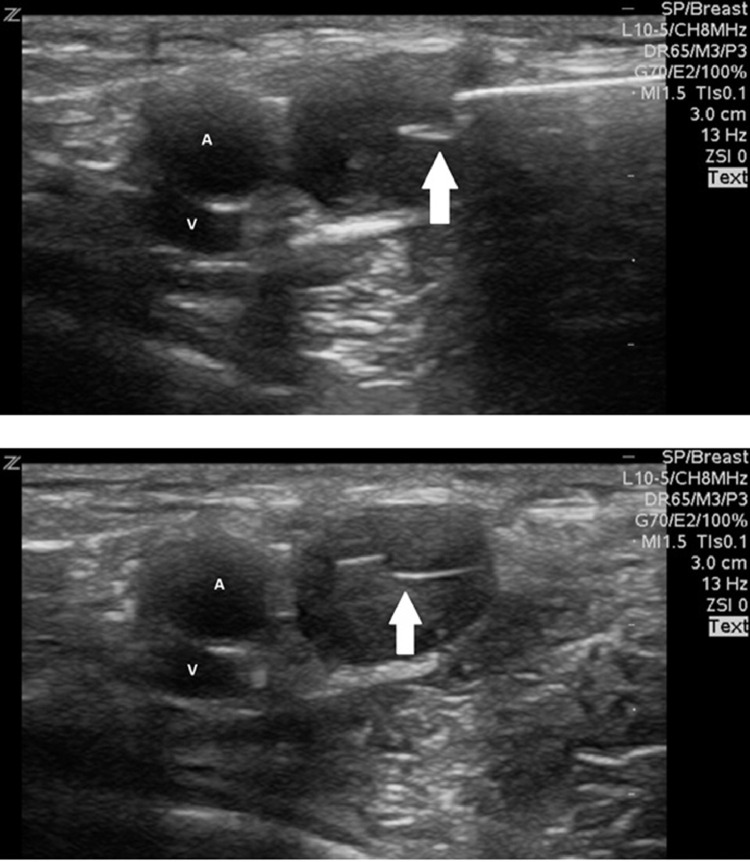
A 79-year-old woman with a newly detected oval circumscribed lump in subcutaneous location on mammography and ultrasound turned out to be a Schwannoma after ultrasound-guided core needle biopsy. A 72-year-old woman with breast cancer in medical history demonstrated a new axillary mass in follow up, initially regarded as a lymph node metastasis. Core needle biopsy did not lead to a sufficient diagnosis. Pathologic examination after intraoperative sampling revealed a Schwannoma. These 2 case reports illustrate the importance of diagnostic imaging and remind to include Schwannomas in the differential diagnosis of breast and axillary masses.

Keywords: Axilla; Breast; MRI; Mammography; Schwannoma; Ultrasound.


Figures

Fig 1 –
Fig. 1
Left mammography in craniocaudal and mediolateral oblique view shows an almost entirely fatty breast. An oval, circumscribed, equal density, subcutaneous mass without microcalcifications, measuring 13 mm is seen at 2-o'clock position and in 1-6 cm distance from the nipple, corresponding to the lump.
Fig 2 –
Fig. 2
Two plane sonography with a 14 MHz ultrasound head demonstrating a 10 × 14 mm oval, circumscribed, hypoechoic mass in parallel orientation, without posterior features and in subcutaneous location, corresponding to the lump.
Fig 3 –
Fig. 3
Pathologic examination showing a totally removed encapsulated mass with spindle cell proliferation arranged in dense (Antoni A tissue) and loosened areas (Antoni B tissue). Areas with nuclear palisading are visible (Verocay bodies). The nuclei show only mild variability in size and shape. No atypia or relevant necrosis are seen.
Fig 4 –
Fig. 4
1,5T MRI: T1 with fat suppression after IV contrast injection showing a 11 × 12 mm oval, circumscribed mass (arrow) with strong homogeneous enhancement in the immediate posterior vicinity of the left axillary artery (A) and vein (V), medial of the proximal humerus (H).
Fig 5 –
Fig. 5
Ultrasound-guided biopsy with a 14-gauge core needle demonstrating the central position of the core needle (arrow) in the target mass next to the axillary artery (A) and vein (V).

References

    1. Das Gupta TK, Brasfield RD, Strong EW, Hajdu SI. Benign solitary Schwannomas (neurilemomas) Cancer. 1969;24(2):355–366. - PubMed
    1. Uchida N, Yokoo H, Kuwano H. Schwannoma of the breast: report of a case. Surg Today. 2005;35(3):238–242. - PubMed
    1. Dialani V, Hines N, Wang Y, Slanetz P. Breast schwannoma. Case Rep Med. 2011;2011 - PMC - PubMed
    1. Takaaki F, Reina Y, Hiroki M, Soichi T, Takayuki A, Hiroyuki K. A Rare Case of Anterior Chest Wall Schwannoma Masquerading as a Breast Tumor. Int Surg. 2014;99(3):196–199. - PMC - PubMed
    1. Qing TT, Esther WLC, Sung HC, Ga SH. Schwannoma: an unexpected diagnosis from a breast lump. J Surg Case Rep. 2014;2014(9):rju085.

Publication types

LinkOut - more resources