Review
. 2021 Jun 15;12(6):730-744.
doi: 10.4239/wjd.v12.i6.730.

Mechanisms linking gut microbial metabolites to insulin resistance

Affiliations
Review

Mechanisms linking gut microbial metabolites to insulin resistance

Hye Rim Jang et al. World J Diabetes. .

Abstract

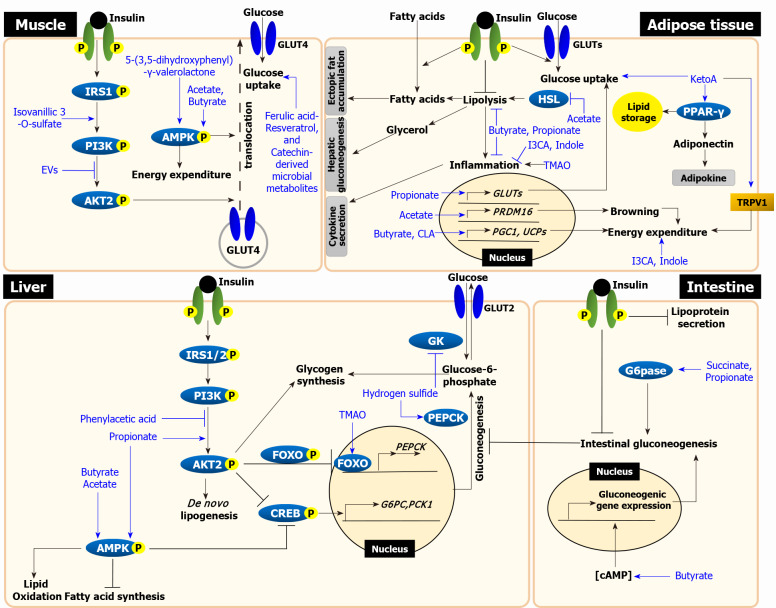
Insulin resistance is the rate-limiting step in the development of metabolic diseases, including type 2 diabetes. The gut microbiota has been implicated in host energy metabolism and metabolic diseases and is recognized as a quantitatively important organelle in host metabolism, as the human gut harbors 10 trillion bacterial cells. Gut microbiota break down various nutrients and produce metabolites that play fundamental roles in host metabolism and aid in the identification of possible therapeutic targets for metabolic diseases. Therefore, understanding the various effects of bacterial metabolites in the development of insulin resistance is critical. Here, we review the mechanisms linking gut microbial metabolites to insulin resistance in various insulin-responsive tissues.

Keywords: Adipose tissue; Gut bacterial metabolites; Insulin resistance; Intestine; Liver; Skeletal muscle.


Conflict of interest statement

Conflict-of-interest statement: The authors declare no conflicts of interest.

Figures

Figure 1
Figure 1
The mechanisms linking microbial metabolites to insulin resistance. In skeletal muscle, insulin stimulates glucose uptake by translocating glucose transporter (GLUT) 4 via insulin receptor substrate (IRS)-phosphoinositide-3-kinase (PI3K)-AKT signaling. Isovanillic 3-O-sulfate increases glucose uptake by activating PI3K-AKT pathway. Gut bacteria-derived extracellular vesicles (EVs) decrease glucose uptake by inhibiting AKT phosphorylation. Glucose uptake is also increased via AMP-activated protein kinase (AMPK) activation, insulin-independent. 5-(3,5-dihydroxyphenyl-γ-valerolactone activates AMPK phosphorylation, which enhances glucose uptake. Although mechanisms have been unknown, other ferulic acid, resveratrol, and catechin-derived microbial metabolites also enhance glucose uptake (left upper panel). In liver, insulin activates glycogen synthesis and de novo lipogenesis and suppresses gluconeogenesis via IRS-PI3K-AKT signaling. Propionate increases the phosphorylation of both AKT and AMPK, which suppresses gluconeogenesis. Hydrogen sulfide stimulates gluconeogenesis via phosphoenolpyruvate carboxykinase activation and reduces glycogen synthesis via the inhibition of glucokinase activity. Trimethylamine N-oxide (TMAO) increases gluconeogenesis via PKR-like ER kinase-FOXO1 pathway. Phenylacetic acid inhibits AKT phosphorylation. All short chain fatty acids (SCFAs), including acetate, propionate, and butyrate, activate AMPK phosphorylation, which lead to decrease lipid accumulation (left lower panel). In adipose tissue, insulin stimulates glucose and fatty acid uptake and suppress lipolysis. Failure to suppress lipolysis in insulin-resistant adipose tissue increases circulating free fatty acids and glycerol, which leads to an increase in ectopic fat accumulation in the liver and muscle and stimulates hepatic gluconeogenesis. 10-oxo-12(Z)-octadecenoic acid (KetoA) increases insulin-stimulated glucose uptake and energy expenditure via TRPV2 activation. KetoA also increases the production and secretion of adiponectin via peroxisome proliferator-activated receptor-γ activation. TMAO increases inflammation in adipocyte. Indole and I3CA have anti-inflammatory effects. Conjugated linoleic acid enhances energy expenditure by increasing the expression of uncoupling proteins (UCPs) genes. All SCFAs inhibit lipolysis. Acetate inhibits lipolysis by suppressing HSL and stimulates also browning by increasing the expression of browning-related genes. Butyrate and propionate attenuate inflammation. Propionate increases glucose uptake by increasing GLUT4 expression. Butyrate enhances energy expenditure by upregulating PPAR-γ coactivator 1 and UCPs genes (right upper panel). The intestine, as discussed in this review, is an organ that actively interacts with gut bacteria and accumulates microbial metabolites. Intestinal lipoprotein secretion and gluconeogenesis are suppressed by insulin. In intestine, propionate and succinate act as gluconeogenic substrate, which activate gluconeogenesis via G6Pase activation. Butyrate increases cyclic adenosine monophosphate levels, which upregulates the expression of gluconeogenic genes and increases gluconeogenesis. Through this mechanisms, increased intestinal gluconeogenesis suppresses hepatic gluconeogenesis (right lower panel). Black lines represent insulin resistance-related events and blue lines represent action of metabolites. Grey boxes represent the effects of adipose tissue on other tissues. EVs: Extracellular vesicles; PEPCK: Phosphoenolpyruvate carboxykinase; GK: Glucokinase; TMAO: Trimethylamine N-oxide; PERK: PKR-like ER kinase; FOXO1: Forkhead box protein O1; cAMP: Cyclic adenosine monophosphate; CREB: cAMP-response element binding protein; G6PC: Glucose 6-phosphatase catalytic subunit; PCK1: Phosphoenolpyruvate carboxykinase 1; TRPV1: Transient receptor potential vanilloid 1; PPAR: Peroxisome proliferator-activated receptor; I3CA: Indole-3-carboxylic acid; CLA: Conjugated linoleic acid; HSL: Hormone-sensitive lipase; PRDM16: PR domain containing 16; PGC1: Peroxisome proliferator-activated receptor-gamma coactivator 1; G6Pase: Glucose 6-phosphatase.

References

    1. Petersen MC, Shulman GI. Mechanisms of Insulin Action and Insulin Resistance. Physiol Rev. 2018;98:2133–2223. - PMC - PubMed
    1. Samuel VT, Shulman GI. Mechanisms for insulin resistance: common threads and missing links. Cell. 2012;148:852–871. - PMC - PubMed
    1. Samuel VT, Shulman GI. The pathogenesis of insulin resistance: integrating signaling pathways and substrate flux. J Clin Invest. 2016;126:12–22. - PMC - PubMed
    1. Erion DM, Park HJ, Lee HY. The role of lipids in the pathogenesis and treatment of type 2 diabetes and associated co-morbidities. BMB Rep. 2016;49:139–148. - PMC - PubMed
    1. Gesta S, Tseng YH, Kahn CR. Developmental origin of fat: tracking obesity to its source. Cell. 2007;131:242–256. - PubMed