Review
. 2021 May 11;12(22):7602-7622.
doi: 10.1039/d1sc00587a.

DNA nanostructure-based nucleic acid probes: construction and biological applications

Affiliations
Review

DNA nanostructure-based nucleic acid probes: construction and biological applications

Dong-Xia Wang et al. Chem Sci. .

Abstract

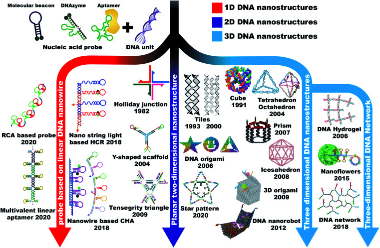
In recent years, DNA has been widely noted as a kind of material that can be used to construct building blocks for biosensing, in vivo imaging, drug development, and disease therapy because of its advantages of good biocompatibility and programmable properties. However, traditional DNA-based sensing processes are mostly achieved by random diffusion of free DNA probes, which were restricted by limited dynamics and relatively low efficiency. Moreover, in the application of biosystems, single-stranded DNA probes face challenges such as being difficult to internalize into cells and being easily decomposed in the cellular microenvironment. To overcome the above limitations, DNA nanostructure-based probes have attracted intense attention. This kind of probe showed a series of advantages compared to the conventional ones, including increased biostability, enhanced cell internalization efficiency, accelerated reaction rate, and amplified signal output, and thus improved in vitro and in vivo applications. Therefore, reviewing and summarizing the important roles of DNA nanostructures in improving biosensor design is very necessary for the development of DNA nanotechnology and its applications in biology and pharmacology. In this perspective, DNA nanostructure-based probes are reviewed and summarized from several aspects: probe classification according to the dimensions of DNA nanostructures (one, two, and three-dimensional nanostructures), the common connection modes between nucleic acid probes and DNA nanostructures, and the most important advantages of DNA self-assembled nanostructures in the applications of biosensing, imaging analysis, cell assembly, cell capture, and theranostics. Finally, the challenges and prospects for the future development of DNA nanostructure-based nucleic acid probes are also discussed.


Conflict of interest statement

There are no conflicts to declare.

Figures

Fig. 1
Fig. 1. Schematic of DNA nanostructure-based nucleic acid probes: construction and biological applications.
Fig. 2
Fig. 2. A brief history of the timeline of 1D, 2D, and 3D DNA nanostructures. 1D DNA nanostructures: RCA based probe. Reproduced from ref. with permission from the American Chemical Society, copyright 2020. Multivalent linear aptamer. Reproduced from ref. with permission from Wiley-VCH, copyright 2020. Nano-string light-based HCR. Reproduced from ref. with permission from the American Chemical Society, copyright 2018. Nanowire based CHA. Reproduced from ref. with permission from the Royal Society of Chemistry, copyright 2018. Reproduced from ref. with permission from the American Chemical Society, copyright 2019. 2D DNA nanostructures: Holliday junction. Reproduced from ref. with permission from Elsevier, copyright 1982. Double-crossover (DX) tiles (left) and triple crossover (TX) tiles (right). Reproduced from ref. with permission from the American Chemical Society, copyright 1993. Reproduced from ref. with permission from the American Chemical Society, copyright 2000. Y-shaped scaffold. Reproduced from ref. with permission from the Nature Publishing Group, copyright 2004. Reproduced from ref. with permission from the Royal Society of Chemistry, copyright 2019. DNA origami. Reproduced from ref. with permission from the Nature Publishing Group, copyright 2006. Tensegrity triangle. Reproduced from ref. with permission from the Nature Publishing Group, copyright 2009. Star pattern. Reproduced from ref. with permission from the Nature Publishing Group, copyright 2020. 3D DNA nanostructures: cube. Reproduced from ref. with permission from the Nature Publishing Group, copyright 1991. Tetrahedron. Reproduced from ref. with permission from the Royal Society of Chemistry, copyright 2004. Octahedron. Reproduced from ref. with permission from the Nature Publishing Group, copyright 2004. Reproduced from ref. with permission from the American Chemical Society, copyright 2018. Prism. Reproduced from ref. with permission from the Royal Society of Chemistry, copyright 2007. Icosahedron. Reproduced from ref. with permission from the National Academy of Sciences of the USA, copyright 2008. 3D DNA origami. Reproduced from ref. with permission from the Nature Publishing Group, copyright 2009. DNA nanorobot. Reproduced from ref. with permission from the American Association for the Advancement of Science, copyright 2012. DNA hydrogel. Reproduced from ref. with permission from the Nature Publishing Group, copyright 2006. Nanoflowers. Reproduced from ref. with permission from the Nature Publishing Group, copyright 2015. DNA network. Reproduced from ref. with permission from the Royal Society of Chemistry, copyright 2019.
Fig. 3
Fig. 3. Typical 1D nanostructure-based probe design strategies. (A) DNA nanowire-based localized CHA reaction for in situ detection of miR-21. Reproduced from ref. with permission from the Royal Society of Chemistry, copyright 2018. (B) Schematic illustration of a responsive DNA nano-string light generated by RCA. Reproduced from ref. with permission from the American Chemical Society, copyright 2018. (C) Schematic illustration of a miRNA triggered DNA “nano-wheel” for visualizing intracellular miRNA. Reproduced from ref. with permission from the American Chemical Society, copyright 2019. (D) Stepwise assembly of a DNA nanowire for cancer-targeted drug delivery. Reproduced from ref. with permission from Wiley-VCH, copyright 2020.
Fig. 4
Fig. 4. Typical 2D nanostructure-based probe design strategies. (A) Illustration of the tripartite Y-shaped DNA probe and bCHA circuit for RNA imaging. Reproduced from ref. with permission from the Royal Society of Chemistry, copyright 2019. (B) Schematics of DNA nanotube self-assembly and proposed control mechanisms. Reproduced from ref. with permission from the Nature Publishing Group, copyright 2019. (C) Schematic illustration of the construction of thrombin aptamer-loaded nanoarrays by DNA origami. Reproduced from ref. with permission from the Nature Publishing Group, copyright 2021.
Fig. 5
Fig. 5. Typical 3D nanostructure-based probe design strategies. (A) Design of a DNA tetrahedron formed by annealing four oligonucleotides. Reproduced from ref. with permission from the American Association for the Advancement of Science, copyright 2005. (B) Construction of a telomerase-responsive DNA icosahedron for platinum delivery. Reproduced from ref. with permission from Wiley-VCH, copyright 2018. (C) Design of an aptamer-gated DNA nanorobot. Reproduced from ref. with permission from the American Association for the Advancement of Science, copyright 2012. (D) Design of self-assembled multifunctional DNA nanoflowers. Reproduced from ref. with permission from the Nature Publishing Group, copyright 2015. (E) Base pairing of multidomain DNA strands to assemble a one-strand DNA hydrogel. Reproduced from ref. with permission from Wiley-VCH, copyright 2016.
Fig. 6
Fig. 6. Connection modes between nucleic acid probes and DNA nanostructures. (A) Multivalent DNA framework-based topological cell sorters. Reproduced from ref. with permission from Wiley-VCH, copyright 2020. (B) Fractal DNA frameworks with precise node numbers and molecular weights encoding quantized fluorescence states. Reproduced from ref. with permission from the Nature Publishing Group, copyright 2020. (C) Click reaction with the designated valences for generating controlled architectures. Reproduced from ref. with permission from the American Chemical Society, copyright 2019.
Fig. 7
Fig. 7. DNA nanostructure applied in biosensing. (A) 3D nanomachine-based electrochemical biosensors for rapid single-step quantitation of miRNA. Reproduced from ref. with permission from the American Chemical Society, copyright 2018. (B) Tetravalent hairpin tetrahedron-mediated hyperbranched HCR for target miR-21 sensing. Reproduced from ref. with permission from the Royal Society of Chemistry, copyright 2019. (C) Nanolantern-based DNA probe and signal amplifier for tumor-related biomarker detection. Reproduced from ref. with permission from the American Chemical Society, copyright 2019. (D) A logic gate nanomachine for cancer cell-targeted imaging. Reproduced from ref. with permission from the American Chemical Society, copyright 2018.
Fig. 8
Fig. 8. DNA nanostructure applied in bioimaging. (A) Environment recognizing DNA-computation circuits for mRNA imaging. Reproduced from ref. with permission from Wiley-VCH, copyright 2020. (B) DNA prism frame structure for sensitive and multiplexed imaging of miRNAs in living cells. Reproduced from ref. with permission from the American Chemical Society, copyright 2020. (C) Mechanism of the entropy-driven 3D DNA amplifier for intracellular mRNA imaging. Reproduced from ref. with permission from the American Chemical Society, copyright 2018. (D) A tripartite DNA probe for RNA imaging in living mice via an HCR circuit. Reproduced from ref. with permission from the Royal Society of Chemistry, copyright 2019. (E) DNA prism nanoprobes for ATP sensing in living cells. Reproduced from ref. with permission from the American Chemical Society, copyright 2017. (F) Programmable pH-responsive DNA tetrahedron for imaging exocytosis and retrieval of synaptic vesicles. Reproduced from ref. with permission from the American Chemical Society, copyright 2020.
Fig. 9
Fig. 9. DNA nanostructure applied in cell assembly and capture. (A) Cell membrane-anchored DNA tetrahedron for cell assembly. Reproduced from ref. with permission from the American Chemical Society, copyright 2019. (B) Programming cell–cell communications assembled with engineered cell origami clusters. Reproduced from ref. with permission from the American Chemical Society, copyright 2020. (C) DNA nanolithography in a microfluidic chip for efficient capture and release of CTCs. Reproduced from ref. with permission from Wiley-VCH, copyright 2020. (D) Discrimination of different cell lines by monitoring DNA tetrahedron-encoded ligand–receptor interactions on the cell membrane. Reproduced from ref. with permission from the American Chemical Society, copyright 2020.
Fig. 10
Fig. 10. DNA nanostructure applied in cancer therapy. (A) Precision-guided missile-like DNA nanostructure for aptamer-based targeted drug delivery into cancer cells. Reproduced from ref. with permission from the American Chemical Society, copyright 2020. (B) The CPT-grafted DNA tetrahedron as a stoichiometric nanomedicine for tumor therapy. Reproduced from ref. with permission from Wiley-VCH, copyright 2019. (C) Self-assembled immunostimulatory CpG oligonucleotide DNA nanostructures for immunotherapy. Reproduced from ref. with permission from the American Chemical Society, copyright 2011. (D) Programming DNA nanoflower for enhanced photodynamic therapy. Reproduced from ref. with permission from Wiley-VCH, copyright 2020. (E) DNA tetrahedron for accurate cancer identification and miRNA silencing induced therapy. Reproduced from ref. with permission from the Royal Society of Chemistry, copyright 2019. (F) RCA-based DNA nanoflower for the delivery of CRISPR/Cas12a RNA to regulate serum cholesterol levels. Reproduced from ref. with permission from the American Association for the Advancement of Science, copyright 2020.

References

    1. Consortium E. P. Nature. 2012;489:57–74. - PubMed
    1. Lacroix A. Sleiman H. F. ACS Nano. 2021;15:3631–3645. - PubMed
    1. Samanta D. Ebrahimi S. B. Mirkin C. A. Adv. Mater. 2020;32:e1901743. - PMC - PubMed
    1. Nicolson F. Ali A. Kircher M. F. Pal S. Adv. Sci. 2020;7:2001669. - PMC - PubMed
    1. Ebrahimi S. B. Samanta D. Mirkin C. A. J. Am. Chem. Soc. 2020;142:11343–11356. - PubMed