. 2021 Apr 29;12(22):7888-7901.
doi: 10.1039/d1sc01266e.

Phosphorus Kβ X-ray emission spectroscopy detects non-covalent interactions of phosphate biomolecules in situ

Affiliations

Phosphorus Kβ X-ray emission spectroscopy detects non-covalent interactions of phosphate biomolecules in situ

Zachary Mathe et al. Chem Sci. .

Abstract

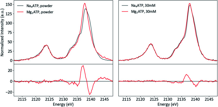
Phosphorus is ubiquitous in biochemistry, being found in the phosphate groups of nucleic acids and the energy-transferring system of adenine nucleotides (e.g. ATP). Kβ X-ray emission spectroscopy (XES) of phosphorus has been largely unexplored, with no previous applications to biomolecules. Here, the potential of P Kβ XES to study phosphate-containing biomolecules, including ATP and NADPH, is evaluated, as is the application of the technique to aqueous solution samples. P Kβ spectra offer a detailed picture of phosphate valence electronic structure, reporting on subtle non-covalent effects, such as hydrogen bonding and ionic interactions, that are key to enzymatic catalysis. Spectral features are interpreted using density functional theory (DFT) calculations, and potential applications to the study of biological energy conversion are highlighted.


Conflict of interest statement

The authors have no conflicts of interest to declare.

Figures

Fig. 1
Fig. 1. Energy level diagrams for 3d transition metal and ligand binding (left) and for phosphorus and oxygen (right) with the VtC region highlighted in green for both.
Fig. 2
Fig. 2. Correlation diagram of orbital symmetries in the Td, C3v and C2v point groups, with those matching the symmetry of the dipole operator of each point group in black and others in gray.
Fig. 3
Fig. 3. MO diagram (left) and calculated Kβ spectrum (right) of PO43−.
Fig. 4
Fig. 4. Calculated Kβ spectra of PO43−, HPO43− and H2PO43− with individual transitions shown as sticks.
Fig. 5
Fig. 5. Powder and solution P Kβ spectra of NaH2PO4, with difference (solution–powder).
Fig. 6
Fig. 6. Calculated spectra of bare H2PO4 compared to acceptor dimers (top) and hydrogen bonding dimers (bottom), with inset plots of the starred transitions' orbitals.
Fig. 7
Fig. 7. Molecular models of ATP (top), ADP (middle), and AMP (bottom).
Fig. 8
Fig. 8. Experimental (top) and calculated (bottom) spectra of ATP, ADP and AMP salts, with differences (AXP–ATP).
Fig. 9
Fig. 9. P Kβ spectra of ATP and ADP in solution, plus a simulated spectrum of the reaction product ADP + Pi, with differences (x-ATP).
Fig. 10
Fig. 10. Powder (left) and solution (right) P Kβ spectra of Na4ATP and Mg2ATP, with differences (Mg2ATP–Na4ATP).
Fig. 11
Fig. 11. Schematic reaction diagram of NADP+ and NADPH, with redox-relevant group circled in red and participating nitrogen shown in blue.
Fig. 12
Fig. 12. Powder and solution spectra of NADP+ and NADPH paired by phase (top) and redox state (bottom), with differences (red-black).

References

    1. Williams R. J. P., in Novartis Foundation Symposia, ed. R. Porter and D. W. Fitzsimons, John Wiley & Sons, Ltd., Chichester, UK, 2008, pp. 95–116
    1. Kholodar S. A. Allen C. L. Gulick A. M. Murkin A. S. The Role of Phosphate in a Multistep Enzymatic Reaction: Reactions of the Substrate and Intermediate in Pieces. J. Am. Chem. Soc. 2015;9:2748–2756. - PMC - PubMed
    1. Xue H.-W. Chen X. Mei Y. Function and regulation of phospholipid signalling in plants. Biochem. J. 2009;421:145–156. - PMC - PubMed
    1. Elston T. Wang H. Oster G. Energy transduction in ATP synthase. Nature. 1998;391:510–513. - PubMed
    1. Miller R. W. Eady R. R. Gormal C. Fairhurst S. A. Smith B. E. Nucleotide binding by the nitrogenase Fe protein: a 31P NMR study of ADP and ATP interactions with the Fe protein of Klebsiella pneumoniae. Biochem. Eng. J. 1998;334:601–607. - PMC - PubMed