. 2021 Oct;156(4):363-375.
doi: 10.1007/s00418-021-02007-7. Epub 2021 Jun 24.

Flow-through isolation of human first trimester umbilical cord endothelial cells

Affiliations

Flow-through isolation of human first trimester umbilical cord endothelial cells

Michael Gruber et al. Histochem Cell Biol. 2021 Oct.

Abstract

Human umbilical vein and artery endothelial cells (HUVEC; HUAEC), placental endothelial cells (fpAEC), and endothelial colony-forming cells (ECFC) from cord blood are a widely used model for researching placental vascular development, fetal and placental endothelial function, and the effect of adverse conditions in pregnancy thereon. However, placental vascular development and angiogenesis start in the first weeks of gestation, and adverse conditions in pregnancy may also affect endothelial function before term, suggesting that endothelial cells from early pregnancy may respond differently. Thus, we established a novel, gentle flow-through method to isolate pure human umbilical endothelial cells from first trimester (FTUEC). FTUEC were characterized and their phenotype was compared to the umbilical endothelium in situ as well as to other fetal endothelial cell models from term of gestation, i.e. HUVEC, fpAEC, ECFC. FTUEC possess a CD34-positive, juvenile endothelial phenotype, and can be expanded and passaged. We regard FTUEC as a valuable tool to study developmental processes as well as the effect of adverse insults in pregnancy in vitro.

Keywords: Endothelial cells; First trimester; HUAEC; HUVEC; Human placenta; Isolation; Umbilical cord.


Conflict of interest statement

The authors declare no competing interests.

Figures

Fig. 1
Fig. 1
Human first trimester umbilical cord cannulation and isolation of endothelial cells. The isolation protocol included separation of the umbilical cord close to the cord insertion, followed by cannulation of the umbilical cord vessels under a stereomicroscope. Enzymatic flow-through digestion was followed by fractionated collection of umbilical cord EC (a). First trimester placenta tissue with umbilical cord (b), and size comparison with a 1 euro cent coin (c). Sequential cannulation of umbilical cord vessels using neonatal umbilical catheters (df). Successful cannulation was confirmed by HE staining of formalin-fixed paraffin-embedded (FFPE) first trimester umbilical cord sections, showing positioning of the catheters (red arrows) inside the cord vessels (g). Scale bar represents 200 µm. Figure parts were created with BioRender.com
Fig. 2
Fig. 2
Isolation and culture of first trimester umbilical cord endothelial cells. After enzymatic flow-through digestion of the umbilical cord endothelium, the FFPE cord was subjected to histological examination for vWF staining at the proximal (a) and distal (b) sides of catheter insertion. At the side proximal to catheter positioning, remnants of tissue marking dye (green) were detected on the endothelial lining (a). At the distal side (b), distinct vWF staining revealed the presence of endothelia in two cord vessels, whereas the third vessel lacked immunostaining due to depletion of EC (red arrow). After 7 days in culture, FTUEC appeared as small colonies (c), which further expanded after 8 (d) and 9 (e) days in culture. Following first passaging, cells achieved appropriate confluence (f). Scale bars represent 100 µm. Figure parts were created with BioRender.com
Fig. 3
Fig. 3
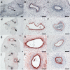
Immunohistochemistry of vWF in human first trimester umbilical cord tissue. Immunohistochemistry for vWF in umbilical cord tissue at gestational age 7+3 (ac), 9+5 (df), 10+3 (gi), and 11+6 (jl) revealed a gestational age-dependent increase in subendothelial and perivascular vWF location in umbilical veins (black arrows, and in higher magnification in b, e, h, and k), while vWF staining was confined to the endothelia of umbilical arteries (open arrows, and in higher magnification in c, f, i, and l). Scale bars represent 100 µm
Fig. 4
Fig. 4
Immunostaining of FTUEC and first trimester umbilical cord tissue. Immunocytochemistry for vWF (a) and CD31 (b) showed strong staining of FTUEC and in situ umbilical cord arteries. CD34 (c) was moderately stained in a proportion of FTUEC, while the endothelium of umbilical arteries was distinctly stained. FTUEC as well as umbilical endothelium was also stained for vimentin (VIM) (d), as was the tunica media and the surrounding Wharton's jelly in umbilical cords. Staining for CD45 (e) and pan cytokeratin (CK wide, f) gave no signals either in cells or in umbilical cord sections. Staining for smooth muscle actin (SMA, g) was negative in FTUEC, and predominantly stained the tunica media of umbilical blood vessels. Negative controls for mouse (h) and rabbit (i) IgG showed no staining. Scale bars represent 100 µm
Fig. 5
Fig. 5
Cellular morphology and size distribution of FTUEC and term pregnancy endothelial cells. FTUEC (a, e), fpAEC (b, f), ECFC (c, g), and HUVEC (d, h) were grown to 80–90% confluence (ad) and subjected to flow cytometric analysis without staining, separated according to size and granularity (eh). Scale bars represent 200 µm
Fig. 6
Fig. 6
Proliferation of FTUEC and term pregnancy endothelial cells. Absolute cell numbers of placenta- and cord blood-derived EC were determined by electric field multichannel cell counting at indicated time points (a). Cell proliferation, based on EdU incorporation, was analyzed by software-based image analysis at experimental start, i.e. after adherence of cells at 0 h, and 24 h of culture (b). Representative images are shown for FTUEC (c), fpAEC (d), ECFC (e), and HUVEC (f) after 24 h of culture. Blue Hoechst® 33342 dye marks all cells; the purple stain is specific for proliferating cells. Scale bar represents 100 µm

References

    1. Blaschitz A, Gauster M, Fuchs D, Lang I, Maschke P, Ulrich D, Karpf E, Takikawa O, Schimek MG, Dohr G, Sedlmayr P. Vascular endothelial expression of indoleamine 2,3-dioxygenase 1 forms a positive gradient towards the feto-maternal interface. PLoS ONE. 2011;6(7):e21774. doi: 10.1371/journal.pone.0021774. - DOI - PMC - PubMed
    1. Carpenter MW. Gestational diabetes, pregnancy hypertension, and late vascular disease. Diabetes Care. 2007;30(Suppl 2):S246–250. doi: 10.2337/dc07-s224. - DOI - PubMed
    1. Charolidi N, Host AJ, Ashton S, Tryfonos Z, Leslie K, Thilaganathan B, Cartwright JE, Whitley GS. First trimester placental endothelial cells from pregnancies with abnormal uterine artery Doppler are more sensitive to apoptotic stimuli. Lab Invest. 2019;99(3):411–420. doi: 10.1038/s41374-018-0139-z. - DOI - PMC - PubMed
    1. Cvitic S, Desoye G, Hiden U. Glucose, insulin, and oxygen interplay in placental hypervascularisation in diabetes mellitus. Biomed Res Int. 2014;2014:145846. doi: 10.1155/2014/145846. - DOI - PMC - PubMed
    1. Cvitic S, Novakovic B, Gordon L, Ulz CM, Muhlberger M, Diaz-Perez FI, Joo JE, Svendova V, Schimek MG, Trajanoski S, Saffery R, Desoye G, Hiden U. Human fetoplacental arterial and venous endothelial cells are differentially programmed by gestational diabetes mellitus, resulting in cell-specific barrier function changes. Diabetologia. 2018;61(11):2398–2411. doi: 10.1007/s00125-018-4699-7. - DOI - PMC - PubMed

LinkOut - more resources