. 2021 Jun 24;13(13):17901-17913.
doi: 10.18632/aging.203196. Epub 2021 Jun 24.

Chloroquine suppresses proliferation and invasion and induces apoptosis of osteosarcoma cells associated with inhibition of phosphorylation of STAT3

Affiliations

Chloroquine suppresses proliferation and invasion and induces apoptosis of osteosarcoma cells associated with inhibition of phosphorylation of STAT3

Chenglong Chen et al. Aging (Albany NY). .

Abstract

Background: Osteosarcoma (OS) is characterized by a high rate of metastasis. It has been found that tumor cells can bypass apoptosis which leads to an uncontrolled proliferation, but chloroquine (CQ) can have an effect on the tumors by inducing apoptosis. We aimed to explore the effects and the hypothetical mechanism of CQ effects on OS.

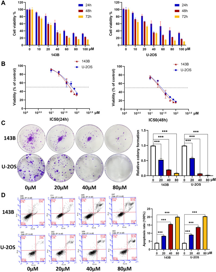
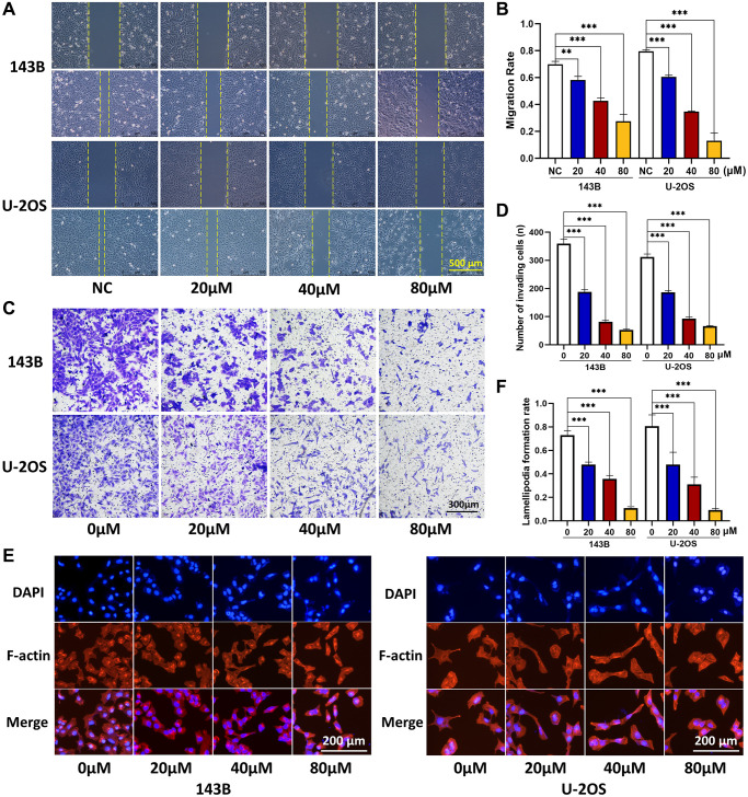
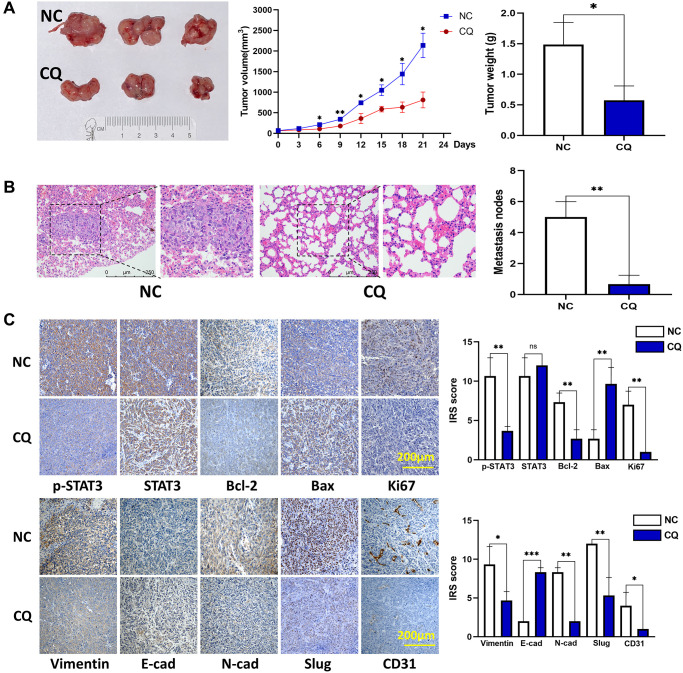
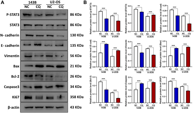
Methods: We first estimated the CQ effects on proliferation, apoptosis, migration, invasion, and lamellipodia formation of OS cells. Mice bearing xenograft model were used to test the anti-tumor growth and lung metastasis effects of CQ in OS. Western blot and immunohistochemistry were used to explore the mechanism of CQ effects and the association between p-STAT3 expression and lung metastasis of OS patients.

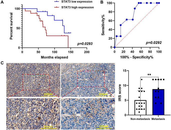
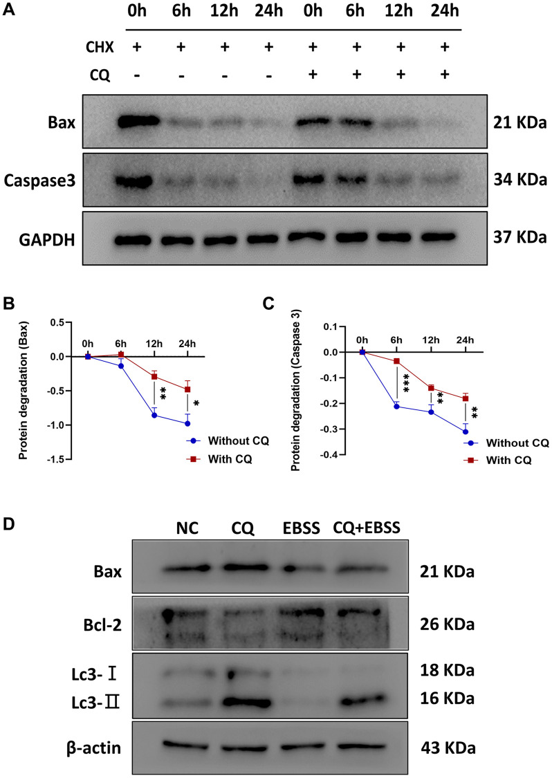
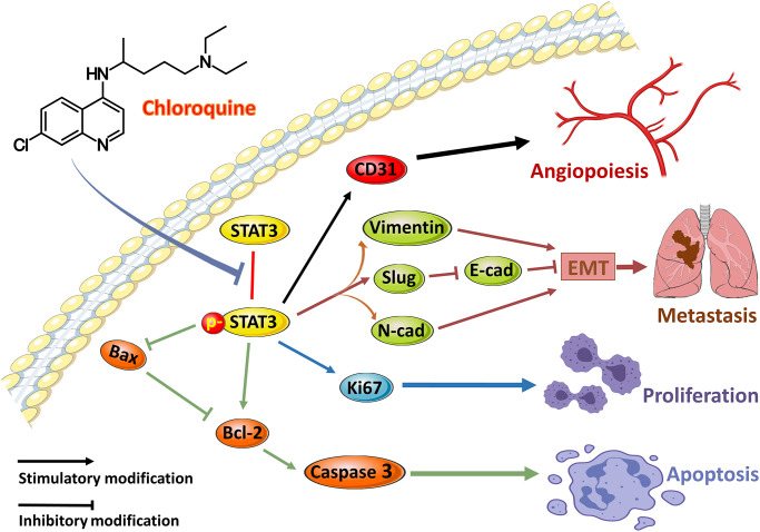
Results: CQ induces the apoptosis and suppressed the viability, proliferation, migration, invasion, and lamellipodia formation of OS cells in vitro. In vivo experiments demonstrated that CQ inhibited tumor growth and lung metastasis. CQ induced apoptosis was dependent on the lysosomal inhibition and inhibition of protein turnover. The lung metastasis was associated with the p-STAT3 expression in OS patients.

Conclusion: CQ inhibited progression of OS cells in vitro, and suppressed tumor growth and lung metastasis in vivo. p-STAT3 can be a predictive biomarker for lung metastasis in osteosarcoma patients.

Keywords: apoptosis; chloroquine; metastasis; osteosarcoma; p-STAT3.


Conflict of interest statement

CONFLICTS OF INTEREST: The authors declare no conflicts of interest related to this study.

Figures

Figure 1
Figure 1
CQ induces the apoptosis and inhibits the viability and proliferation of OS cells. (A) The OS cell viability at 24 h, 48 h, and 72 h after treated with CQ (n = 3). (B) The IC50 value at 24 h and 48 h in 143B and U-2OS cells (n = 3). (C) The colony formation assay showed that the proliferation of OS cells was inhibited gradually by CQ with the increasing of concentration (***p < 0.001; n = 3). (D) Flow cytometry showed that CQ could regulate apoptosis of OS cells (***p < 0.001; n = 3). All the data were expressed as the mean ± SD.
Figure 2
Figure 2
CQ suppress the OS cell migration, invasion, and lamellipodia formation. (A, B) Migration ability of OS cells was inhibited gradually by CQ with the increasing of concentration (**P < 0.01; ***p < 0.001; n = 3). (C, D) Invasion ability of OS cells was inhibited gradually by CQ with the increasing of concentration (***p < 0.001; n = 3). (E, F) Cytoskeletal assay showed that the lamellipodia formation of OS cells was inhibited gradually by CQ with the increasing of concentration (***p < 0.001; n = 3). The data were expressed as the mean ± SEM.
Figure 3
Figure 3
CQ suppression of mouse OS cell xenograft growth and lung metastasis in vivo. (A) Xenograft photographs, growth curves of tumor, and tumor weight showed that CQ suppressed xenograft growth in nude mice (*p < 0.05; n = 3). (B) HE staining of mouse lung tissues. The results showed suppression of lung metastasis in vivo (**p < 0.01; n = 3). (C) Representative pictures from IHC assay. p-STAT3, STAT3, Bcl-2, Bax, Ki67, Vimentin, E-cadherin, N-cadherin, Slug, and CD31 expression in the tumors collected from different groups were determined using IHC (ns p > 0.05; *p < 0.05; **p < 0.01; ***p < 0.001; n = 3). The data were expressed as the mean ± SD.
Figure 4
Figure 4
The expression of E-cadherin, Bax, Caspase3, and CD31 were up-regulated, and p-STAT3, N-cadherin, Vimentin, Bcl-2, Ki67, Slug were downregulated by CQ treatment in OS cells. (A) Representative pictures from tumor cell western blot assay. (B) The graph is the quantified data of the western blot, and β-actin was used as a load control (**p < 0.01; ***p < 0.001 n = 3). The data were expressed as the mean ± SD.
Figure 5
Figure 5
p-STAT3 expression was elevated by IHC in OS and associated with poor prognosis. (A) Kaplan–Meier curves showed the p-STAT3 expression on overall survival in 30 OS patients (*p < 0.05; n = 30). (B) ROC curve of predictive value of p-STAT3 expression on lung metastasis of OS patients (*p < 0.05; n = 30). (C) IHC staining of p-STAT3 in OS samples (**p < 0.01; n = 30). The data were expressed as the mean ± SD.
Figure 6
Figure 6
CQ reduced the protein degradation of Bax and Caspase3, and CQ-induced apoptosis of OS cells was dependent on the lysosomal inhibition. The (A) Representative pictures from tumor cell western blot assay. (B) The graph is the quantified data of the western blot for the expression of BAX, and GAPDH was used as a load control (*p < 0.05; **p < 0.01; n = 3). (C) The graph is the quantified data of the western blot for the expression of Caspase3, and GAPDH was used as a load control (**p < 0.01; ***p < 0.001; n = 3). (D) Representative pictures from 143B cells western blot assay and the cells were treated with PBS, 20 μM CQ, EBSS, or the combination of CQ and EBSS. The data were expressed as the mean ± SD.
Figure 7
Figure 7
The schematic of the hypothetic mechanisms of CQ effects on p-STAT3/Bcl-2/Caspase3 pathway. CQ inhibited the phosphorylation of STAT3 and then by downregulating the expression of p-STAT3 to suppress the CD31 to inhibit angiopoiesis, and suppress N-cadherin, Vimentin, and Slug as well as downregulate E-cadherin to inhibit EMT and metastasis, and suppress Ki67 to inhibit proliferation, as well as upregulate Bax and Caspase3 and downregulate Bcl-2 to induce apoptosis of OS cells.

References

    1. Siegel RL, Miller KD, Jemal A. Cancer statistics, 2018. CA Cancer J Clin. 2018; 68:7–30. 10.3322/caac.21442 - DOI - PubMed
    1. Han Y, Guo W, Ren T, Huang Y, Wang S, Liu K, Zheng B, Yang K, Zhang H, Liang X. Tumor-associated macrophages promote lung metastasis and induce epithelial-mesenchymal transition in osteosarcoma by activating the COX-2/STAT3 axis. Cancer Lett. 2019; 440–441:116–25. 10.1016/j.canlet.2018.10.011 - DOI - PubMed
    1. Lussier DM, Johnson JL, Hingorani P, Blattman JN. Combination immunotherapy with α-CTLA-4 and α-PD-L1 antibody blockade prevents immune escape and leads to complete control of metastatic osteosarcoma. J Immunother Cancer. 2015; 3:21. 10.1186/s40425-015-0067-z - DOI - PMC - PubMed
    1. Mohammad RM, Muqbil I, Lowe L, Yedjou C, Hsu HY, Lin LT, Siegelin MD, Fimognari C, Kumar NB, Dou QP, Yang H, Samadi AK, Russo GL, et al. Broad targeting of resistance to apoptosis in cancer. Semin Cancer Biol. 2015. (Suppl); 35:S78–103. 10.1016/j.semcancer.2015.03.001 - DOI - PMC - PubMed
    1. Lakhter AJ, Sahu RP, Sun Y, Kaufmann WK, Androphy EJ, Travers JB, Naidu SR. Chloroquine promotes apoptosis in melanoma cells by inhibiting BH3 domain-mediated PUMA degradation. J Invest Dermatol. 2013; 133:2247–54. 10.1038/jid.2013.56 - DOI - PMC - PubMed

Publication types

MeSH terms