. 2021 Jun 25;16(6):e0253456.
doi: 10.1371/journal.pone.0253456. eCollection 2021.

The effect of human amnion epithelial cells on lung development and inflammation in preterm lambs exposed to antenatal inflammation

Affiliations

The effect of human amnion epithelial cells on lung development and inflammation in preterm lambs exposed to antenatal inflammation

Paris Clarice Papagianis et al. PLoS One. .

Abstract

Background: Lung inflammation and impaired alveolarization are hallmarks of bronchopulmonary dysplasia (BPD). We hypothesize that human amnion epithelial cells (hAECs) are anti-inflammatory and reduce lung injury in preterm lambs born after antenatal exposure to inflammation.

Methods: Pregnant ewes received either intra-amniotic lipopolysaccharide (LPS, from E.coli 055:B5; 4mg) or saline (Sal) on day 126 of gestation. Lambs were delivered by cesarean section at 128 d gestation (term ~150 d). Lambs received intravenous hAECs (LPS/hAECs: n = 7; 30x106 cells) or equivalent volumes of saline (LPS/Sal, n = 10; or Sal/Sal, n = 9) immediately after birth. Respiratory support was gradually de-escalated, aimed at early weaning from mechanical ventilation towards unassisted respiration. Lung tissue was collected 1 week after birth. Lung morphology was assessed and mRNA levels for inflammatory mediators were measured.

Results: Respiratory support required by LPS/hAEC lambs was not different to Sal/Sal or LPS/Sal lambs. Lung tissue:airspace ratio was lower in the LPS/Sal compared to Sal/Sal lambs (P<0.05), but not LPS/hAEC lambs. LPS/hAEC lambs tended to have increased septation in their lungs versus LPS/Sal (P = 0.08). Expression of inflammatory cytokines was highest in LPS/hAECs lambs.

Conclusions: Postnatal administration of a single dose of hAECs stimulates a pulmonary immune response without changing ventilator requirements in preterm lambs born after intrauterine inflammation.


Conflict of interest statement

The University of Western Australia (via JJP) has consultancy agreements with Chiesi Farmaceutici S.p.A. unrelated to the subject of this study. Fisher & Paykel Healthcare have material transfer agreements with Hudson Research Institute that are also unrelated to this work. There are no other relevant interests relating to employment, consultancy, patents, or products in development or marketed products to declare. These material transfer agreements do not alter our adherence to PLOS ONE policies on sharing data and materials. None of the authors have conflicts of interest to disclose.

Figures

Fig 1
Fig 1. pH, PaCO2, PaO2, HCO3 and BE of Sal/Sal lambs (open circles), LPS/Sal lambs (orange circles) and LPS/hAEC lambs (blue circles).
Cord blood gases were analysed separately to postnatal blood gases days 1–6 (D1-D6). ^Signifies P<0.05 between Sal/Sal and LPS/Sal. *Signifies P<0.05 between Sal/Sal and LPS/hAECs &Time signifies P<0.05 change over 1–6 days, not between treatment groups. Data are mean ± SD.
Fig 2
Fig 2. Heat maps depicting intensity of ventilator support in preterm lambs.
Types of respiratory support are graded according to decreasing invasivity and intensity of support: Red—intubated and mechanically ventilated (MV); orange–intubated with continuous positive airway pressure (ET CPAP); green–nasal CPAP (nCPAP); light blue—nasal humidified high flow (nHHF) and blue—no support (none). Lambs within Sal/Sal, LPS/Sal and LPS/hAEC groups are represented in left-to-right in the order of delivery. The first 6 days of respiratory support are presented as columns, from left-to-right, as the proportion of time spend on any mode of respiratory support on that day.
Fig 3
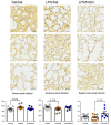
Fig 3. Representative images depicting variability in lung parenchyma of preterm lambs within the same treatment group.
Quantification of tissue, airspace and septal crest areal fractions in the lungs of preterm lambs on day 7 of life. Elastin is stained in black and tissue in yellow. All images are taken at 20X magnification. Scale is 50 μm.
Fig 4
Fig 4. Proliferating Ki67+ cells within the tissue and airspace of the lungs, within the tissue only, or within the airspaces only of preterm lambs on day 7 of life.
Representative images of the lungs of Sal/Sal, LPS/Sal and LPS/hAEC treated preterm lambs. Clumps of proliferating cells were often noted in the airspaces of animals exposed to antenatal LPS. Line represents mean. Scale is 50 μm.
Fig 5
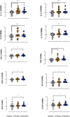
Fig 5. mRNA expression in the lungs of Sal/Sal (clear circles), LPS/Sal (orange circles) and LPS/hAEC (blue circles) treated preterm lambs.
Data represent calibrated normalized relative quantity (CNRQ). Sal/Sal group is normalized to approximately 0.00 using qBase+ software. LPS/Sal and LPS/hAEC groups are expressed as a fold change from Sal/Sal. Open circles are Sal/Sal, yellow circles are LPS/Sal and blue circles are LPS/hAECs. *Signifies P<0.05 between Sal/Sal and Sal/hAEC. ^Signifies P<0.05 between Sal/Sal and LPS/Sal. Data are mean ± SD in duplicates.
Fig 6
Fig 6. hAEC conditioned media at 33°C, 37°C or 39°C does not influence mouse macrophage phagocytic activity.
Immortalized mouse macrophages were incubated with standard media or with hAEC-conditioned media from temperatures 33°C, 37°C and 39°C (n = 9, performed in triplicate). Percentage represents proportion of cells that phagocytosed FITC-marked fluorescent beads. Open circles are representative of hAECs cultured at 33°C, grey circles are hAECs cultured at 37°C and black circles are hAECs cultured at 39°C.

Similar articles

Cited by

References

    1. Been JV, Rours IG, Kornelisse RF, Jonkers F, de Krijger RR, Zimmermann LJ. Chorioamnionitis alters the response to surfactant in preterm infants. J Pediatr. 2010;156(1):10–5 e1. doi: 10.1016/j.jpeds.2009.07.044 - DOI - PubMed
    1. Lahra MM, Beeby PJ, Jeffery HE. Maternal versus fetal inflammation and respiratory distress syndrome: a 10-year hospital cohort study. Arch Dis Child Fetal Neonatal Ed. 2009;94(1):F13–6. doi: 10.1136/adc.2007.135889 - DOI - PubMed
    1. Kallapur SG, Kramer BW, Nitsos I, Pillow JJ, Collins JJ, Polglase GR, et al.. Pulmonary and systemic inflammatory responses to intra-amniotic IL-1alpha in fetal sheep. Am J Physiol Lung Cell Mol Physiol. 2011;301(3):L285–95. doi: 10.1152/ajplung.00446.2010 - DOI - PMC - PubMed
    1. Viscardi RM, Muhumuza CK, Rodriguez A, Fairchild KD, Sun CC, Gross GW, et al.. Inflammatory markers in intrauterine and fetal blood and cerebrospinal fluid compartments are associated with adverse pulmonary and neurologic outcomes in preterm infants. Pediatr Res. 2004;55(6):1009–17. doi: 10.1203/01.pdr.0000127015.60185.8a - DOI - PubMed
    1. Choi CW, Kim BI, Kim HS, Park JD, Choi JH, Son DW. Increase of interleukin-6 in tracheal aspirate at birth: a predictor of subsequent bronchopulmonary dysplasia in preterm infants. Acta Paediatr. 2006;95(1):38–43. - PubMed

Publication types

Substances