

Gastric cancer - histopathological correlations between tumor and non-tumor gastric mucosa changes
- PMID: 34171062
- PMCID: PMC8343497
- DOI: 10.47162/RJME.61.4.15
Gastric cancer - histopathological correlations between tumor and non-tumor gastric mucosa changes
Abstract
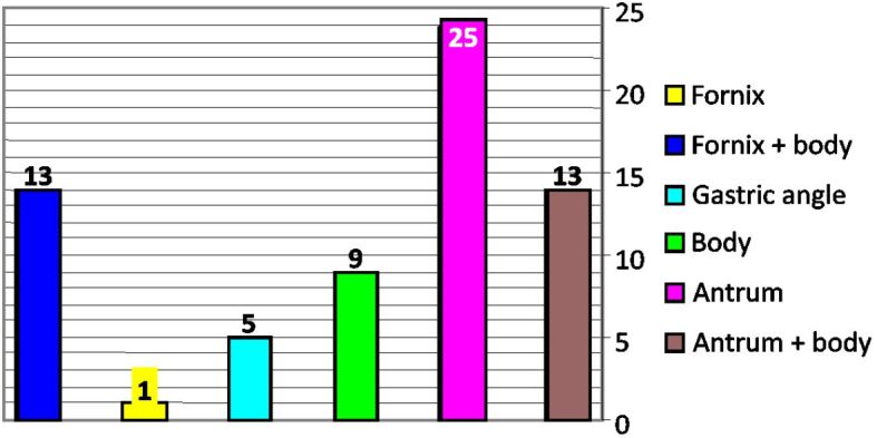
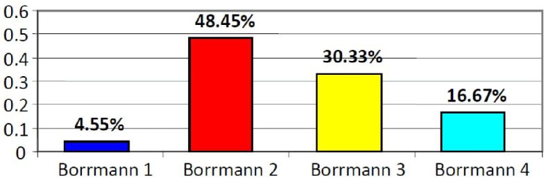
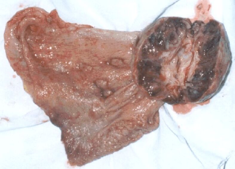
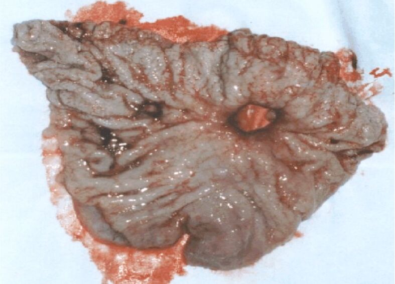
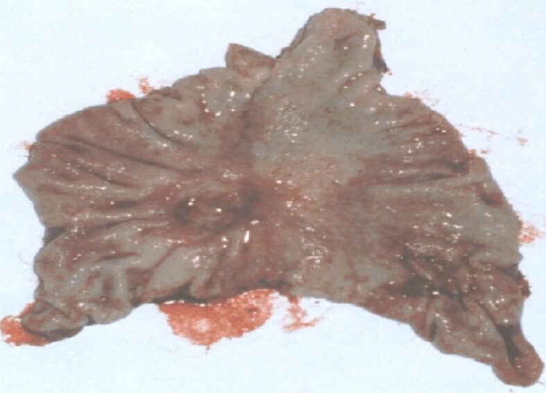
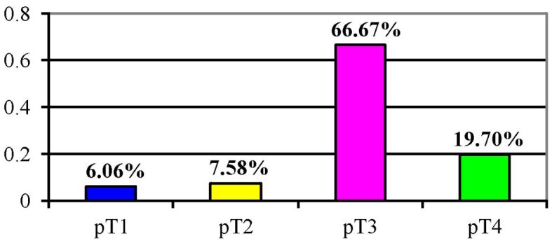
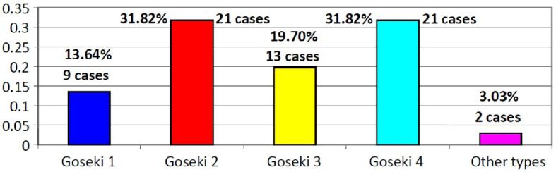
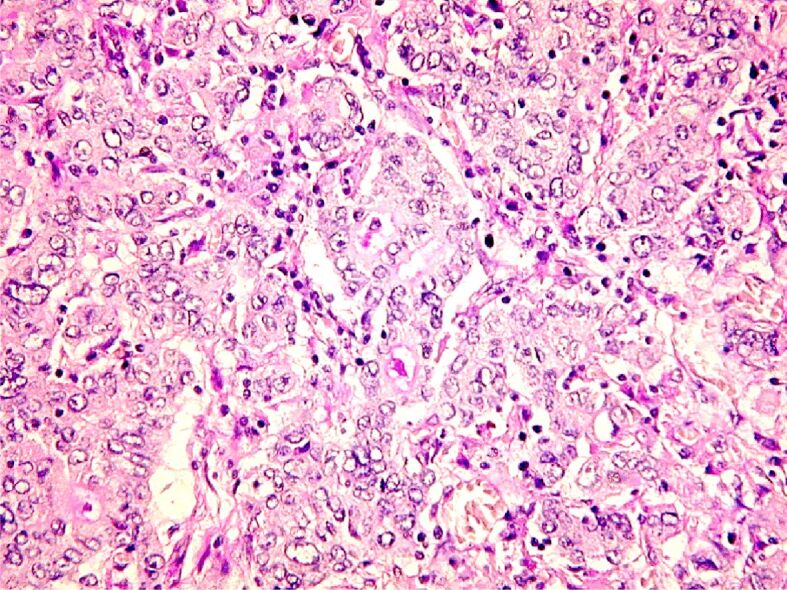
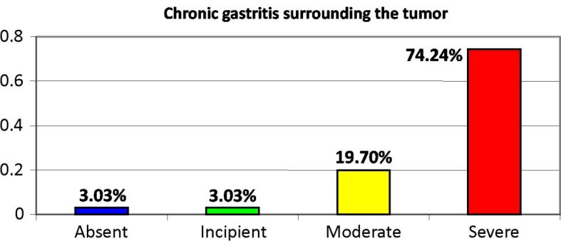
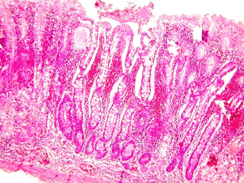
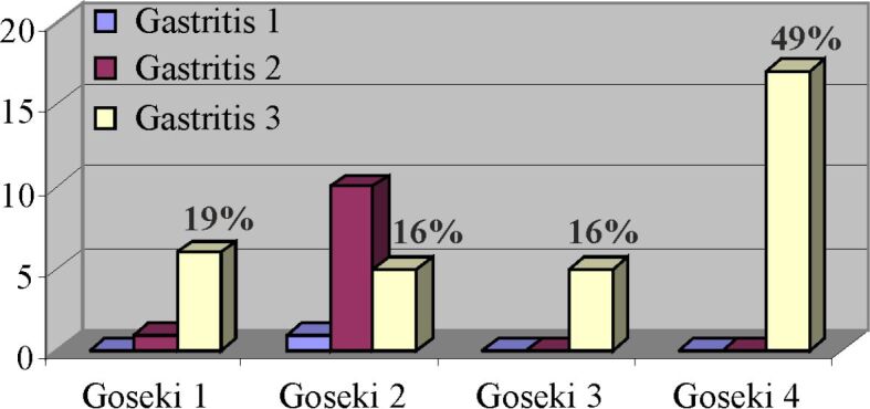
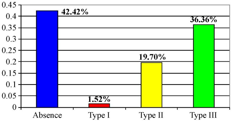
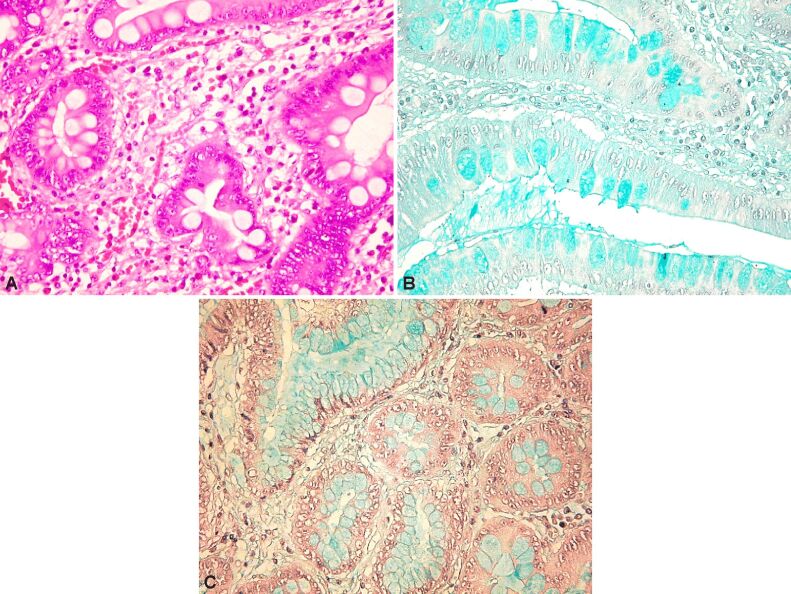
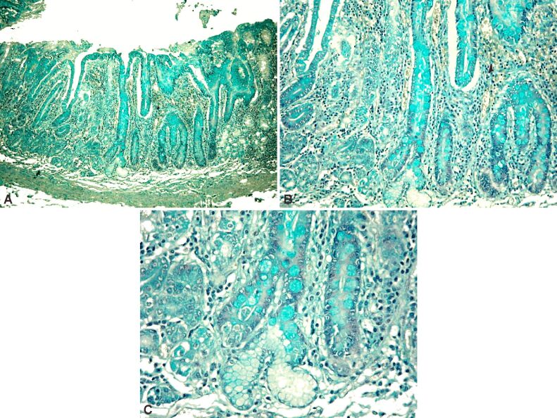
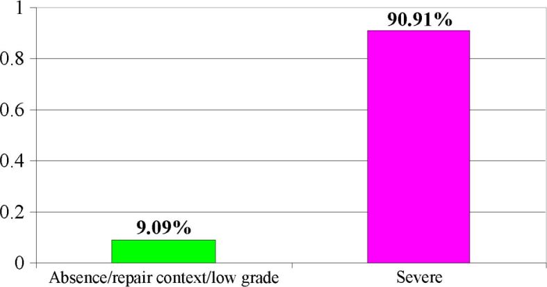
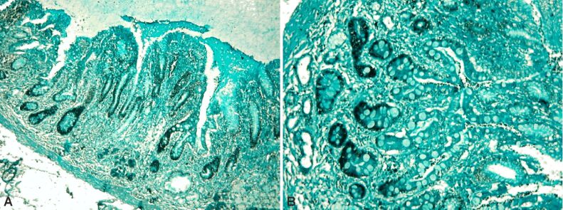
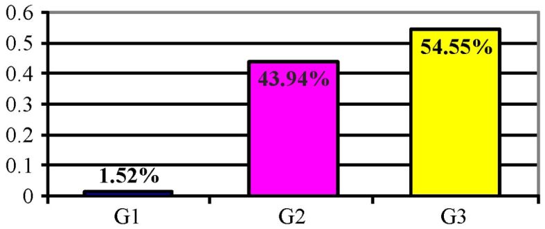
Gastric cancer is a widely geographically distributed malignancy with high prevalence, therefore being a serious health problem that needs standardized methods for early diagnosis and treatment. The aim of the study was to evaluate the correlation of some epidemiological and clinical data with the histological features. The study group was made up of 66 patients that underwent surgical removal of the gastric neoplasm, and the pathological exam showed the morphological features of the tumor, as well as the ones of the unaffected mucosal tissue. Topographically, the highest incidence of the tumor was registered in the gastric antrum, but in recent years, an increased incidence of the superior gastric pole localization was recorded. The macroscopic aspects reveal that the ulcerated type 2 Borrmann is the most frequent, and alongside type 3 Borrmann, the ulcer-infiltrative type represents most of the gastric antrum cancers. The analysis of the tumor invasion showed that most carcinomas underwent surgery when the tumor invaded the serosa (pT3) or even the perigastric tissues (pT4). In our research, we chose Goseki's microscopic classification because of its best coverage of the histological heterogeneity of the gastric carcinomas, providing information about the percentage of the cellular and secretory differentiation with direct impact on the invasion of the tumor. In more than 70% of the cases, the patients showed lesions of severe chronic atrophic gastritis of the non-tumor mucosa. Lately, the incidence of Helicobacter pylori has been 5.5%, lower than indicated by mainstream literature. We observed that the incidence of type 3 incomplete intestinal metaplasia, as the most commonly involved factor in the etiopathogenesis of gastric neoplasms, was encountered in 36.3% of the cases, this percentage rising proportionally with age and being frequently associated with antrum tumors. In conclusion, the permanent analysis of the relation between epidemiological data and some histological features might be relevant for the characterization of the tumoral process or the non-tumor gastric mucosa, leading to an evaluation of the prognosis.
Conflict of interest statement
The authors declare no conflict of interests.
Figures

Similar articles
-
Comparison of Helicobacter pylori infection and gastric mucosal histological features of gastric ulcer patients with chronic gastritis patients.World J Gastroenterol. 2005 Feb 21;11(7):976-81. doi: 10.3748/wjg.v11.i7.976. World J Gastroenterol. 2005. PMID: 15742399 Free PMC article.
-
Antral-type mucosa in the gastric incisura, body, and fundus (antralization): a link between Helicobacter pylori infection and intestinal metaplasia?Am J Gastroenterol. 2000 Jan;95(1):114-21. doi: 10.1111/j.1572-0241.2000.01609.x. Am J Gastroenterol. 2000. PMID: 10638568
-
Role of Helicobacter pylori in gastric carcinoma.Natl Med J India. 1995 Mar-Apr;8(2):58-60. Natl Med J India. 1995. PMID: 7735060
-
Atrophy-metaplasia-dysplasia-carcinoma sequence in the stomach: a reality or merely an hypothesis?Best Pract Res Clin Gastroenterol. 2001 Dec;15(6):983-98. doi: 10.1053/bega.2001.0253. Best Pract Res Clin Gastroenterol. 2001. PMID: 11866488 Review.
-
Gastric atrophy, metaplasia, and dysplasia: a clinical perspective.J Clin Gastroenterol. 2003 May-Jun;36(5 Suppl):S29-36; discussion S61-2. doi: 10.1097/00004836-200305001-00006. J Clin Gastroenterol. 2003. PMID: 12702963 Review.
Cited by
-
Expression of M3 muscarinic acetylcholine receptors in gastric cancer.Rom J Morphol Embryol. 2021 Oct-Dec;62(4):1001-1010. doi: 10.47162/RJME.62.4.12. Rom J Morphol Embryol. 2021. PMID: 35673819 Free PMC article.
-
Hawthorn with "homology of medicine and food": a review of anticancer effects and mechanisms.Front Pharmacol. 2024 Jun 10;15:1384189. doi: 10.3389/fphar.2024.1384189. eCollection 2024. Front Pharmacol. 2024. PMID: 38915462 Free PMC article. Review.
References
-
- Parkin DM, Bray F, Ferlay J, Pisani P. Global cancer statistics, 2002. CA Cancer J Clin. 2005;55(2):74–108. - PubMed
-
- Zhang X, Li M, Chen S, Hu J, Guo Q, Liu R, Zheng H, Jin Z, Yuan Y, Xi Y, Hua B. Endoscopic screening in Asian countries is associated with reduced gastric cancer mortality: a meta-analysis and systematic review. Gastroenterology. 2018;155(2):347–354e9. - PubMed
MeSH terms
LinkOut - more resources
Full Text Sources
Medical
