. 2020 Oct-Dec;61(4):1163-1171.
doi: 10.47162/RJME.61.4.18.

Fortuitous discovery of melanomas in the ENT Department - a histopathological and immunohistochemical study

Affiliations

Fortuitous discovery of melanomas in the ENT Department - a histopathological and immunohistochemical study

Cornelia Marina Trandafir et al. Rom J Morphol Embryol. 2020 Oct-Dec.

Abstract

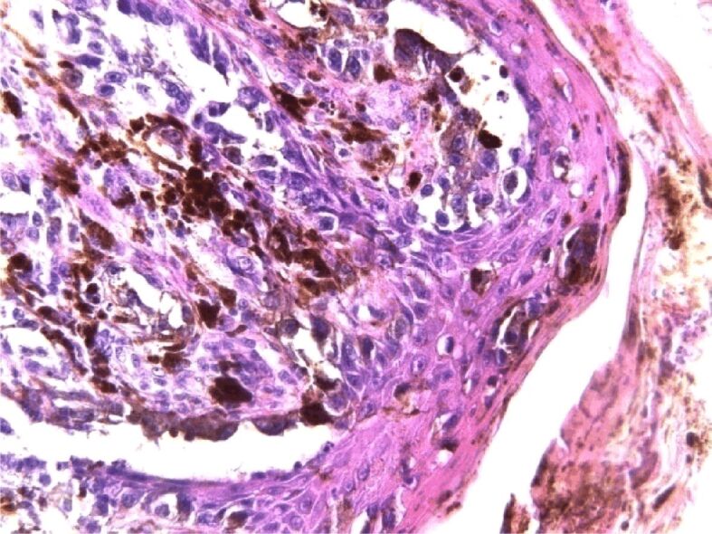
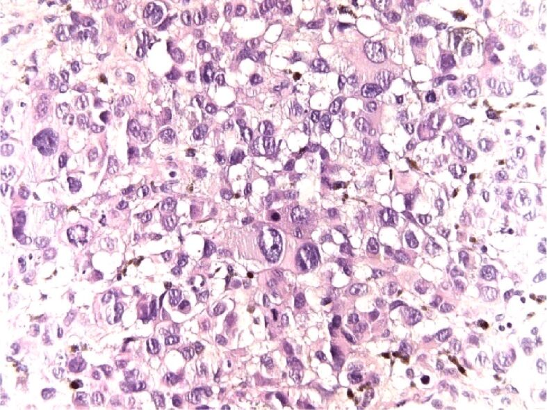
The melanoma, having its origin in the melanocyte cells, is one of the most aggressive forms of skin cancer in the world with one of the highest rates of brain metastasis. The incidence of cutaneous melanoma in the Mediterranean countries varies from three to five cases∕100 000 people∕year. Its prognosis is based on an early diagnosis. Sinonasal mucosal melanoma (SNMM) is an extremely rare tumor, accounting for 0.3-2% of all melanomas. The non-specific symptomatology is often delaying the presentation of the patient at the hospital and therefore the diagnosis. The SNMM is a highly aggressive tumor, and the presence of metastasis at the diagnosis usually implies a poor prognosis. The management of the melanomas requires a precise pre-therapeutic assessment and a multidisciplinary approach for the diagnosis, with surgical treatment or radiotherapy required in order to ensure a better a quality of life. In this paper, we retrospectively analyzed two cases of mucosal melanoma and one case of cutaneous melanoma of the nose.


Conflict of interest statement

The authors declare that they have no conflict of interests.

Figures

Figure 1
Figure 1
Tumor cell at dermo–epidermal junction and organized in nests in the superficial dermis. The tumor is not ulcerated. Cutaneous superficial spreading melanoma. HE staining, ×400. HE: Hematoxylin–Eosin
Figure 2
Figure 2
Tumoral melanocytes rise in the epidermis among the cell of the granular layer. Epithelioid melanocytes with rich intracytoplasmic melanin pigment, condensed chromatin on the nuclear membrane that gives the nucleus a vesicular appearance and eosinophilic macronucleoli. Epidermal invasion. HE staining, ×400
Figure 3
Figure 3
In the dermis are nests of epithelioid and fusiform melanocytes, inflammatory cells consisting of lymphocytes and melanophages arranged in spots, around and in between the tumor cells islands. Superficial spreading melanoma, nests in dermis. HE staining, ×400
Figure 4
Figure 4
Along the sheath of the hair follicle, the tumor cells expand lentiginous; in the surrounding dermis are lymphocytes and melanophages. Lentiginous growth along the hair follicle. HE staining, ×400
Figure 5
Figure 5
Epithelioid cells with eosinophilic cytoplasm, highly pleomorphic vesicular nuclei and eosinophilic macronucleolus; some of the cells are binucleated or multinucleated. Mucosal melanoma with epithelioid cells. HE staining, ×400
Figure 6
Figure 6
Nests of epithelioid tumor cells with clear cytoplasm and minimal amount of brown pigment located near small blood vessels, inflammatory cell such as lymphocytes, plasma cell and macrophages with brown pigment. Mucosal melanoma. HE staining, ×400
Figure 7
Figure 7
Intensely positive HMB45 on tumor cell, cytoplasmic immunoreaction. Anti-HMB45 antibody immunomarking, ×400. HMB45: Human melanoma black 45
Figure 8
Figure 8
Diffuse intense S100 positivity in tumor cell, cytoplasmic immunoreaction. Anti-S100 antibody immunomarking, ×400
Figure 9
Figure 9
Intensely positive Melan A cytoplasmic immunoreaction. Anti-Melan A antibody immunomarking, ×400
Figure 10
Figure 10
A few tumor cells with Ki67 positive nuclear immunoreaction. Anti-Ki67 antibody immunomarking, ×400
Figure 11
Figure 11
Large tumor cell with reduced eosinophilic cytoplasm, vesicular nuclei with unevenly distributed chromatin pattern, multiple eosinophilic macronucleoli and numerous atypical mitoses. Mucosal amelanotic melanoma. HE staining, ×400
Figure 12
Figure 12
Dyscohesive tumor cell, atypical mitoses and red blood cells extravasation among tumor cell. Mucosal amelanotic melanoma. HE staining, ×400
Figure 13
Figure 13
Intense Melan A positivity in tumor cells of cytoplasmic immunoreaction. Anti-Melan A antibody immunomarking, ×400
Figure 14
Figure 14
Cytoplasmic and nuclear immunoreaction in some tumor cell for S100 protein. Anti-S100 antibody immunomarking, ×400
Figure 15
Figure 15
Intensely and homogenous positive nuclear immunoreaction on tumor cell for BAP-1. Anti-BAP-1 antibody immunomarking, ×400. BAP-1: Breast cancer-associated protein-1
Figure 16
Figure 16
Cytoplasmic immunoreaction in a few tumor cells for HMB45. Anti-HMB45 antibody immunomarking, ×400

Similar articles

Cited by

References

    1. Elder DE, Elenitsas R, Murphy GF, Xu X. Benign pigmented lesions and malignant melanoma. In: Elder DE, Elenitsas R, Rosenbach M, Murphy GF, Rubin AI, Xu X, editors. Lever’s histopathology of the skin. 11. Philadelphia USA: Wolters Kluwer; 2014. pp. 853–968.
    1. Salerni G, Terán T, Puig S, Malvehy J, Zalaudek I, Argenziano G, Kittler H. Meta-analysis of digital dermoscopy follow-up of melanocytic skin lesions: a study on behalf of the International Dermoscopy Society. J Eur Acad Dermatol Venereol. 2013;27(7):805–814. - PubMed
    1. Garbe C, Büttner P, Weiss J, Soyer HP, Stocker U, Krüger S, Roser M, Weckbecker J, Panizzon R, Bahmer F, Tilgen W, Guggenmoos-Holzmann I. Associated factors in the prevalence of more than 50 common melanocytic nevi, atypical melanocytic nevi, and actinic lentigines: multicenter case-control study of the Central Malignant Melanoma Registry of the German Dermatological Society. J Invest Dermatol. 1994;102(5):700–705. - PubMed
    1. Bolognia JB, Jorizzo JL, Schaffer JV, editors. Dermatology. 3. Philadelphia: Elsevier–Saunders; 2012. https://www.worldcat.org/title/dermatology/oclc/751834750
    1. Dickson PV, Gershenwald JE. Staging and prognosis of cutaneous melanoma. Surg Oncol Clin N Am. 2011;20(1):1–17. - PMC - PubMed