Prenatal findings and pregnancy outcome in fetuses with right and double aortic arch. A 10-year experience at a tertiary center
- PMID: 34171066
- PMCID: PMC8343474
- DOI: 10.47162/RJME.61.4.19
Prenatal findings and pregnancy outcome in fetuses with right and double aortic arch. A 10-year experience at a tertiary center
Abstract
Objective: Our objective was to evaluate the accuracy of the prenatal diagnosis and the relation between the type of right aortic arch (RAA) with other intra- or extracardiac (EC) and chromosomal anomalies.
Methods: A retrospective, observational study was conducted between 2011-2020 in a Romanian tertiary center. All RAA cases, including double aortic arch (DAA), were extracted from the databases and studied thoroughly.
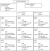
Results: We detected 18 RAA cases: five (27.78%) type I (mirror image, "V" type), 11 (61.12%) type II ("U" type), and two (11.10%) DAA cases. Heart anomalies were associated in 38.89% (overall), 60% (type I), 36.37% (type II), and 0% (DAA) cases. Tetralogy of Fallot represented the most prevalent cardiac malformation (in 22.23% of cases). EC anomalies were present in 44.44% of fetuses (20% of type I, 54.55% of type II, and 50% of DAA cases). Genetic abnormalities were found in 41.17% of pregnancies, with 22q11.2 deletion in 23.53%. 55.55% of the cases had a good neonatal evolution and 44.45% of the pregnancies were terminated. An overall good outcome of pregnancy was noted in 40% of type I RAA, 63.64% of type II RAA, and 50% of DAA cases. All RAA cases examined in the first trimester were correctly diagnosed.
Conclusions: RAA can be accurately diagnosed and classified by means of prenatal ultrasound since early pregnancy. A detailed anatomy scan and genetic testing, including 22q11 deletion, should be offered to all pregnancies when RAA is discovered. When isolated, RAA associates a good outcome, indifferently the anatomical type.
Conflict of interest statement
The authors declare that they have no conflict of interests.
Figures


Similar articles
-
Prenatal echocardiographic assessment of right aortic arch.Ultrasound Obstet Gynecol. 2019 Jul;54(1):96-102. doi: 10.1002/uog.20098. Epub 2019 Jun 10. Ultrasound Obstet Gynecol. 2019. PMID: 30125417
-
Prenatal Diagnosis of the Right Aortic Arch: Change in Detection Rate, the Status of Associated Anomalies, and Perinatal Outcomes in 137 Fetuses.Pediatr Cardiol. 2022 Dec;43(8):1888-1897. doi: 10.1007/s00246-022-02929-6. Epub 2022 May 14. Pediatr Cardiol. 2022. PMID: 35568727
-
Prenatal diagnosis of right aortic arch: associated anomalies and fetal prognosis according to different subtypes.J Perinat Med. 2024 Jan 29;52(3):304-309. doi: 10.1515/jpm-2023-0410. Print 2024 Mar 25. J Perinat Med. 2024. PMID: 38281095
-
[Isolated right aortic arch: prenatal diagnosis characteristics, pregnancy outcomes and systematic review].Gynecol Obstet Fertil Senol. 2019 Oct;47(10):726-731. doi: 10.1016/j.gofs.2019.09.002. Epub 2019 Sep 5. Gynecol Obstet Fertil Senol. 2019. PMID: 31494313 French.
-
Incidence of chromosomal anomalies in fetuses with isolated right aortic arch: A meta-analysis.Prenat Diagn. 2020 Feb;40(3):294-300. doi: 10.1002/pd.5606. Epub 2019 Dec 19. Prenat Diagn. 2020. PMID: 31736147 Review.
Cited by
-
Analysis of prognostic risk factors and development of a predictive model for fetuses with right aortic arch with mirror-image branching: a prognostic evaluation method based on ultrasound parameter features.Quant Imaging Med Surg. 2024 Sep 1;14(9):6869-6881. doi: 10.21037/qims-23-1648. Epub 2024 Apr 29. Quant Imaging Med Surg. 2024. PMID: 39281135 Free PMC article.
-
Prenatal diagnosis in fetal right aortic arch using chromosomal microarray analysis and whole exome sequencing: a Chinese single-center retrospective study.Mol Cytogenet. 2024 Sep 27;17(1):22. doi: 10.1186/s13039-024-00691-3. Mol Cytogenet. 2024. PMID: 39334424 Free PMC article.
References
-
- Achiron R, Rotstein Z, Heggesh J, Bronshtein M, Zimand S, Lipitz S, Yagel S. Anomalies of the fetal aortic arch: a novel sonographic approach to in-utero diagnosis. Ultrasound Obstet Gynecol. 2002;20(6):553–557. - PubMed
-
- Galindo A, Nieto O, Nieto MT, Rodríguez-Martín MO, Herraiz I, Escribano D, Granados MA. Prenatal diagnosis of right aortic arch: associated findings, pregnancy outcome, and clinical significance of vascular rings. Prenat Diagn. 2009;29(10):975–981. - PubMed
-
- McElhinney DB, Clark BJ, Weinberg PM, Kenton ML, McDonald-McGinn D, Driscoll DA, Zackai EH, Goldmuntz E. Association of chromosome 22q11 deletion with isolated anomalies of aortic arch laterality and branching. J Am Coll Cardiol. 2001;37(8):2114–2119. - PubMed
-
- Cavoretto PI, Sotiriadis A, Girardelli S, Spinillo S, Candiani M, Amodeo S, Farina A, Fesslova V. Postnatal outcome and associated anomalies of prenatally diagnosed right aortic arch with concomitant right ductal arch: a systematic review and meta-analysis. Diagnostics (Basel) 2020;10(10):831–831. - PMC - PubMed
Publication types
MeSH terms
LinkOut - more resources
Full Text Sources

