. 2020 Oct-Dec;61(4):1193-1212.
doi: 10.47162/RJME.61.4.21.

Peripheral adenopathies in children - an attempt of clinical morphological profile

Affiliations

Peripheral adenopathies in children - an attempt of clinical morphological profile

Georgeta Ligia Stănescu et al. Rom J Morphol Embryol. 2020 Oct-Dec.

Abstract

Aim: The authors have proposed to assess peripheral adenopathies in a series of hospitalized children in order to identify and define clinical and morphological profiles of different types of lymph node (LN) diseases.

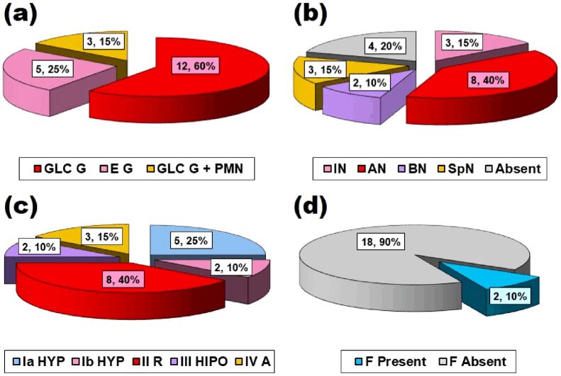
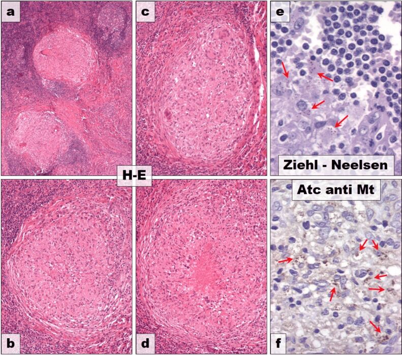
Materials and methods: The studied group consisted of 58 patients less than 18 years of age. The investigation algorithm included: gender, age, site, involvement, side, extension and histopathological (HP) type of LN lesions. Tissue fragments were processed using classical histological techniques (formalin fixation and paraffin embedment) and stained with Hematoxylin-Eosin (HE). In some cases (tuberculous lesions and lymphomas), special stainings (Ziehl-Neelsen) and immunohistochemistry were used. Stratification scales of cases were defined according to each parameter in order to compare the data. All obtained data were assessed individually, compared to each other and with similar data from the literature with the help of a statistical apparatus [χ² (chi-squared) test and analysis of variance (ANOVA) test] in some cases.

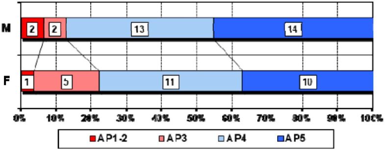
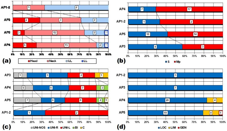
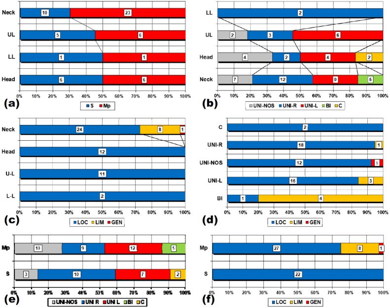
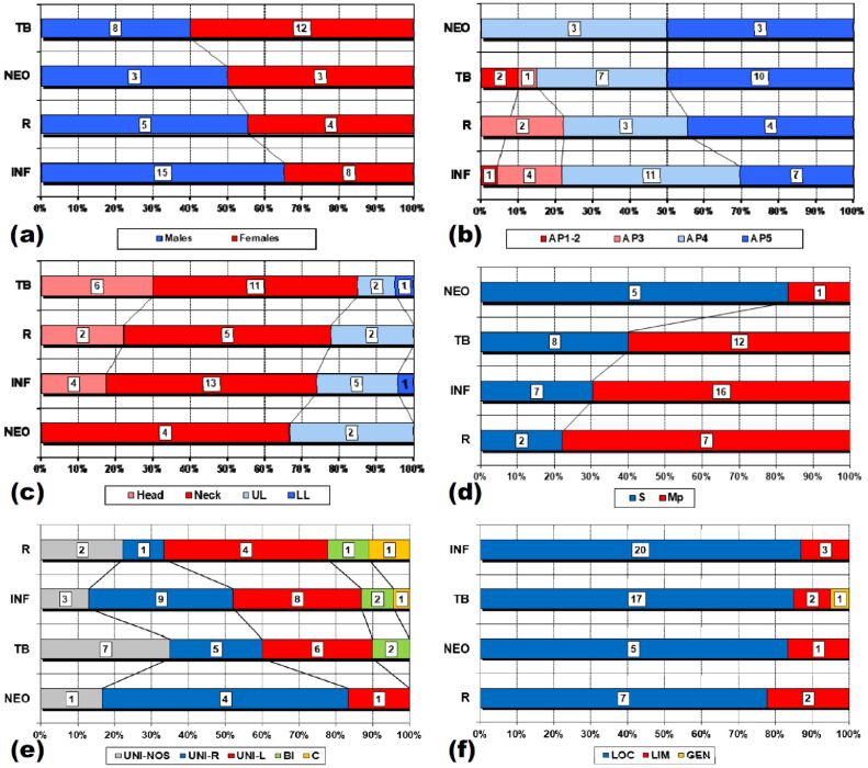
Results: The young patients were slightly more frequently boys, of all ages but with a mean age of 10 and half years. The affected LNs belonged most often to neck region, either on the left or on the right side but sometimes bilateral or even on the midline; usually, more than one LN was involved in the area. In most of the cases, the lesions were localized in only one LN area. HP picture was dominated by the inflammatory processes, firstly the nonspecific ones, followed by tuberculosis.

Discussions: Our observations fitted, for each parameter, with the wide ranges found in the literature. Comparisons between parameters' variations revealed differences, sometimes significant that we tried to organize in clinical and morphological profiles.

Conclusions: The assessment of our data allowed us to define some clinical and morphological profiles of different types of adenopathy that, by improvement on studies including larger series, could be of real use in daily pediatric practice.


Conflict of interest statement

The authors declare that they have no conflict of interests.

Figures

Figure 1
Figure 1
Reactive lymph node with prominent follicular hyperplasia. HE staining: (a) ×40; (b) ×100
Figure 2
Figure 2
Suppurated lymphadenitis: (a) General view; (b) Detail – numerous neutrophils. HE staining: (a) ×40; (b) ×200
Figure 3
Figure 3
“Cat scratch” disease: (a) General view; (b) Detail – central necrosis (orange arrows), palisading histiocytes (blue arrows). HE staining: (a) ×40; (b) ×100
Figure 4
Figure 4
Chronic lymphadenitis: (a) Follicular hyperplasia; (b) Intrafollicular fibrosis; (c) Detail of previous image; (d) Follicular and sinus fibrosis; (e) General view; (f) Adipose degeneration. HE staining: (a and f) ×100; (b, d and e) ×40; (c) ×200
Figure 5
Figure 5
Tuberculous lymphadenitis: (a) Granuloma cellularity; (b) Types of necrosis; (c) Types of granulomas; (d) Presence of fibrosis. A: Areactive; AN: Acidophilic necrosis; BN: Basophilic necrosis; E G: Epithelioid granuloma; F: Fibrosis; GLC G: Giant Langhans cell granuloma; HYP: Hyperplastic; Hipo: Hyporeactive; IN: Incipient necrosis; R: Reactive; SpN: Suppurated necrosis
Figure 6
Figure 6
Tuberculous lymphadenitis: (a) General view; (b) Type Ia hyperplastic granuloma; (c) Type Ib hyperplastic granuloma; (d) Type II reactive granuloma; (e) Acid fast Mt in mononuclear macrophages (red arrows); (f) Anti-Mt + antibody (red arrows), ×200. HE staining: (a) ×40; (b–d) ×100. Ziehl–Neelsen staining: (e) ×200. Mt: Mycobacterium tuberculosis
Figure 7
Figure 7
Diffuse large B-cell lymphoma [×40 and ×200 (detail frame)], immunohistochemical panel of positive antibodies used: (a) HE staining; (b) CD20+; (c) CD79a+; (d) Bcl-2+; (e) CD30+; (f) Ki67 index. Bcl-2: B-cell lymphoma 2; CD: Cluster of differentiation; HE: Hematoxylin–Eosin
Figure 8
Figure 8
Gender distribution related to age. AP1-2: 0–2 years; AP3: 3–5 years; AP4: 6–11 years; AP5: 12–18 years
Figure 9
Figure 9
Correlations between gender and gross morphological parameters: (a) Gender–site correlation; (b) Gender–LN involvement correlation; (c) Gender–LN side correlation; (d) Gender–LN extension correlation. BI: Bilateral; C: Center; GEN: Generalized; LIM: Limited; LL: Lower limb; LN: Lymph node; LOC: Localized; Mp: Multiple; S: Solitary; U-NOS: Unilateral – not otherwise specified; U-L: Unilateral – left; U-R: Unilateral – right; UL: Upper limb
Figure 10
Figure 10
Correlations between age and gross morphological parameters. (a) Age–site correlation; (b) Age–LN involvement correlation; (c) Age–LN side correlation; (d) Age–LN extension correlation. AP1-2: 0–2 years; AP3: 3–5 years; AP4: 6–11 years; AP5: 12–18 years; BI: Bilateral; C: Center; GEN: Generalized; LIM: Limited; LL: Lower limb; LN: Lymph node; LOC: Localized; Mp: Multiple; S: Solitary; U-NOS: Unilateral – not otherwise specified; U-L: Unilateral – left; U-R: Unilateral – right; UL: Upper limb
Figure 11
Figure 11
Correlations between gross morphological parameters. (a) Site–involvement correlation; (b) Site–side correlation; (c) Site–extension correlation; (d) Side–extension correlation; (e) Involvement–side correlation; (f) Involvement–extension correlation. BI: Bilateral; C: Center; GEN: Generalized; LIM: Limited; LL: Lower limb; LOC: Localized; M: Multiple; S: Solitary; U-NOS: Unilateral – not otherwise specified; U-L: Unilateral – left; U-R: Unilateral – right; UL: Upper limb
Figure 12
Figure 12
Correlations between histological type and all other parameters: (a) HP type–gender correlation; (b) HP type–age correlation; (c) HP type–site correlation; (d) HP type–involvement correlation; (e) HP type–side correlation; (f) HP type–extension correlation. AP1-2: 0–2 years; AP3: 3–5 years; AP4: 6–11 years; AP5: 12–18 years; BI: Bilateral; C: Center; GEN: Generalized; LIM: Limited; INF: Inflammatory process; LL: Lower limb; LOC: Localized; Mp: Multiple; NEO: Neoplasia; R: Reactive; S: Solitary; TB: Tuberculosis; U-NOS: Unilateral – not otherwise specified; U-L: Unilateral – left; U-R: Unilateral – right; UL: Upper limb
Figure 13
Figure 13
Graphical representation of age distribution in the four groups. INF: Inflammatory process; NEO: Neoplastic lesion; TB: Tuberculosis; R: Reactive
Figure 14
Figure 14
Comparison of male/female ratio with other studies. OS: Our study. Blue dotted line: Peripheral lymphadenopathy (P-LAP) – 1: [36]; 2: [37]; 3: [7]; 4: [40]; 5: [39]; 6: [42]; 7: [29]; 8: [38]; 9: [32]; 10: [23]; 11: [9]; 12: [41]; 13: [10]; 14: [31]. Red dotted line: Cervical lymphadenopathy (C-LAP) – 15: [6]; 16: [47]; 17: [8]; 18: [43]; 19: [44]; 20: [45]; 21: [46]
Figure 15
Figure 15
Comparison of mean age and age range with other studies. OS: Our study; Peripheral lymphadenopathy (P-LAP) – 1: [38]; 2: [10]; 3: [23]; 4: [35]; 5: [39]; 6: [41]; 7: [42]; Cervical lymphadenopathy (C-LAP) – 8: [45]; 9: [47]; 10: [43]; 11): [46]. AV: Average; VMAX: Maximum value; VMIN: Minimum value
Figure 16
Figure 16
Comparison of LAP site with other studies. OS: Our study; Peripheral lymphadenopathy (P-LAP) – 1: [29]; 2: [9]; 3: [32]; 4: [37]; 5: [23]; 6: [30]; 7: [36]; Head and neck lymphadenopathy (H&N-LAP) – 8: [6]. GEN: Generalized; LL: Lower limb; UL: Upper limb
Figure 17
Figure 17
Comparison of LAP involvement with other studies. OS: Our study; P-LAP: Peripheral lymphadenopathy – 1: [9]; C-LAP: Cervical lymphadenopathy – 2: [8]
Figure 18
Figure 18
Comparison of LAP extension with other studies. OS: Our study; P-LAP: Peripheral lymphadenopathy – 1: [40]; 2: [38]; 3: [35]; 4: [34]; 5: [10]; C-LAP: Cervical lymphadenopathy – 6: [8]
Figure 19
Figure 19
Comparison of HP types’ distribution with other studies. OS: Our study; P-LAP: Peripheral lymphadenopathy – 1: [30]; 2: [36]; 3: [38]; 4: [23]; 5: [33]; 6: [37]; 7: [7]; 8: [35]; 9: [40]; 10: [32]; 11: [31]; 12: [9]; C-LAP: Cervical lymphadenopathy – 13: [8]; 14: [47]; 15: [44]; H&N-LAP: Head and neck lymphadenopathy – 16: [6]; 17: [29]; (1): Primary sarcomas or carcinomas were excluded; (2): Age range – one year to 80 years; *: Reactive + Sinus histiocytosis; **: Inflammation (acute, chronic, granulomatous) + Sarcoidosis; ***: Lymphoma + other. HP: Histopathological

Similar articles

References

    1. Mohseni S, Shojaiefard A, Khorgami Z, Alinejad S, Ghorbani A, Ghafouri A. Peripheral lymphadenopathy: approach and diagnostic tools. Iran J Med Sci. 2014;39(2 Suppl):158–170. - PMC - PubMed
    1. Allhiser JN, McKnight TA, Shank JC. Lymphadenopathy in a family practice. J Fam Pract. 1981;12(1):27–32. - PubMed
    1. Williamson HA. Lymphadenopathy in a family practice: a descriptive study of 249 cases. J Fam Pract. 1985;20(5):449–452. - PubMed
    1. Bazemore AW, Smucker DR. Lymphadenopathy and malignancy. Am Fam Physician. 2002;66(11):2103–2110. - PubMed
    1. Ferrer R. Lymphadenopathy: differential diagnosis and evaluation. Am Fam Physician. 1998;58(6):1313–1320. - PubMed