. 2020 Oct-Dec;61(4):1259-1278.
doi: 10.47162/RJME.61.4.27.

Analysis of the distribution and expression of some tumor invasiveness markers in palate squamous cell carcinomas

Affiliations

Analysis of the distribution and expression of some tumor invasiveness markers in palate squamous cell carcinomas

Adrian Pătru et al. Rom J Morphol Embryol. 2020 Oct-Dec.

Abstract

Oral cancer remains an important global health issue and despite recent diagnostic and therapeutic advances, it continues to have an unfavorable prognostic and decreased survival. Although palatal tumors represent one of the rarest locations of oral squamous cell carcinomas (SCCs), they are among the most aggressive local tumors, leaving behind important morpho-functional disabilities. In order to explain such local aggressiveness, the present study aims to investigate the immunohistochemical expression in palate SCCs of some markers known to be involved in the process of tumor invasiveness, such as Wiskott-Aldrich syndrome like (WASL), Claudin-1 (CLDN1), Integrin beta-6 (ITGB6) and c-Mesenchymal to epithelial transition protein (c-Met). We have found here a higher tumor WASL and CLDN1 reactivity in well-differentiated (G1) palate SCCs, and regardless the histological type, degree of differentiation or tumor topography, an overexpression at the invasion front, and in those palate' SCC cases with muscular invasiveness and with lymph node (LN) dissemination. ITGB6 and c-Met had a higher reactivity in moderately differentiated (G2) palate SCCs, especially at the periphery of tumor proliferations, at the invasion front and in those high invasive cases and as well as in those that associated LN dissemination. All four investigated markers were also positive at the level of LN metastatic proliferations. None of the markers could statistically stratify on age group and pain, and on bone and perineural invasion while all of them statistically stratified on survival and grading. We concluded that these markers have a prognostic role allowing the identification of those cases with an unfavorable clinical evolution and decreased survival.


Conflict of interest statement

The authors declare that there is no conflict of interests regarding the publication of this paper. All authors read and approved the final manuscript.

Figures
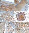
Figure 1
Figure 1
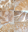
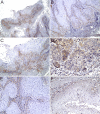
WASL reactivity: (A) In cytoplasm of basal and suprabasal keratinocytes of normal palatine epithelium adjacent to the tumors; (B) Cytoplasmic and even nuclear reactivity extended towards the upper layers in the hyperplastic and dysplastic epithelium; (C) Cytoplasmic reactivity in the minor salivary glands with higher intensity in the excretory ducts compared with glandular acini; (D) Reactivity at the level of adipose tissue and in the lymphoid tissue especially in the germinal centers; (E) Reactivity in the striated muscle tissue; (F) Reactivity at the level of vascular endothelial cells, fibroblasts and macrophages. Anti-WASL antibody immunolabeling: (A–C and F) ×200; (D and E) ×100. WASL: Wiskott–Aldrich syndrome like
Figure 2
Figure 2
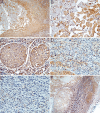
WASL reactivity in tumor samples of palate SCC: (A) The most reactive specimens were those from the patients with verrucous palate SCC subtype; (B) The reactivity was more obvious at the invasive front compared with superficial part of the tumor; (C) A slightly lower reactivity was recorded in well-differentiated (G1) palate SCC, especially at the periphery of tumor proliferations; (D) These cases were followed as reactivity by the papillary palate SCC subtype; (E and F) In these cases, reactivity was more obvious at the invasive front compared with superficial tumor part. Anti-WASL antibody immunolabeling: (A) ×50; (B, C, E and F) ×200; (D) ×100. SCC: Squamous cell carcinoma; WASL: Wiskott–Aldrich syndrome like
Figure 3
Figure 3
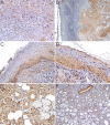
WASL reactivity in tumor samples of palate SCC: (A) A slightly lower reactivity was recorded in the acantholytic SCC cases and it was more obvious at the level of the tumor acantholysis zones; (B) Both cytoplasmic and nuclear reactivity in the discohesive tumor cells; (C) A nuclear reactivity present in the basaloid palate SCC cases; (D and E) Weak cytoplasmic and nuclear reactivity or no reactivity in the undifferentiated areas of palate G3 SCC cases; (F) Weak reactivity at the level of tumor proliferations disseminated in the lymph nodes. Anti-WASL antibody immunolabeling: (A and F) ×100; (B–E) ×400. G3: Poorly differentiated tumor; SCC: Squamous cell carcinoma; WASL: Wiskott–Aldrich syndrome like
Figure 4
Figure 4
WASL distribution on localization: Class A groups the IRS scores ranging from 1 to 6; Class B groups the IRS scores higher than 6. IRS: Immunoreactive score; WASL: Wiskott–Aldrich syndrome like
Figure 5
Figure 5
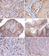
CLDN1 reactivity: (A) Keratinocytes reactivity from the lower half of the normal adjacent epithelium with predominantly membranous pattern; (B) A cytoplasmic positive reaction in the lower half of the hyperplastic and dysplastic epithelium and even a nuclear pattern in the basal and parabasal epithelial cells; (C) A strong reaction in the excretory ducts from minor salivary glands; (D) A strong reaction with membranous prevalent pattern in the palate G2 SCC cases; (E) A reduction in tumor reactivity was noticed in the periphery of neoplastic proliferations, especially in those cases with 1 and 2 invasive pattern; (F) A strong reaction was noticed in all invasive cells from those palate SCC with 3 and 4 invasive pattern. Anti-CLDN1 antibody immunolabeling: (A–C, E and F) ×200; (D) ×100. CLDN1: Claudin-1; G2: Moderately differentiated tumor; SCC: Squamous cell carcinoma
Figure 6
Figure 6
CLDN1 reactivity: (A) A progressive reactivity decreasing in the acantholytic variant with no reactivity towards the lumen proliferations; (B) Inside these lumens, tumor acantholytic cells temporarily retain a granular cytoplasmic reactivity; (C) A lower reactivity was noticed in the G1 SCC cases with either absent or a faint thin concentric ring pattern at the level of keratin pearls; (D) In the papillary palate SCC cases was observed a membranous reactivity pattern more obvious at the level of the neoplastic keratinocytes from inside proliferations than the basaloid neoplastic cells from periphery; (E) A slightly weaker reactivity was observed in the case of palate verrucous SCC; (F) In the basaloid palate SCC cases, we recorded a weak reactivity restricted to center of the tumor islands, with a cytoplasmic prevalent pattern. Anti-CLDN1 antibody immunolabeling: (A and C) ×100; (B and F) ×400; (D and E) ×50. CLDN1: Claudin-1; G1: Well-differentiated tumor; SCC: Squamous cell carcinoma
Figure 7
Figure 7
CLDN1 reactivity: (A) In G3 palate SCCs, we noticed a peculiar weak nuclear pattern in the poorly differentiated cells and a higher cytoplasmic reaction in those more differentiated neoplastic cells; (B) The reactivity was also present at the level of lymph node metastases but with a lower intensity then in the primary tumors. Anti-CLDN1 antibody immunolabeling: (A) ×200; (B) ×50. CLDN1: Claudin-1; G3: Poorly differentiated tumor; SCC: Squamous cell carcinoma. ITGB6 reactivity: (C) Reactivity was present in the lower part of the tumor adjacent epithelium, with membranous and cytoplasmic pattern; (D) A nuclear pattern was observed in the atypical cells from hyperplastic and dysplastic epithelium; (E) Reactivity at the level of minor salivary glands, with serous acinar cells as the most reactive, followed by the ductal epithelial cells and as the least reactive were the mucous acinar cells; (F) An intense positive reaction was observed at the level of smooth muscle fibers and at the level of endothelial cells from intra- and peritumoral blood vessels. Anti-ITGB6 antibody immunolabeling: (C and F) ×200; (D and E) ×400. ITGB6: Integrin beta-6
Figure 8
Figure 8
ITGB6 reactivity: (A) The highest ITGB6 reactivity was recorded in the G2 palate SCCs, with a cytoplasmic and nuclear dominant pattern; (B) The reactivity was more obvious at the periphery than inside of neoplastic proliferation and also at the invasive front compared with tumor superficial part; (C) In the palate papillary SCCs, the reaction was more intense in the neoplastic epithelium that rim the papillae cores and at the invasion front, with a cytoplasmic prevalent pattern; (D) In the verrucous palate SCC, the reactivity was most cytoplasmic and more intense in the basal and parabasal neoplastic cells; (E) A decrease in reactivity was recorded in G3 palate SCCs, where the dominant pattern was the nuclear one; (F) A similar reactivity was observed in the basaloid palate SCCs, with the nuclear staining more prevalent at the invasive front and at the periphery of neoplastic proliferations. Anti-ITGB6 antibody immunolabeling: (A and E) ×400; (B and F) ×200; (C and D) ×50. G2: Moderately differentiated tumor; G3: Poorly differentiated tumor; ITGB6: Integrin beta-6; SCC: Squamous cell carcinoma
Figure 9
Figure 9
ITGB6 reactivity: (A) In the acantholytic carcinomas, the prevalent reactivity was at the membrane level, especially in those neoplastic cells that rim the pseudoglandular lumens; (B) The slightest reactivity was observed in G1 palate SCCs, with membranous and cytoplasmic pattern; (C) The neoplastic proliferation from the lymph node metastasis kept their reactivity. Anti-ITGB6 antibody immunolabeling: (A) ×200; (B) ×100; (C) ×50. G1: Well-differentiated tumor; ITGB6: Integrin beta-6; SCC: Squamous cell carcinoma. c-Met reactivity: (D) Either absent or weak cytoplasmic positive reaction in the basal and parabasal layers of normal adjacent epithelium; (E) A slightly more intense reactivity was noticed in the minor salivary glands and especially in the epithelium of the excretory ducts, with a dual cytoplasmic and membranous pattern; (F) A weak c-Met reactivity, with a predominantly cytoplasmic pattern was also noticed in fibroblasts, cells from the stromal inflammatory infiltrate and endothelial cells form intra- and peritumoral blood vessels. Anti-c-Met antibody immunolabeling: (D) ×100; (E) ×200; (F) ×400. c-Met: c-Mesenchymal to epithelial transition protein
Figure 10
Figure 10
c-Met reactivity: (A and B) The highest reactivity was noticed in the G2 palate SCCs, with a prevalent membranous pattern in the superficial part of tumors, while at the invasion front, the prevalent pattern was a cytoplasmic one; (C) Regardless tumor topography, the reactivity was higher at neoplastic cells from the periphery, with a dual cytoplasmic and membranous pattern, while those inside the proliferations had lower reactivity and predominantly with membranous pattern; (D) A slighter reactivity was noticed in the papillary palate SCCs cases; (E) The reactivity was more obvious at the surface of papillary projections, with both cytoplasmic and membranous pattern; (F) On the contrary, at the invasive front, the reactivity was weak and with cytoplasmic pattern. Anti-c-Met antibody immunolabeling: (A) ×100; (B, E and F) ×200; (C) ×400; (D) ×50. c-Met: c-Mesenchymal to epithelial transition protein; G2: Moderately differentiated tumor; SCC: Squamous cell carcinoma
Figure 11
Figure 11
c-Met reactivity: (A) A slightly lower reactivity was presented in the verrucous palate SCC case; (B) The reactivity was the same as in the papillary palate SCCs cases, with the tumor superficial part as more reactive than the invasion front; (C) In the G3 palate SCC cases, the reactivity was weak and with cytoplasmic prevalent pattern; (D) In the basaloid palate SCC cases, the reactivity was also weak, with a prevalent cytoplasmic pattern that was more obvious at the neoplastic cells from the periphery of basaloid proliferations; (E) In the G1 palate SCC cases, the reactivity was present at the periphery of tumor islands, with cytoplasmic prevalent pattern, and the keratin pearls being devoid of reactivity; (F) The least reactive were acantholytic carcinomas, where we noticed a cytoplasmic pattern in the outermost layers of tumor proliferations, while in the acantholytic tumor cells, we did not observe any reactivity. Anti-c-Met antibody immunolabeling: (A) ×50; (B and C) ×100; (D) ×400; (E and F) ×20. c-Met: c-Mesenchymal to epithelial transition protein; G1: Well-differentiated tumor; G3: Poorly differentiated tumor; SCC: Squamous cell carcinoma

Similar articles

Cited by

References

    1. Bray F, Ferlay J, Soerjomataram I, Siegel RL, Torre LA, Jemal A. Global cancer statistics 2018: GLOBOCAN estimates of incidence and mortality worldwide for 36 cancers in 185 countries. CA Cancer J Clin. 2018;68(6):394–424. - PubMed
    1. Gupta N, Gupta R, Acharya AK, Patthi B, Goud V, Reddy S, Garg A, Singla A. Changing trends in oral cancer - a global scenario. Nepal J Epidemiol. 2016;6(4):613–619. - PMC - PubMed
    1. Rivera C. Essentials of oral cancer. Int J Clin Exp Pathol. 2015;8(9):11884–11894. - PMC - PubMed
    1. Omura K. Current status of oral cancer treatment strategies: surgical treatments for oral squamous cell carcinoma. Int J Clin Oncol. 2014;19(3):423–430. - PubMed
    1. Oskam IM, Verdonck-de Leeuw IM, Aaronson NK, Witte BI, de Bree R, Doornaert P, Langendijk JA, Leemans CR. Prospective evaluation of health-related quality of life in long-term oral and oropharyngeal cancer survivors and the perceived need for supportive care. Oral Oncol. 2013;49(5):443–448. - PubMed

Substances