

Gingival proliferative growth - stress and cytoarchitecture related with fixed and mobile orthodontic therapy
- PMID: 34171076
- PMCID: PMC8343617
- DOI: 10.47162/RJME.61.4.29
Gingival proliferative growth - stress and cytoarchitecture related with fixed and mobile orthodontic therapy
Abstract
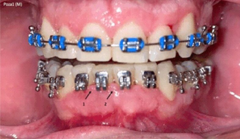
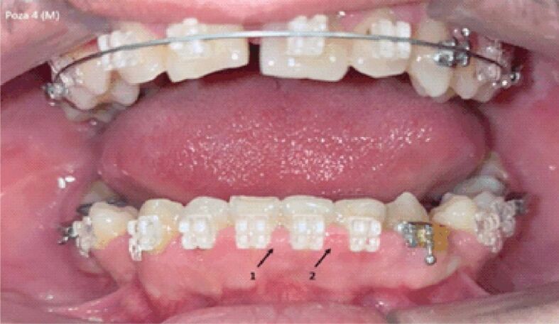
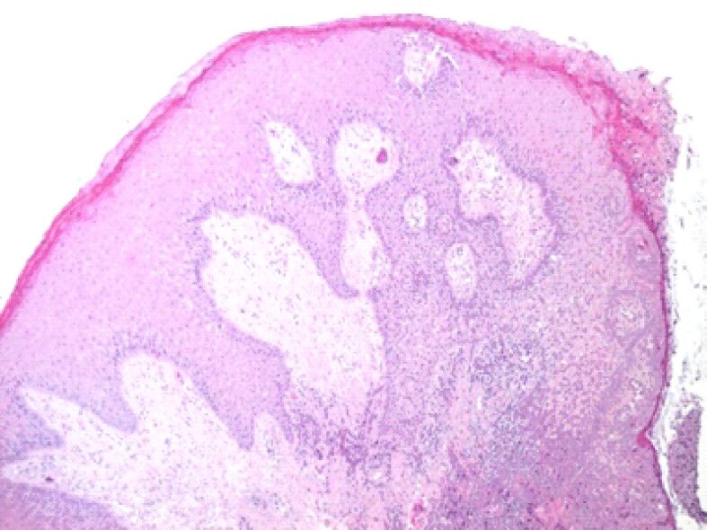
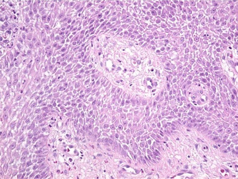
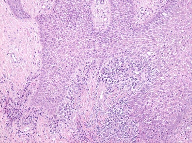
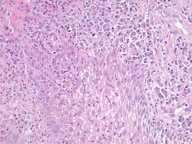
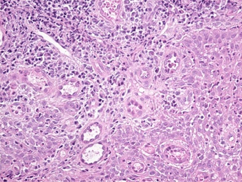
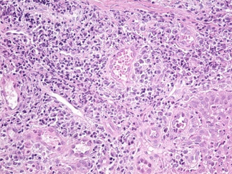
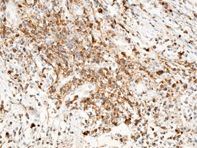
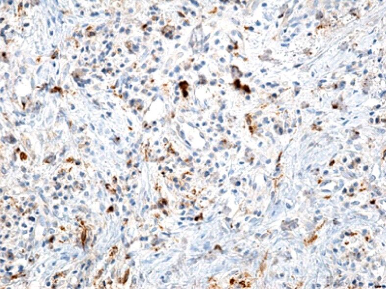
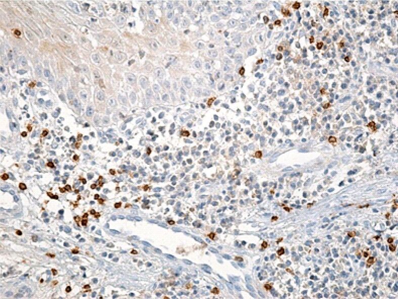
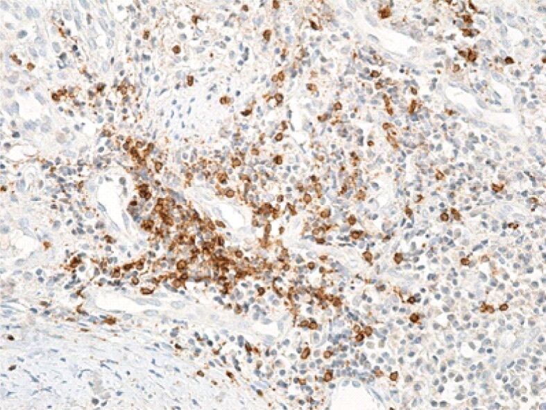
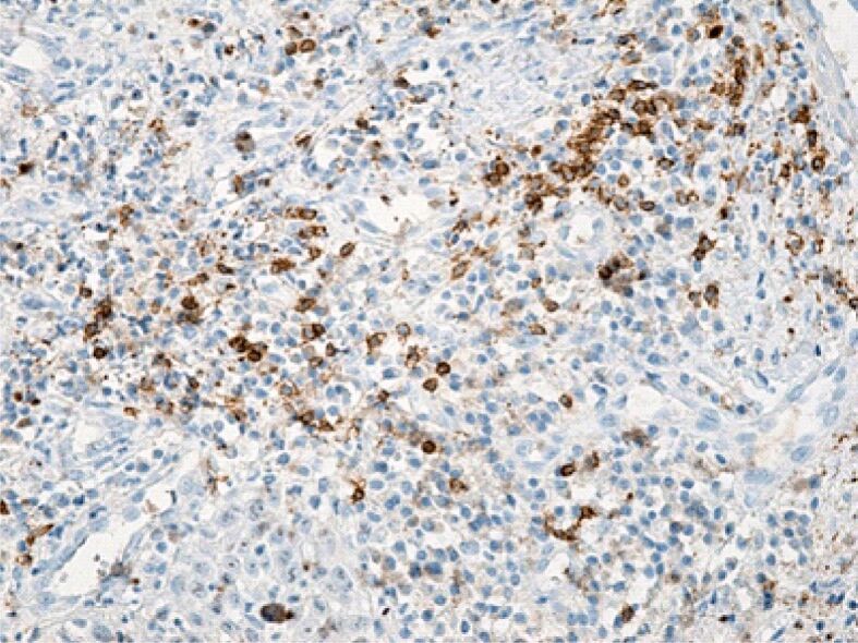
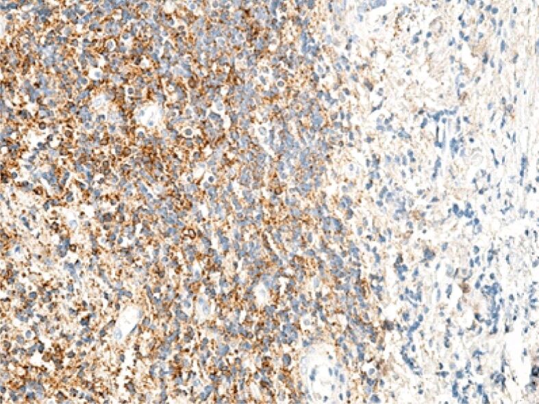
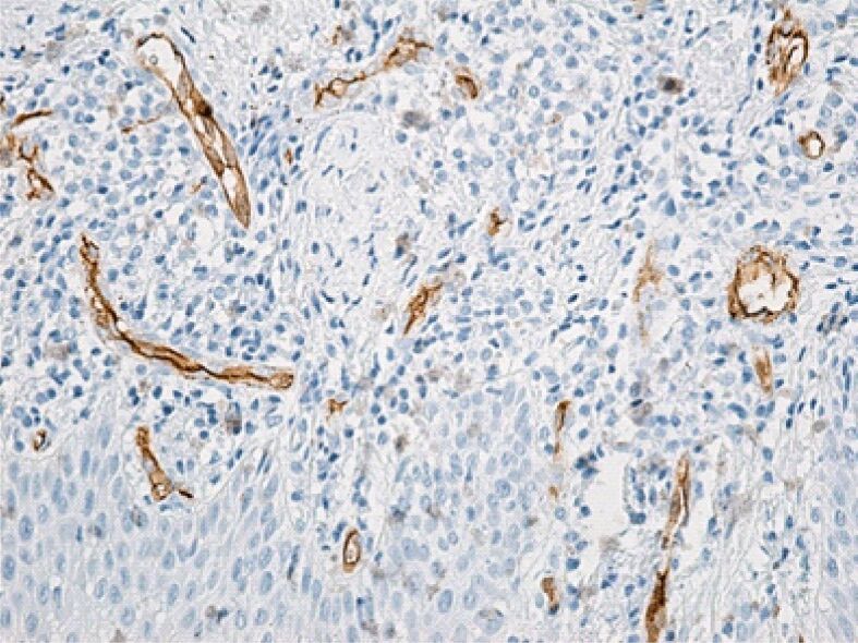
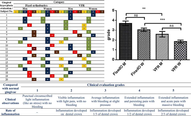
The fixed orthodontic measures taken induce significant stress to the gingival growth process during arch wire maneuvers of aligning and leveling. We observed, for a period of one to four years, fixed orthodontic devices in 80 human subjects. From these, we selected 44 subjects (22 women and 22 men) where the inflammatory process exhibited following the orthodontic fixed treatment, and with vacuum-formed orthodontic retainers (VFR) succeeding to fixed treatment. Samples were collected from each patient and histological and immunohistochemical (IHC) methodology was made to analyze the cytoarchitecture. Statistics were made after one-way analysis of variance (ANOVA), with the Bonferroni's correction. The IHC examination performed in the early stage revealed the presence in the inflammatory infiltrate of CD8-type T-lymphocytes, and of dendritic cells in large numbers. The examination performed in the late stage revealed the presence in the inflammatory infiltrate of CD20-type B-lymphocytes, which are mature cells capable of immunoglobulin synthesis, their activation being an important step in the maturation of the antibody response. The stress generated by arch wires in both genders was significantly higher than in the case of VFR. This observation was pointed out also by the cytohistological investigation outcome but was also based on an original scale conceived by our research team, following gingival hyperplasia evaluation. Also, with statistical significance, the comparative obtained values for men (p=0.01) and for women (p=0.001) illustrate clinical observations, allowing to affirm that, in our case, men were more stressed in bearing arch wire devices (AWD) and VFR, in comparison with women.
Conflict of interest statement
The authors declare no conflict of interests.
Figures
Similar articles
-
Comparison of Stability of the Results of Orthodontic Treatment and Gingival Health between Hawley and Vacuum-formed Retainers.J Contemp Dent Pract. 2018 Apr 1;19(4):443-449. J Contemp Dent Pract. 2018. PMID: 29728551 Clinical Trial.
-
The origin and evolution of the Hawley retainer for the effectiveness to maintain tooth position after fixed orthodontic treatment compare to vacuum-formed retainer: A systematic review of RCTs.Int Orthod. 2020 Jun;18(2):225-236. doi: 10.1016/j.ortho.2020.02.008. Epub 2020 Mar 19. Int Orthod. 2020. PMID: 32201168
-
Bonded versus vacuum-formed retainers: a randomized controlled trial. Part 1: stability, retainer survival, and patient satisfaction outcomes after 12 months.Eur J Orthod. 2018 Jul 27;40(4):387-398. doi: 10.1093/ejo/cjx058. Eur J Orthod. 2018. PMID: 29059289 Clinical Trial.
-
Vacuum-formed retainers and bonded retainers for dental stabilization-a randomized controlled trial. Part II: patients' perceptions 6 and 18 months after orthodontic treatment.Eur J Orthod. 2021 Apr 3;43(2):136-143. doi: 10.1093/ejo/cjaa039. Eur J Orthod. 2021. PMID: 32613244 Clinical Trial.
-
Epidemiologic study of orthodontic retention procedures.Am J Orthod Dentofacial Orthop. 2018 Apr;153(4):496-504. doi: 10.1016/j.ajodo.2017.08.013. Am J Orthod Dentofacial Orthop. 2018. PMID: 29602341 Review.
References
-
- Redlich M, Shoshan S, Palmon A. Gingival response to orthodontic force. Am J Orthod Dentofacial Orthop. 1999;116(2):152–158. - PubMed
-
- Ramadan AAF. Effect of nickel and chromium on gingival tissues during orthodontic treatment: a longitudinal study. World J Orthod. 2004;5(3):230–234; discussion 235. - PubMed
-
- Øilo M, Bakken V. Biofilm and dental biomaterials. Materials (Basel) 2015;8(6):2887–2900.
MeSH terms
LinkOut - more resources
Full Text Sources
Research Materials
