

Prenatal diagnosis of syndromic alobar holoprosencephaly associated with digynic triploidy fetus
- PMID: 34171079
- PMCID: PMC8343603
- DOI: 10.47162/RJME.61.4.32
Prenatal diagnosis of syndromic alobar holoprosencephaly associated with digynic triploidy fetus
Abstract
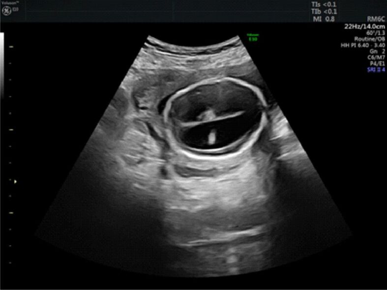
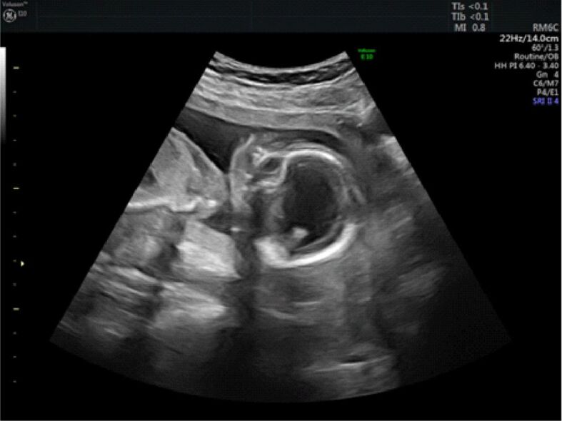
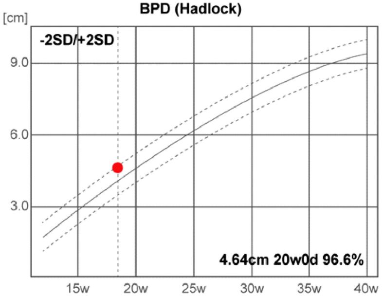
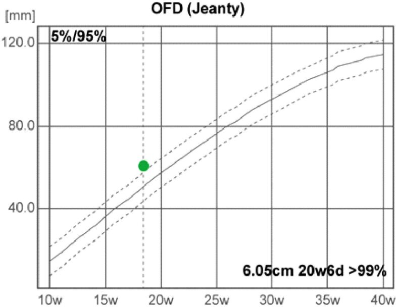
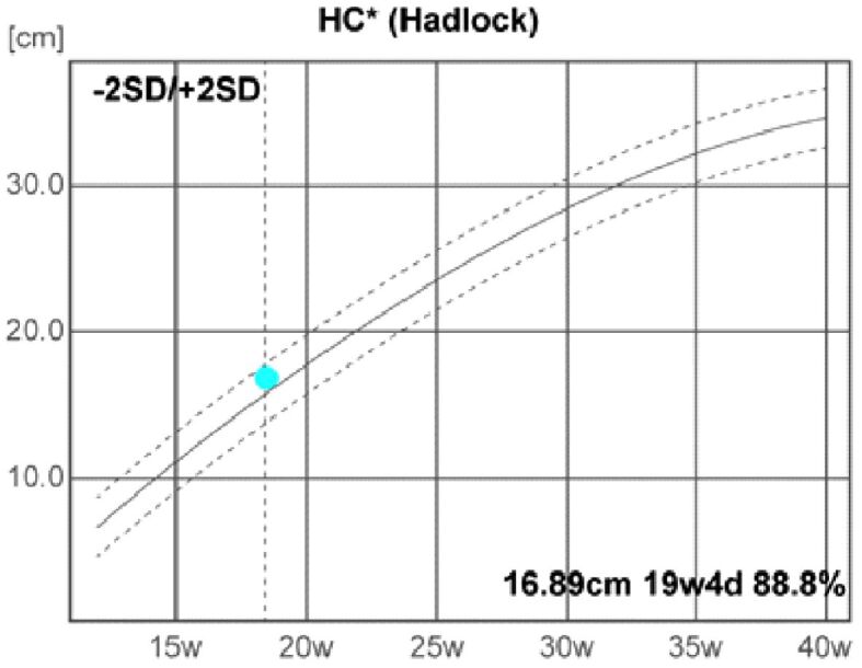
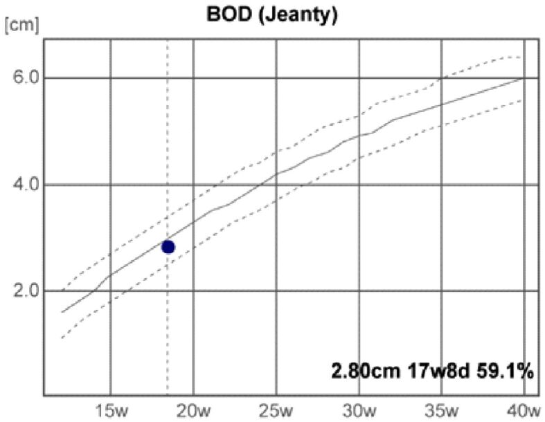
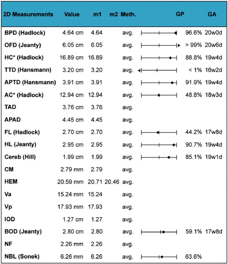
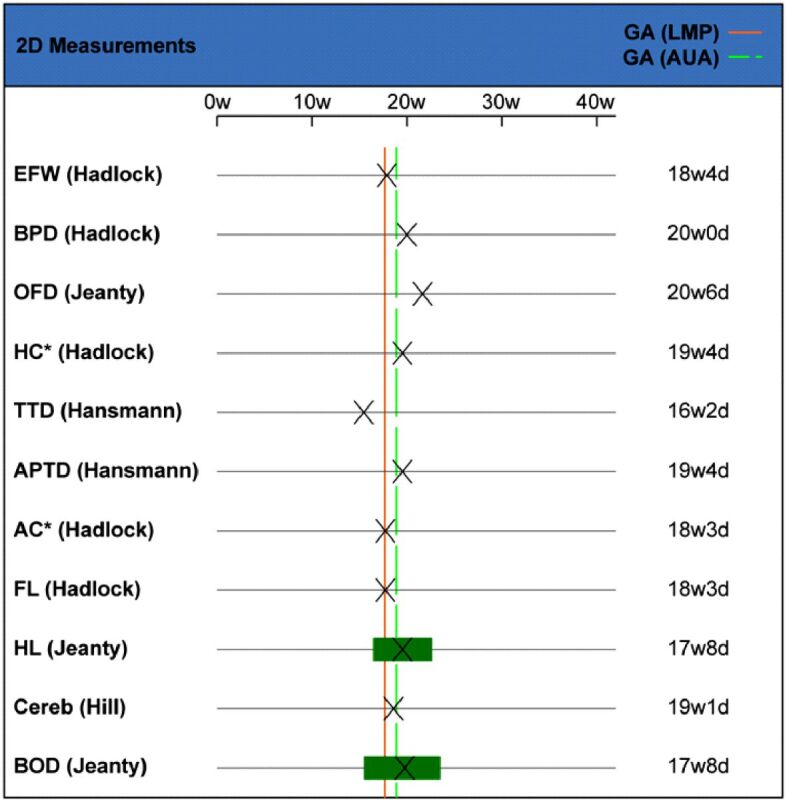
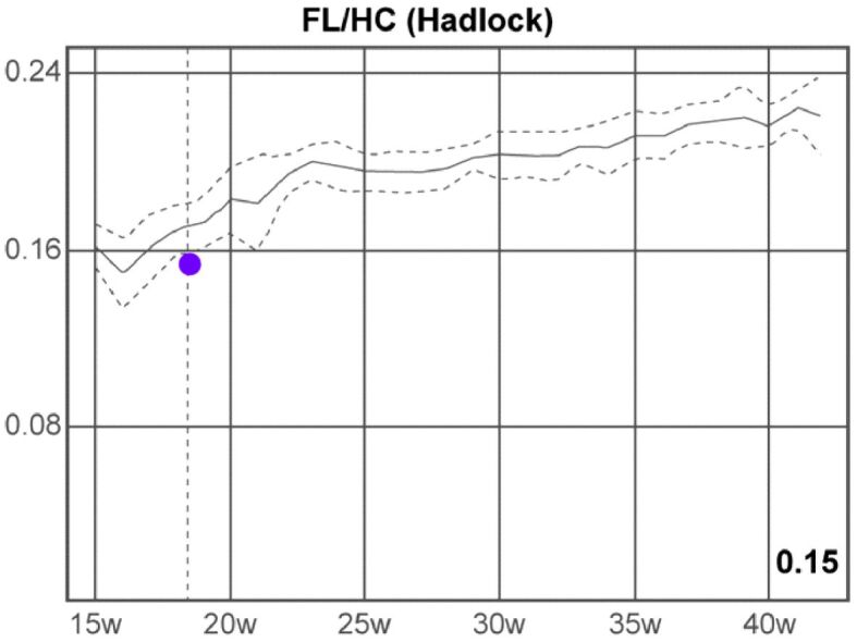
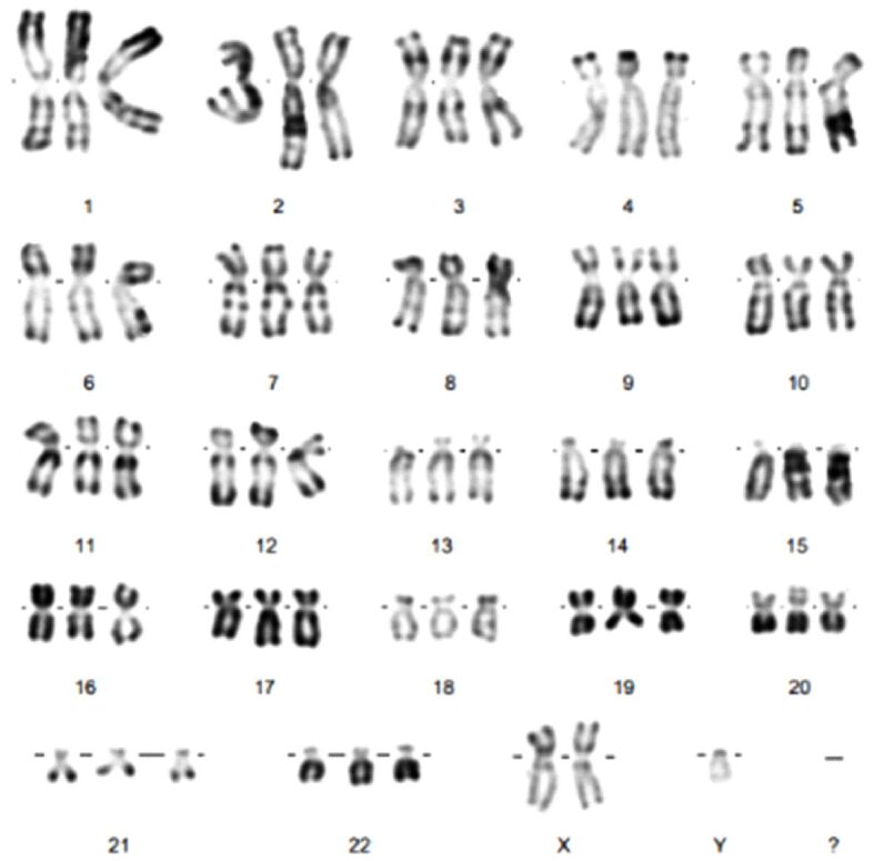
Holoprosencephaly (HPE) is a dramatic human brain malformation sequence with an extreme variable phenotypic spectrum and genetic heterogeneity, variable degree of severity and unknown etiology, in many cases. HPE is classified into syndromic, chromosomal, and non-syndromic, non-chromosomal. The most cases of HPE are syndromic. We present an atypical case of syndromic alobar HPE associated with digynic triploidy fetus, prenatally diagnosed, early at 18 weeks of gestation, by ultrasound (US) and complex genetic investigations. The US examination was performed with a specialized US machine, General Electric Voluson E10 OLED BT18, using two-dimensional (2D) scanning, three-dimensional (3D) image reconstruction, four-dimensional (4D) spatiotemporal image methodology and the highest power Doppler US technology. A detailed US examination of the fetus revealed several major abnormalities of the fetal head and severe facial malformations. Based on the antenatal US findings, the fetus was diagnosed with alobar HPE. After a careful examination and genetic counseling, additional cytogenetic investigations and molecular genetic analyses were performed, which revealed an abnormal number of 69 chromosomes, digynic triploidy (69,XXY). Two days later, the parents choose to interrupt the current gestation because of major fetal malformations. The pathological examination of the embryo reaffirmed the antenatal diagnostics.
Conflict of interest statement
The authors declare that they have no conflict of interests.
Figures

Similar articles
-
Digynic triploidy in a fetus presenting with semilobar holoprosencephaly.Taiwan J Obstet Gynecol. 2018 Dec;57(6):881-884. doi: 10.1016/j.tjog.2018.11.001. Taiwan J Obstet Gynecol. 2018. PMID: 30545546
-
Prenatal ultrasound findings of holoprosencephaly spectrum: Unusual associations.Prenat Diagn. 2020 Apr;40(5):565-576. doi: 10.1002/pd.5649. Epub 2020 Feb 10. Prenat Diagn. 2020. PMID: 31955448
-
Prenatal diagnosis of triploidy associated with holoprosencephaly: a case report and review of the literature.Am J Perinatol. 2009 Aug;26(7):479-83. doi: 10.1055/s-0029-1214248. Epub 2009 Apr 27. Am J Perinatol. 2009. PMID: 19399707 Review.
-
Prenatal ultrasound diagnosis in 51 cases of holoprosencephaly: craniofacial anatomy, associated malformations, and genetics.Cleft Palate Craniofac J. 2010 Jan;47(1):15-21. doi: 10.1597/08-036.1. Cleft Palate Craniofac J. 2010. PMID: 19860496
-
Alobar holoprosencephaly, mobile proboscis and trisomy 13 in a fetus with maternal gestational diabetes mellitus: a 2D ultrasound diagnosis and review of the literature.Arch Gynecol Obstet. 2007 May;275(5):385-7. doi: 10.1007/s00404-006-0264-6. Epub 2006 Oct 18. Arch Gynecol Obstet. 2007. PMID: 17047972 Review.
Cited by
-
Micrognathia as a Diagnosis Marker for the Prenatal Identification of Edwards Syndrome.Biomedicines. 2025 Feb 25;13(3):573. doi: 10.3390/biomedicines13030573. Biomedicines. 2025. PMID: 40149550 Free PMC article.
References
-
- Nyberg DA, Mack LA, Bronstein A, Hirsch J, Pagon RA. Holoprosencephaly: prenatal sonographic diagnosis. AJR Am J Roentgenol. 1987;149(5):1051–1058. - PubMed
-
- Măluţan AM, Dudea M, Ciortea R, Mureşan M, Bucuri CE, Mihu C, Mihu D. Cyclopia and proboscis - the extreme end of holoprosencephaly. Rom J Morphol Embryol. 2017;58(4):1555–1559. - PubMed
-
- Bendavid C, Rochard L, Dubourg C, Seguin J, Gicquel I, Pasquier L, Vigneron J, Laquerrière A, Marcorelles P, Jeanne-Pasquier C, Rouleau C, Jaillard S, Mosser J, Odent S, David V. Array-CGH analysis indicates a high prevalence of genomic rearrangements in holoprosencephaly: an updated map of candidate loci. Hum Mutat. 2009;30(8):1175–1182. - PubMed
-
- Golden JA. Holoprosencephaly. In: Squire LR, editor. Encyclopedia of Neuroscience. Academic Press; 2009. pp. 1181–1187.
Publication types
MeSH terms
LinkOut - more resources
Full Text Sources
