. 2021 Jul 14;29(7):1124-1136.e11.
doi: 10.1016/j.chom.2021.06.006. Epub 2021 Jun 15.

SARS-CoV-2 spike L452R variant evades cellular immunity and increases infectivity

Affiliations

SARS-CoV-2 spike L452R variant evades cellular immunity and increases infectivity

Chihiro Motozono et al. Cell Host Microbe. .

Abstract

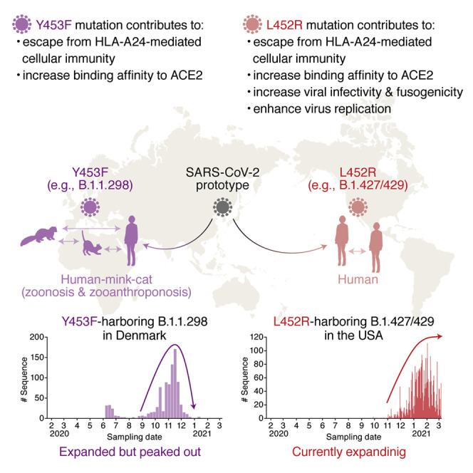
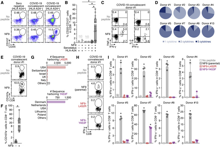
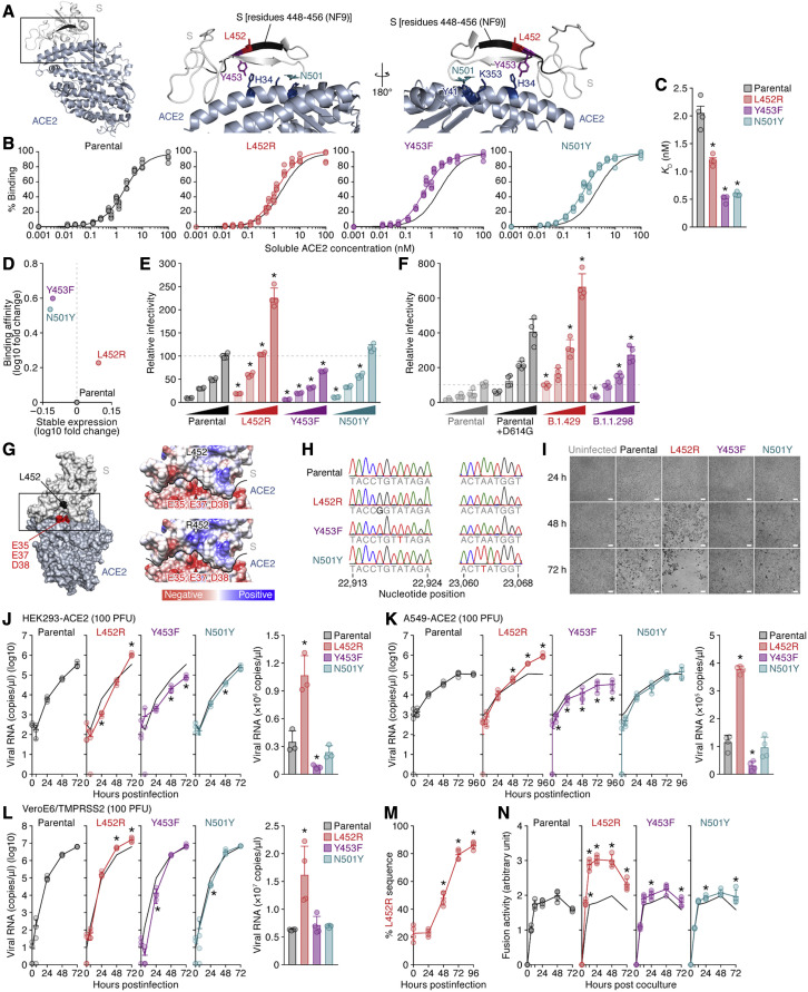
Many SARS-CoV-2 variants with naturally acquired mutations have emerged. These mutations can affect viral properties such as infectivity and immune resistance. Although the sensitivity of naturally occurring SARS-CoV-2 variants to humoral immunity has been investigated, sensitivity to human leukocyte antigen (HLA)-restricted cellular immunity remains largely unexplored. Here, we demonstrate that two recently emerging mutations in the receptor-binding domain of the SARS-CoV-2 spike protein, L452R (in B.1.427/429 and B.1.617) and Y453F (in B.1.1.298), confer escape from HLA-A24-restricted cellular immunity. These mutations reinforce affinity toward the host entry receptor ACE2. Notably, the L452R mutation increases spike stability, viral infectivity, viral fusogenicity, and thereby promotes viral replication. These data suggest that HLA-restricted cellular immunity potentially affects the evolution of viral phenotypes and that a further threat of the SARS-CoV-2 pandemic is escape from cellular immunity.

Keywords: B.1.1.298; B.1.427/429; COVID-19; L452R; SARS-CoV-2; Y453F; cellular immunity; naturally occurring variants; receptor-binding motif; spike protein.


Conflict of interest statement

Declaration of interests The authors declare no competing interests.

Figures

None
Graphical abstract
Figure 1
Figure 1
Escape of two naturally occurring SARS-CoV-2 mutations from S RBM-specific CD8+ T cells (A and B) Detection of HLA-A24-restricted NF9-specific CTLs. HLA-A24-positive CTL lines of 3 seronegative donors and 16 COVID-19 convalescent donors as well as 6 HLA-A24-negative COVID-19 convalescent donors (listed in Table S2) were stimulated with or without 1 μM NF9 peptide (NYNYLYRLF, residues 448–456 of the SARS-CoV-2 S protein). (A) Representative FACS plots showing surface expression of CD25 and CD137 on the CD8+ T cell subset (i.e., CD3+CD8+ cells) of an HLA-A24-positive seronegative donor (left), an HLA-A24-negative COVID-19 convalescent donor (middle), and an HLA-A24-positive COVID-19 convalescent donor #1 (right). (B) The median of the percentage of CD25+CD137+ cells among CD8+ T cells. (C and D) Multifunctionality of HLA-A24-restricted NF9-specific CTLs. HLA-A24-positive CTL lines of eight COVID-19 convalescent patients were stimulated with or without 10 nM NF9 peptide. Representative FACS plots showing intracellular expression of IFN-γ, TNF-α, and IL-2 in the CD8+ T cell subset of COVID-19 convalescent donor #1 (C) and pie charts showing the proportion of cytokine-positive cells in each convalescent sample (D) are shown. (E and F) Potential killing activity of HLA-A24-restricted NF9-specific CTLs. HLA-A24-positive CTL lines of eight COVID-19 convalescent donors were stimulated with C1R-A2402 cells pulsed with or without 10 nM NF9 peptide. Representative FACS plots showing surface expression of CD107a in the CD8+ T cell subset of a COVID-19 convalescent donor #1 (E) and the median of the percentage of CD107a+ cells in CD8+ T cells (F) are shown. (G) Distribution of the L452R and Y453F mutants during the pandemic. Shown are the top five countries where variants harboring the L452R (top) and Y453F (bottom) mutations are found. The raw data are summarized in Table S3. (H and I) Mutations conferring escape from HLA-A24-restricted NF9-specific CTLs. HLA-A24-positive CTL lines of eight COVID-19 convalescent donors were stimulated with 1 nM NF9 peptide or its derivatives: NF9-L452R (NYNYRYRLF) and NF9-Y453F (NYNYLFRLF). Representative FACS plots showing intracellular expression of IFN-γ in the CD8+ T cell subset of a COVID-19 convalescent donor #1 (H) and the mean percentage of IFN-γ+ cells in CD8+ T cells (I) are shown. In (A, E, and H), the numbers in the FACS plot represent the percentage of gated cells among CD8+ T cells. In (C), the number represents the percentage of cells in each quadrant. In (B), a statistically significant difference (p < 0.05) is determined by the Wilcoxon signed-rank test. NS, no statistical significance. In (F), each symbol of the COVID-19 convalescent data represents the mean of technical triplicates. Statistically significant differences (p < 0.05) between COVID-19 convalescent samples with and without the NF9 peptide are determined by the Wilcoxon signed-rank test. In (I), the assay was performed in triplicate, and the means are shown with the SD. Statistically significant differences (p < 0.05) versus “no peptide” are determined by ANOVA, with multiple comparisons by Bonferroni correction. See also Figure S1 and Tables S1, S2, and S3.
Figure 2
Figure 2
Increase in the binding affinity to ACE2, viral infectivity, viral fusogenicity, and viral replication capacity by the L452 mutation (A) Location of the NF9 peptide (residues 448–456) in the cocrystal structure of the SARS-CoV-2 S and human ACE2 proteins (PDB: 6M17) (Yan et al., 2020). An overview (left), an enlarged view of the boxed area in the left panel (middle), and a view of the middle panel rotated 180° on the y axis (right) are shown. Residues 448–456 of SARS-CoV-2 S (corresponding to the NF9 peptide) are shown in black. (B–D) Binding affinity of SARS-CoV-2 S RBD to ACE2 by yeast surface display. The percentage of the binding of the SARS-CoV-2 S RBD expressed on yeast to soluble ACE2 (B) and the KD values (C) are shown. Assays were performed in quadruplicate. (D) The level of stable expression of the SARS-CoV-2 RBD on yeast (x axis) and the binding affinity toward ACE2 (y axis) compared to the parental RBD. In (B), the fitting curve of parental RBD is shown as black lines in all panels. (E and F) Pseudovirus assay. The HIV-1-based reporter virus pseudotyped with the parental SARS-CoV-2 S or its derivatives (E, L452R, Y453F, and N501Y; F, D614G, B.1.429 [S13I/W152C/L452R/D614G], and B.1.1.298 [HV69-70del/Y453F/D614G]) was inoculated into HEK293 cells transiently expressing human ACE2 and TMPRSS2 at four different doses (1, 3, 5, and 10 ng p24 antigens). Percentages of infectivity compared to the virus pseudotyped with parental S (10 ng p24 antigen) are shown. (G) Gain of electrostatic complementarity by the L452R substitution. Left: the surface structure of SARS-CoV-2 S and ACE2 (PDB: 6M17) (Yan et al., 2020). Residue 452 of SARS-CoV-2 S and the negatively charged patch on ACE2 (residues E35, E37, and D38) are indicated in black and red. The boxed area is enlarged in the upper right panel. Right: Coulombic surface coloring of the structures of SARS-CoV-2 S and ACE2 (PDB: 6M17) (Yan et al., 2020) (top) and a model of the L452R substitution (bottom). The black line indicates the border between SARS-CoV-2 S and ACE2. (H) Chromatograms of the mutated regions of SARS-CoV-2 viruses artificially generated by reverse genetics. Chromatograms of nucleotide positions 22,913–22,924 (left) and 23,060–23,068 (right) of parental SARS-CoV-2 (strain WK-521; GISAID ID: EPI_ISL_408667) and the L452R (T22917G in nucleotide), Y453F (A22920T in nucleotide), and N501Y (A23063T in nucleotide) mutants are shown. (I–L) Growth kinetics of parental SARS-CoV-2 and SARS-CoV-2 mutants. Parental SARS-CoV-2 and the L452R, Y453F, and N501Y mutants (100 plaque-forming units [PFU]) were inoculated into HEK293-ACE2 cells (I and J), A549-ACE2 cells (K), and VeroE6/TMPRSS2 cells (L), and the copy number of viral RNA in the culture supernatant was quantified by real-time PCR. (I) Representative bright-field images of HEK293-ACE2 cells uninfected or infected with the viruses indicated at 24, 48, or 72 h post-infection are also shown. Bars, 200 μm. (J–L) Left: the growth curve of the viruses inoculated. The result for the parental virus is shown in all panels as a black line. Right: the amount of viral RNA in the culture supernatant at 72 h post-infection. Assays were performed in triplicate (J) or quadruplicate (K and L). (M) Competition assay. Parental virus and the L452R mutant were mixed at a 1:1 ratio based on PFU, and the mixture was inoculated into HEK293-ACE2 cells. The percentage of L452R mutant at each time point was analyzed as described in STAR Methods. The data are shown as the average of four biological replicates. (N) SARS-CoV-2 S-based fusion assay. Effector cells (S-expressing cells) and target cells (ACE2-expressing cells) were prepared, and the fusion activity was measured as described in STAR Methods. Assays were performed in quadruplicate, and fusion activity (arbitrary unit) is shown. In (C), statistically significant differences (p < 0.05) versus parental S are determined by the Mann-Whitney U test. In (E and F), statistically significant differences (p < 0.05) versus parental S (E) and the D614G mutant (F) at the same dose were determined by Student’s t test. In (J–L and N), statistically significant differences (p < 0.05) versus parental virus (J–L) or parental S (N) were determined by Student’s t test. In (M), values at respective time points were compared with those at the last time point using a two-tailed, paired Student’s t test, and an asterisk denotes familywise error rate <0.05 using the Holm test. See also Figure S1.
Figure 3
Figure 3
Epidemic dynamics of the B.1.1.298 and B.1.427/429 lineages during the pandemic The PANGO lineages harboring L452R (A and B) and Y453F (C and D) and their epidemic dynamics are summarized. (A and C) Distribution of the L452R and Y453F mutants during the pandemic. The top five PANGO lineages (https://cov-lineages.org/index.html) that harbor the L452R (A) and Y453F (C) mutations are shown. The raw data are summarized in Table S3. (B and D) Epidemic dynamics of the L452R-harboring B.1.427/429 lineage in California, USA (B, top) and the USA (B, bottom), and the Y453F-harboring B.1.1.298 lineage in Denmark (D). The numbers of sequences harboring mutations per day (left y axis, bars) and the numbers of total sequences per day (right y axis, dots) from January 22, 2020 to March 6, 2021 are summarized. Note that an L452R variant isolated from gorillas and three Y453F variants isolated from cats are not included. See also Figure S2 and Tables S4 and S5.

References

    1. Anderson B.D., Ikeda T., Moghadasi S.A., Martin A.S., Brown W.L., Harris R.S. Natural APOBEC3C variants can elicit differential HIV-1 restriction activity. Retrovirology. 2018;15:78. - PMC - PubMed
    1. Andreatta M., Nielsen M. Gapped sequence alignment using artificial neural networks: application to the MHC class I system. Bioinformatics. 2016;32:511–517. - PMC - PubMed
    1. Banerjee A., Mossman K., Baker M.L. Zooanthroponotic potential of SARS-CoV-2 and implications of reintroduction into human populations. Cell Host Microbe. 2021;29:160–164. - PMC - PubMed
    1. Baum A., Fulton B.O., Wloga E., Copin R., Pascal K.E., Russo V., Giordano S., Lanza K., Negron N., Ni M., et al. Antibody cocktail to SARS-CoV-2 spike protein prevents rapid mutational escape seen with individual antibodies. Science. 2020;369:1014–1018. - PMC - PubMed
    1. Bayarri-Olmos R., Rosbjerg A., Johnsen L.B., Helgstrand C., Bak-Thomsen T., Garred P., Skjoedt M.-O. The SARS-CoV-2 Y453F mink variant displays a pronounced increase in ACE-2 affinity but does not challenge antibody neutralization. J. Biol. Chem. 2021;296:100536. - PMC - PubMed

Publication types