

Cardiac sequelae after coronavirus disease 2019 recovery: a systematic review
- PMID: 34171458
- PMCID: PMC8220864
- DOI: 10.1016/j.cmi.2021.06.015
Cardiac sequelae after coronavirus disease 2019 recovery: a systematic review
Abstract
Background: Coronavirus disease 2019 (COVID-19) has been implicated in a wide spectrum of cardiac manifestations following the acute phase of the disease.
Objectives: To assess the range of cardiac sequelae after COVID-19 recovery.
Data sources: PubMed, Embase, Scopus (inception through 17 February 2021) and Google scholar (2019 through 17 February 2021).
Study eligibility criteria: Prospective and retrospective studies, case reports and case series.
Participants: Adult patients assessed for cardiac manifestations after COVID-19 recovery.
Exposure: Severe acute respiratory syndrome coronavirus 2 infection diagnosed by PCR.
Methods: Systematic review.
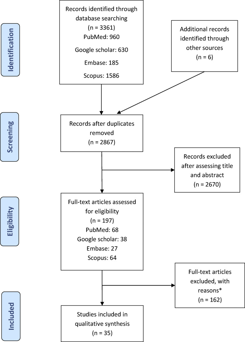
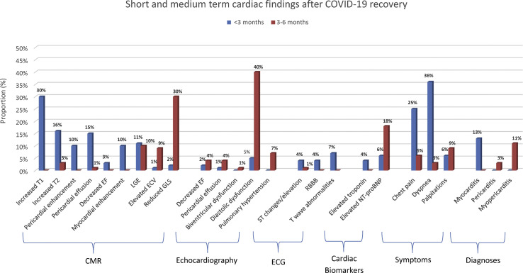
Results: Thirty-five studies (fifteen prospective cohort, seven case reports, five cross-sectional, four case series, three retrospective cohort and one ambidirectional cohort) evaluating cardiac sequelae in 52 609 patients were included. Twenty-nine studies used objective cardiac assessments, mostly cardiac magnetic resonance imaging (CMR) in 16 studies, echocardiography in 15, electrocardiography (ECG) in 16 and cardiac biomarkers in 18. Most studies had a fair risk of bias. The median time from diagnosis/recovery to cardiac assessment was 48 days (1-180 days). Common short-term cardiac abnormalities (<3 months) included increased T1 (proportion: 30%), T2 (16%), pericardial effusion (15%) and late gadolinium enhancement (11%) on CMR, with symptoms such as chest pain (25%) and dyspnoea (36%). In the medium term (3-6 months), common changes included reduced left ventricular global longitudinal strain (30%) and late gadolinium enhancement (10%) on CMR, diastolic dysfunction (40%) on echocardiography and elevated N-terminal proB-type natriuretic peptide (18%). In addition, COVID-19 survivors had higher risk (risk ratio 3; 95% CI 2.7-3.2) of developing heart failure, arrythmias and myocardial infarction.
Conclusions: COVID-19 appears to be associated with persistent/de novo cardiac injury after recovery, particularly subclinical myocardial injury in the earlier phase and diastolic dysfunction later. Larger well-designed and controlled studies with baseline assessments are needed to better measure the extent of cardiac injury and its clinical impact.
Keywords: COVID-19; Cardiac sequelae; Coronavirus; Heart injury; Long COVID-19; Post-acute COVID-19; SARS-CoV-2.
Copyright © 2021 European Society of Clinical Microbiology and Infectious Diseases. Published by Elsevier Ltd. All rights reserved.
Figures
References
-
- World Health Organization . 2021. WHO coronavirus disease (COVID-19) dashboard.https://covid19.who.int/ [Webpage] (updated 5/12/2021). Available from:
-
- Eiros R., Barreiro-Perez M., Martin-Garcia A., Almeida J., Villacorta E., Perez-Pons A., et al. Pericarditis and myocarditis long after SARS-CoV-2 infection: a cross-sectional descriptive study in health-care workers. medRxiv. 2020 Preprint.
Publication types
MeSH terms
LinkOut - more resources
Full Text Sources
Medical
Miscellaneous
