

A deep-learning based artificial intelligence (AI) approach for differentiation of clear cell renal cell carcinoma from oncocytoma on multi-phasic MRI
- PMID: 34171743
- PMCID: PMC9990181
- DOI: 10.1016/j.clinimag.2021.06.016
A deep-learning based artificial intelligence (AI) approach for differentiation of clear cell renal cell carcinoma from oncocytoma on multi-phasic MRI
Abstract
Purpose: To investigate the diagnostic performance of a deep convolutional neural network for differentiation of clear cell renal cell carcinoma (ccRCC) from renal oncocytoma.
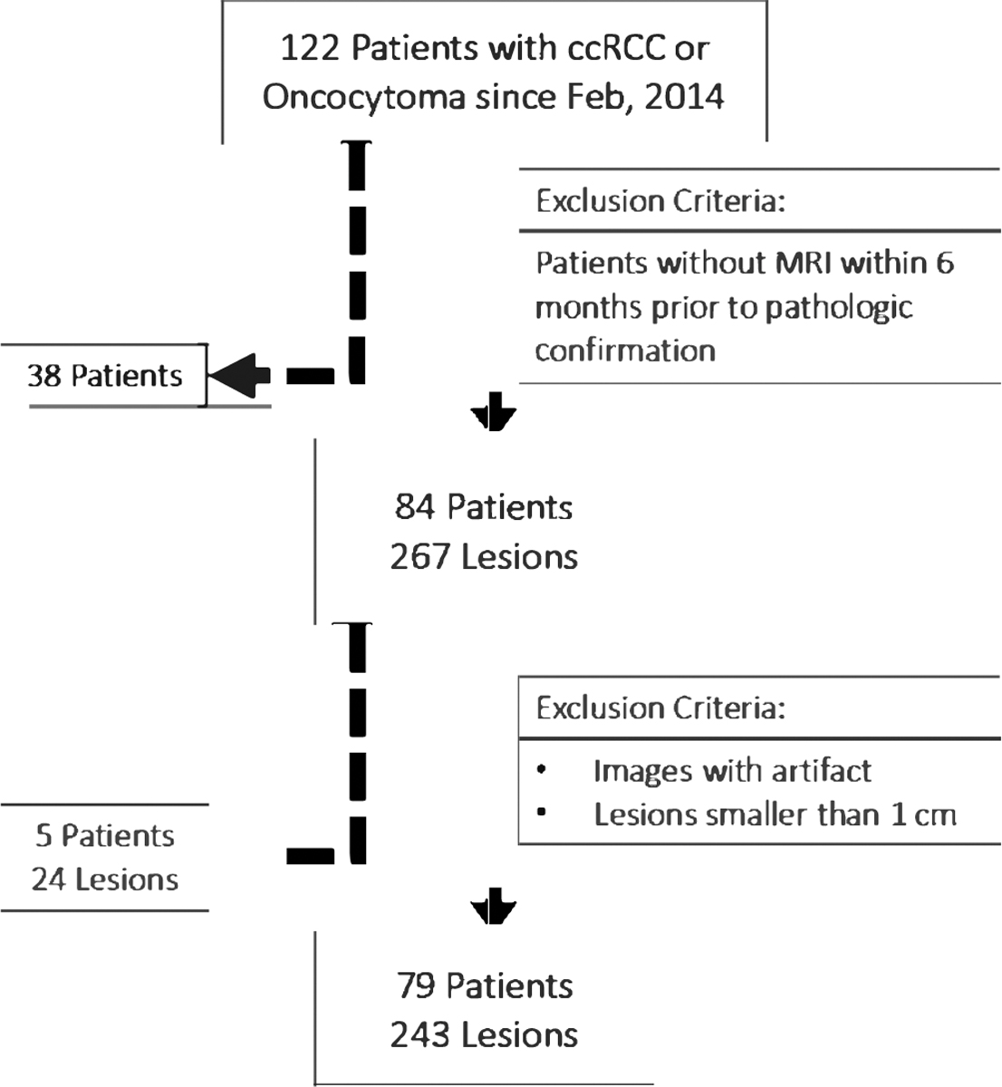
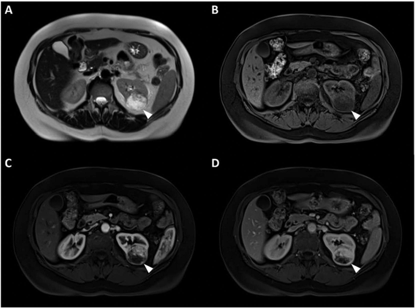
Methods: In this retrospective study, 74 patients (49 male, mean age 59.3) with 243 renal masses (203 ccRCC and 40 oncocytoma) that had undergone MR imaging 6 months prior to pathologic confirmation of the lesions were included. Segmentation using seed placement and bounding box selection was used to extract the lesion patches from T2-WI, and T1-WI pre-contrast, post-contrast arterial and venous phases. Then, a deep convolutional neural network (AlexNet) was fine-tuned to distinguish the ccRCC from oncocytoma. Five-fold cross validation was used to evaluate the AI algorithm performance. A subset of 80 lesions (40 ccRCC, 40 oncocytoma) were randomly selected to be classified by two radiologists and their performance was compared to the AI algorithm. Intra-class correlation coefficient was calculated using the Shrout-Fleiss method.
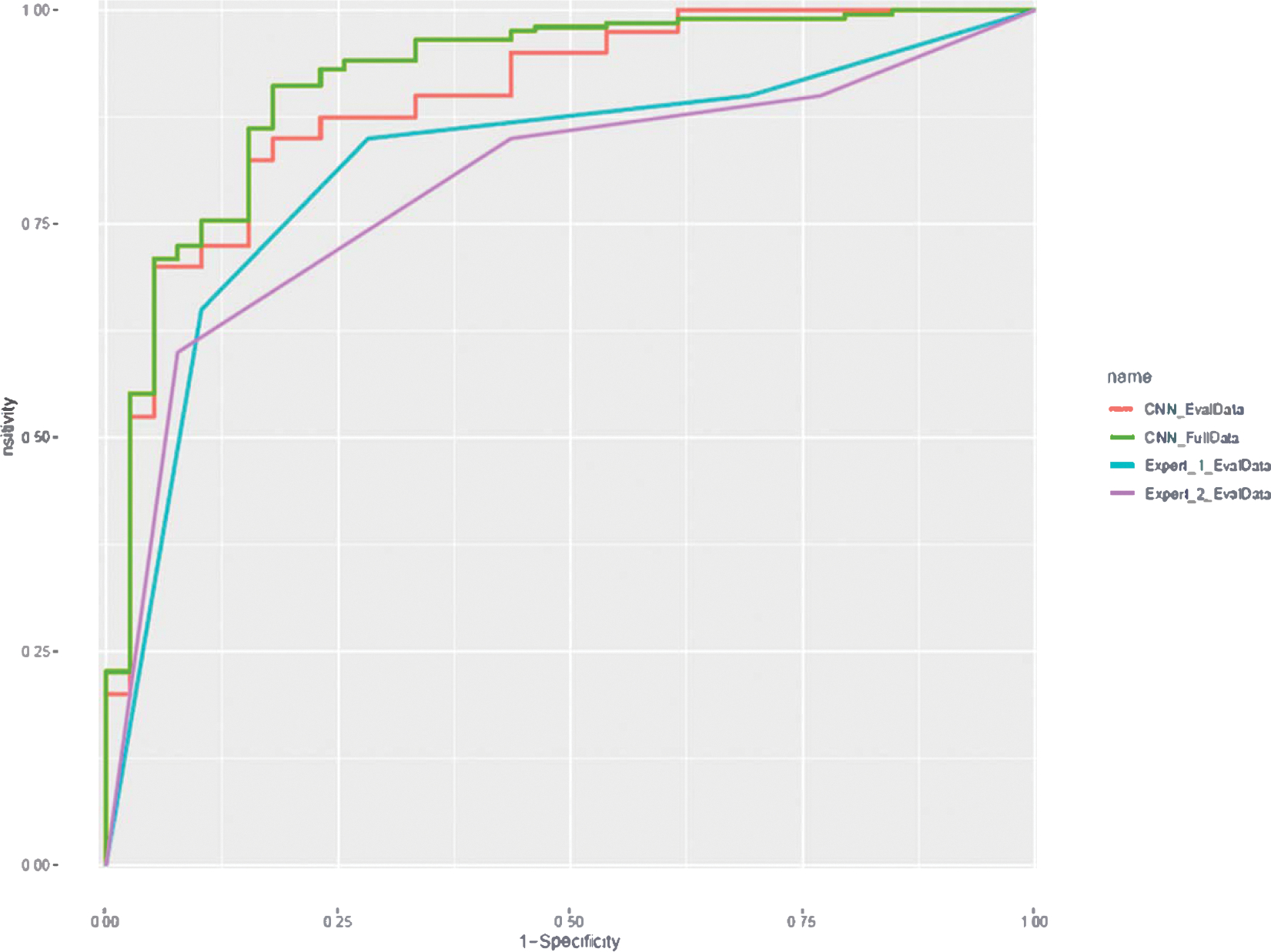
Results: Overall accuracy of the AI system was 91% for differentiation of ccRCC from oncocytoma with an area under the curve of 0.9. For the observer study on 80 randomly selected lesions, there was moderate agreement between the two radiologists and AI algorithm. In the comparison sub-dataset, classification accuracies were 81%, 78%, and 70% for AI, radiologist 1, and radiologist 2, respectively.
Conclusion: The developed AI system in this study showed high diagnostic performance in differentiation of ccRCC versus oncocytoma on multi-phasic MRIs.
Keywords: Clear cell renal cell carcinoma; Deep learning; Multi-phasic MRI; Oncocytoma; Radiomics.
Copyright © 2021. Published by Elsevier Inc.
Conflict of interest statement
Declaration of competing interest
None.
Figures


References
-
- Duchene DA, Lotan Y, Cadeddu JA, Sagalowsky AI, Koeneman KS. Histopathology of surgically managed renal tumors: analysis of a contemporary series. Urology 2003;62(5):827 30. - PubMed
-
- Tsui KH, Shvarts 0, Smith RB, Figlin R, de Kemion JB, Belldegrun A. Renal cell carcinoma: prognostic significance of incidentally detected tumors. J Urol 2000; 163(2):426–30. - PubMed
-
- Gill IS, Aron M, Gervais DA, Jewett MA. Small renal mass. N Engl J Med 2010;362(7):624–34. - PubMed
-
- Capitanio U, Montorsi F. Renal cancer. Lancet 2016;387(10021):894 906. - PubMed
MeSH terms
Grants and funding
LinkOut - more resources
Full Text Sources
Medical
