

Prolonged exposure to traffic-related particulate matter and gaseous pollutants implicate distinct molecular mechanisms of lung injury in rats
- PMID: 34172050
- PMCID: PMC8235648
- DOI: 10.1186/s12989-021-00417-y
Prolonged exposure to traffic-related particulate matter and gaseous pollutants implicate distinct molecular mechanisms of lung injury in rats
Abstract
Background: Exposure to air pollution exerts direct effects on respiratory organs; however, molecular alterations underlying air pollution-induced pulmonary injury remain unclear. In this study, we investigated the effect of air pollution on the lung tissues of Sprague-Dawley rats with whole-body exposure to traffic-related PM1 (particulate matter < 1 μm in aerodynamic diameter) pollutants and compared it with that in rats exposed to high-efficiency particulate air-filtered gaseous pollutants and clean air controls for 3 and 6 months. Lung function and histological examinations were performed along with quantitative proteomics analysis and functional validation.
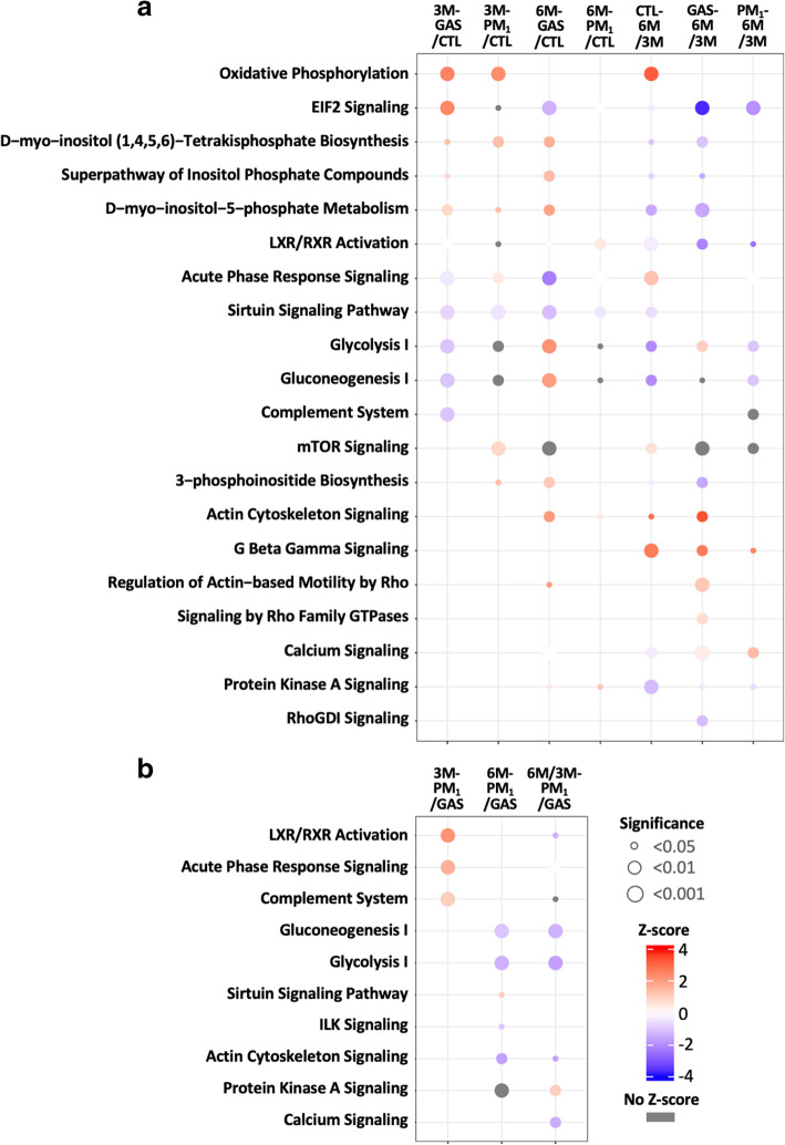
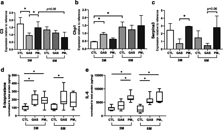
Results: Rats in the 6-month PM1-exposed group exhibited a significant decline in lung function, as determined by decreased FEF25-75% and FEV20/FVC; however, histological analysis revealed earlier lung damage, as evidenced by increased congestion and macrophage infiltration in 3-month PM1-exposed rat lungs. The lung tissue proteomics analysis identified 2673 proteins that highlighted the differential dysregulation of proteins involved in oxidative stress, cellular metabolism, calcium signalling, inflammatory responses, and actin dynamics under exposures to PM1 and gaseous pollutants. The presence of PM1 specifically enhanced oxidative stress and inflammatory reactions under subchronic exposure to traffic-related PM1 and suppressed glucose metabolism and actin cytoskeleton signalling. These factors might lead to repair failure and thus to lung function decline after chronic exposure to traffic-related PM1. A detailed pathogenic mechanism was proposed to depict temporal and dynamic molecular regulations associated with PM1- and gaseous pollutants-induced lung injury.
Conclusion: This study explored several potential molecular features associated with early lung damage in response to traffic-related air pollution, which might be used to screen individuals more susceptible to air pollution.
Keywords: Gaseous pollutant; Lung injury; Molecular mechanism; Particulate matter; Proteomics; Traffic-related air pollution.
Conflict of interest statement
The authors declare that they have no competing interests.
Figures


Similar articles
-
Chronic pulmonary exposure to traffic-related fine particulate matter causes brain impairment in adult rats.Part Fibre Toxicol. 2018 Nov 9;15(1):44. doi: 10.1186/s12989-018-0281-1. Part Fibre Toxicol. 2018. PMID: 30413208 Free PMC article.
-
Effects of antibiotics and metals on lung and intestinal microbiome dysbiosis after sub-chronic lower-level exposure of air pollution in ageing rats.Ecotoxicol Environ Saf. 2022 Nov;246:114164. doi: 10.1016/j.ecoenv.2022.114164. Epub 2022 Oct 13. Ecotoxicol Environ Saf. 2022. PMID: 36244167
-
Respiratory Effects of Traffic-Related Air Pollution: A Randomized, Crossover Analysis of Lung Function, Airway Metabolome, and Biomarkers of Airway Injury.Environ Health Perspect. 2023 May;131(5):57002. doi: 10.1289/EHP11139. Epub 2023 May 4. Environ Health Perspect. 2023. PMID: 37141245 Free PMC article. Clinical Trial.
-
Outdoor air pollution and asthma.Lancet. 2014 May 3;383(9928):1581-92. doi: 10.1016/S0140-6736(14)60617-6. Lancet. 2014. PMID: 24792855 Free PMC article. Review.
-
Lung toxicity of particulates and gaseous pollutants using ex-vivo airway epithelial cell culture systems.Environ Pollut. 2022 Jul 15;305:119323. doi: 10.1016/j.envpol.2022.119323. Epub 2022 Apr 18. Environ Pollut. 2022. PMID: 35447256 Review.
Cited by
-
The Effects and Pathogenesis of PM2.5 and Its Components on Chronic Obstructive Pulmonary Disease.Int J Chron Obstruct Pulmon Dis. 2023 Apr 6;18:493-506. doi: 10.2147/COPD.S402122. eCollection 2023. Int J Chron Obstruct Pulmon Dis. 2023. PMID: 37056681 Free PMC article. Review.
-
Chitosan Nanoparticle-Encapsulated Cordyceps militaris Grown on Germinated Rhynchosia nulubilis Reduces Type II Alveolar Epithelial Cell Apoptosis in PM2.5-Induced Lung Injury.Int J Mol Sci. 2025 Jan 27;26(3):1105. doi: 10.3390/ijms26031105. Int J Mol Sci. 2025. PMID: 39940873 Free PMC article.
-
Deficiency of interleukin-6 receptor ameliorates PM2.5 exposure-induced pulmonary dysfunction and inflammation but not abnormalities in glucose homeostasis.Ecotoxicol Environ Saf. 2022 Dec 1;247:114253. doi: 10.1016/j.ecoenv.2022.114253. Epub 2022 Nov 4. Ecotoxicol Environ Saf. 2022. PMID: 36343449 Free PMC article.
-
Lagged Effects of Exposure to Air Pollutants on the Risk of Pulmonary Tuberculosis in a Highly Polluted Region.Int J Environ Res Public Health. 2022 May 9;19(9):5752. doi: 10.3390/ijerph19095752. Int J Environ Res Public Health. 2022. PMID: 35565147 Free PMC article.
-
Data on lung and intestinal microbiome after air pollution exposure in ageing rats.Data Brief. 2023 Feb 23;47:109004. doi: 10.1016/j.dib.2023.109004. eCollection 2023 Apr. Data Brief. 2023. PMID: 36909015 Free PMC article.
References
-
- Network GBoDC . Global Burden of Disease Study 2016 (GBD 2016) Results. Institute for Health Metrics and Evaluation (IHME) Seattle, United States. 2017.
-
- Vignal C, Pichavant M, Alleman LY, Djouina M, Dingreville F, Perdrix E, Waxin C, Ouali Alami A, Gower-Rousseau C, Desreumaux P, Body-Malapel M. Effects of urban coarse particles inhalation on oxidative and inflammatory parameters in the mouse lung and colon. Part Fibre Toxicol. 2017;14(1):46. doi: 10.1186/s12989-017-0227-z. - DOI - PMC - PubMed
Publication types
MeSH terms
Substances
LinkOut - more resources
Full Text Sources
Medical
