. 2021 Jun 25;20(1):93.
doi: 10.1186/s12943-021-01372-0.

Circ3823 contributes to growth, metastasis and angiogenesis of colorectal cancer: involvement of miR-30c-5p/TCF7 axis

Affiliations

Circ3823 contributes to growth, metastasis and angiogenesis of colorectal cancer: involvement of miR-30c-5p/TCF7 axis

Yaxin Guo et al. Mol Cancer. .

Abstract

Background: Colorectal cancer (CRC) is one of the most common malignant tumours. The recurrence and metastasis of CRC seriously affect the survival rate of patients. Angiogenesis is an extremely important cause of tumour growth and metastasis. Circular RNAs (circRNAs) have been emerged as vital regulators for tumour progression. However, the regulatory role, clinical significance and underlying mechanisms still remain largely unknown.

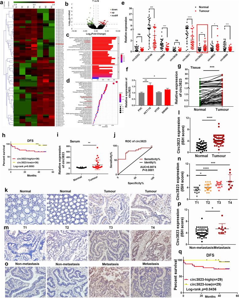
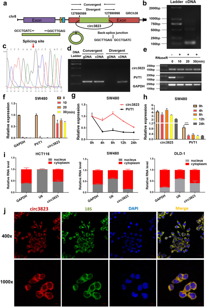
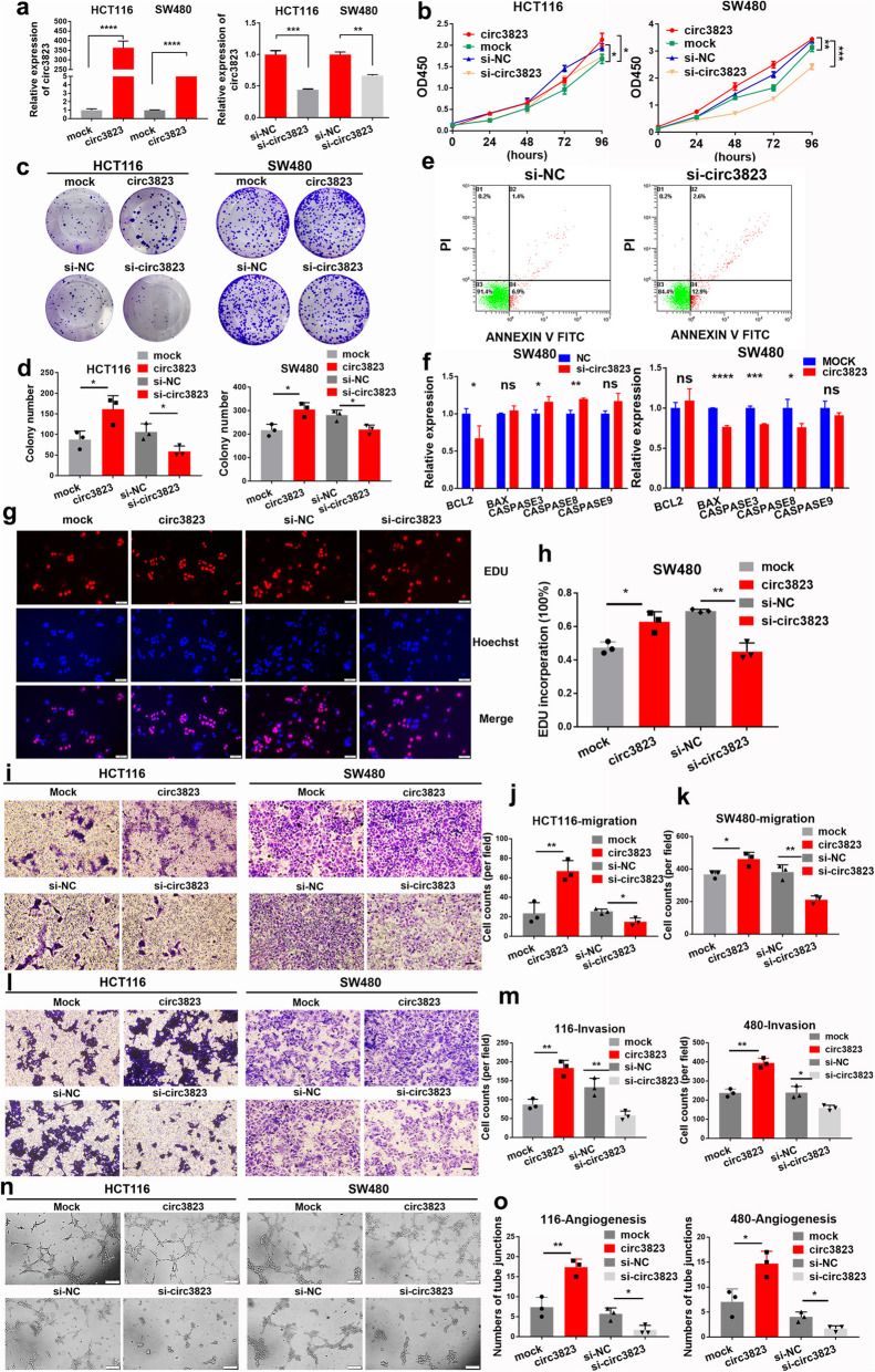
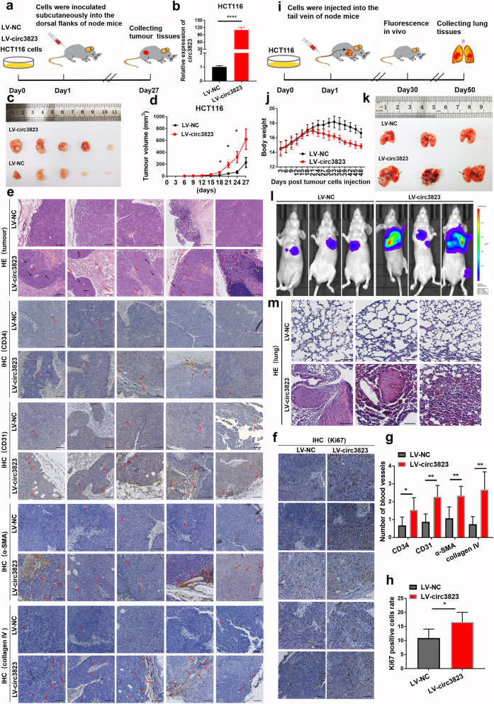
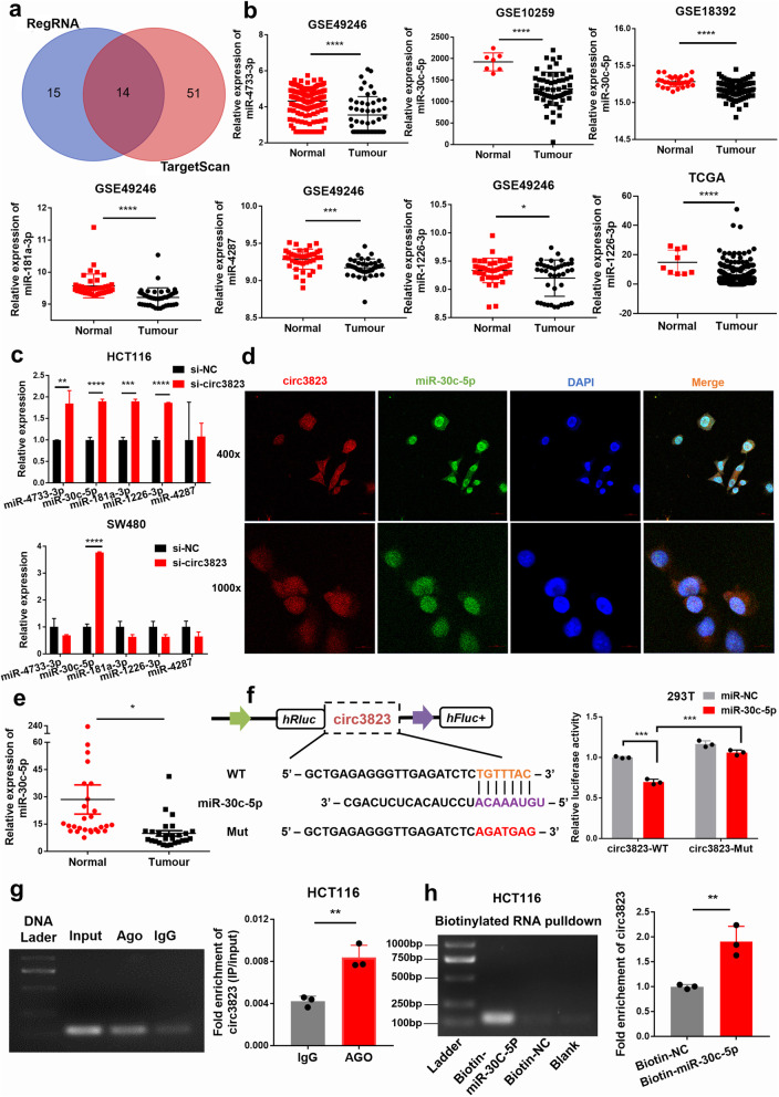
Methods: High-throughput sequencing was used to analyse differential circRNAs expression in tumour and non-tumour tissues of CRC. In situ hybridization (ISH) and qRT-PCR were used to determine the level of circ3823 in CRC tissues and serum samples. Then, functional experiments in vitro and in vivo were performed to investigate the effects of circ3823 on tumour growth, metastasis and angiogenesis in CRC. Sanger sequencing, RNase R and Actinomycin D assay were used to verify the ring structure of circ3823. Mechanistically, dual luciferase reporter assay, fluorescent in situ hybridization (FISH), RNA immunoprecipitation (RIP) and RNA pull-down experiments were performed to confirm the underlying mechanisms of circ3823.

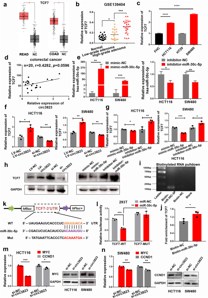
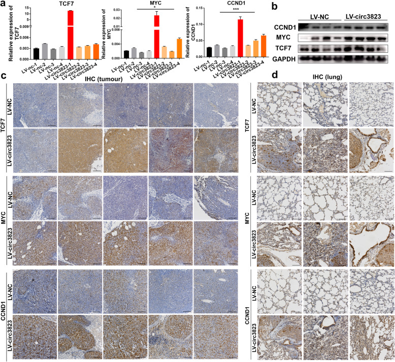
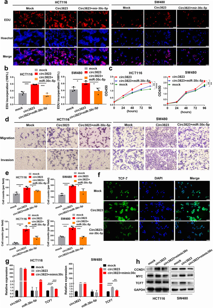
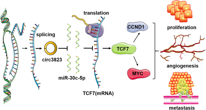
Results: Circ3823 was evidently highly expressed in CRC and high circ3823 expression predicted a worse prognosis of CRC patients. Receiver operating characteristic curves (ROCs) indicated that the expression of circ3823 in serum showed high sensitivity and specificity for detecting CRC which means circ3823 have the potential to be used as diagnostic biomarkers. Functional experiments in vitro and in vivo indicated that circ3823 promote CRC cell proliferation, metastasis and angiogenesis. Mechanism analysis showed that circ3823 act as a competing endogenous RNA of miR-30c-5p to relieve the repressive effect of miR-30c-5p on its target TCF7 which upregulates MYC and CCND1, and finally facilitates CRC progression. In addition, we found that N6-methyladenosine (m6A) modification exists on circ3823. And the m6A modification is involved in regulating the degradation of circ3823.

Conclusions: Our findings suggest that circ3823 promotes CRC growth, metastasis and angiogenesis through circ3823/miR-30c-5p/TCF7 axis and it may serve as a new diagnostic marker or target for treatment of CRC patients. In addition, m6A modification is involved in regulating the degradation of circ3823.

Keywords: Angiogenesis; Colorectal cancer (CRC); N6-methyladenosine (m6A); Tumour progression; circ3823.


Conflict of interest statement

The authors declare that they have no competing interests.

Figures

Fig. 1
Fig. 1
CircRNA expression profiling in CRC tissues and normal tissues and validation of the circRNAs expression in serums and tissues (T: tumour, N: normal). a Hierarchical clustering of differentially expressed circRNA in two groups. b Volcano plot showed the expression profile between tumour tissue and normal tissue. Conditions for screening differences:│Fold Change│ > 2; p < 0.05. The red points in the plot indicated significantly up-regulated circRNAs, and the green points indicated significantly down-regulated circRNAs. c GO function analysis of differentially expressed circRNAs. d KEGG pathway analysis of differentially expressed circRNAs. e The expression of circ2253, circ2749, circ2990, circ3038, circ3651, circ3823, circ4953 and circ8080 was detected by qRT-PCR in 28 CRC tissues and matched normal tissues. f The expression of circ3823 was measured by qRT-PCR in CRC cells and FHC. g The level of circ3823 in 72 CRC tissues and matched normal tissues were analyzed by qRT-PCR. h The disease-free survival analysis of 72 CRC patients was performed based on qRT-PCR data. Log-rank test was used to estimate the significance. i The expression of circ3823 was measured by qRT-PCR in 28 serum samples from CRC patients and 28 serum samples from healthy people. GAPDH was used as an internal reference. j ROC curves based on the expression of circ3823 in serum. k, l ISH images and ISH scores statistical analysis of circ3823 in CRC tissues and normal tissues. m, n ISH images and ISH scores statistical analysis of circ3823 in T1, T2, T3, T4 CRC tissues. o, p ISH images and ISH scores statistical analysis of circ3823 in lymph node metastasis tissues and non-lymph node metastasis tissues (magnification, × 200, scale bar, 50 μm). q The disease-free survival analysis of 58 CRC patients based on circ3823 ISH scores. Log-rank test was used to estimate the significance. Data were indicated as mean ± SD, ns P ≥ 0.05, *P < 0.05, **P < 0.01, ***P < 0.001, ****P < 0.0001
Fig. 2
Fig. 2
Verification of the closed loop structure of circ3823 and its subcellular location. a The formation process of circ3823 which was produced at the PVT1 gene located on chromosome8. b PCR product of circ3823 in agarose gel electrophoresis. c The back-splice junction of circ3823 was identified by Sanger sequencing. d Convergent and divergent primers were used to verification of the closed loop structure. e, f Verification of the closed loop structure of circ3823 through RNase R. g, h The abundance of circ3823 and PVT1 were detected by qRT-PCR in SW480 cells treated with Actinomycin D at the indicated time point. i qRT-PCR was used to measure the level of circ3823 in the nuclear and cytoplasmic of HCT116, SW480, DLD-1 cells. j FISH was performed to observe the cellular location of circ3823 (red) and 18S (green) in cells (magnification, × 400, scale bar, 20 μm and magnification, × 1000, scale bar, 10 μm). Data were indicated as mean ± SD (n = 3), ns P ≥ 0.05, *P < 0.05, **P < 0.01, ***P < 0.001, ****P < 0.0001
Fig. 3
Fig. 3
Circ3823 promotes CRC cell proliferation, migration, invasion and increases the tube junction forming ability of HUVEC in vitro. a Expression levels of circ3823 in HCT116 and SW480 cells treated with circ3823 plasmid and circ3823 siRNA. b, c, d, g, h Cell proliferation detection of CRC cells were measured by CCK-8, clone formation and EDU assay (magnification, × 200, scale bar, 50 μm). e 1 × 104 SW480 cells were counted per experiment to determine apoptosis rate by flow cytometry via Annexin V-FITC/PI. f Apoptosis-related molecules were detected using qRT-PCR in SW480 cells. i, j, k, l, m Cell migration and invasion abilities were determined by transwell assays (magnification, × 100, scale bar, 100 μm). n, o Tube junction forming ability of HUVEC were determined by incubate with HCT116, SW480 cell culture supernatant after transfection (magnification, × 100, scale bar, 200 μm). Data were indicated as mean ± SD, ns P ≥ 0.05, *P < 0.05, **P < 0.01, ***P < 0.001, ****P < 0.0001
Fig. 4
Fig. 4
Circ3823 facilitates tumourigenesis, metastasis and angiogenesis in vivo. a, b The process of constructing subcutaneous xenograft tumour in nude mice. CRC cell line stably overexpressing circ3823 was constructed by lentivirus and the expression of circ3823 was detected by qRT-PCR. c Images of xenograft tumours of each group (n = 5). d Growth curves of xenograft tumours measured every 3 days. e, g Pathology of the tumour tissue was detected by HE staining. IHC staining of CD34/31, α-SMA and collagen IV to investigate angiogenesis (mitotic phase: black arrow, angiogenesis: red arrow, inflammatory cell infiltration: yellow arrow) (magnification, × 100, scale bar, 100 μm). f, h IHC staining of ki-67 in each slide to evaluate cell cycle (magnification, × 100, scale bar, 100 μm). i The process of constructing metastasis xenograft tumour in nude mice. j The body weight of the two groups of nude mice. k After injection of stably overexpressing circ3823 HCT116 cells into the tail vein of nude mice at 50 days, the mice were euthanized, and metastatic lung nodules were marked with black arrow. l Bioluminescent images of lungs for each experimental group at 7 weeks. m HE staining of lung sections displayed metastatic nodules of the lungs (magnification, × 100, scale bar, 100 μm). Data were indicated as mean ± SD, ns P ≥ 0.05, *P < 0.05, **P < 0.01, ***P < 0.001, ****P < 0.0001
Fig. 5
Fig. 5
Circ3823 functions as a sponge for miR-30c-5p. a The microRNA binding on circ3823 predicted by targetScan and RegRNA. b MiR-4733-3p, miR-30c-5p, miR-181a-3p, miR-4287 and miR-1226-3p was significantly down-regulated in CRC according to GEO and TCGA. c MiR-30c-5p was detected using qRT-PCR after downregulation of circ3823 in HCT116 and SW480. d FISH was performed to observe the cellular location of circ3823 (red) and miR-30c-5p (green) in cells (magnification, × 400, scale bar, 20 μm and magnification, × 1000, scale bar, 10 μm). e Relative expression of miR-30c-5p in CRC tissues and adjacent non-tumour tissues was determined by qRT-PCR (n = 27). f Schematic illustration of circ3823-WT and circ3823-Mut luciferase reporter vectors. The relative luciferase activities were detected in 293 T cells after transfection with circ3823-WT or circ3823-Mut and miR-30c-5p mimics or mimic-NC, respectively. g Anti-AGO2 RIP was executed in HCT116 cells, followed by qRT-PCR and nucleic acid electrophoresis to detect the enrichment ability of AGO2 on circ3823 compared with IgG. h RNA pull-down was executed in HCT116 cells, followed by qRT-PCR and nucleic acid electrophoresis to detect the enrichment of circ3823. Data were indicated as mean ± SD, ns P ≥ 0.05, *P < 0.05, **P < 0.01, ***P < 0.001, ****P < 0.0001
Fig. 6
Fig. 6
TCF7 is directly targeted by miR-30c-5p and indirectly regulated by circ3823. a TCF7 was significantly up-regulated in both rectal cancer and colon cancer according to GEPIA. b GEO data show that TCF7 is closely related to the histopathological grade of colon cancer. c The expression of TCF7 in CRC cells compared with FHC. d Pearson correlation analysis of circ3823 and TCF7 expression in 20 CRC tissues. e The mimic and inhibitor efficiency of hsa-miR-30c-5p in HCT116 and SW480. f, g, h Relative mRNA and protein levels of TCF7 were detected in cells after transfected with LV-NC, LV-circ3823, si-NC, si-circ3823, mimic-NC, mimic-miR-30c-5p, inhibitor-NC and inhibitor-miR-30c-5p using qRT-PCR and western blot, respectively. i, j RNA pull-down was executed in HCT116 cells, followed by qRT-PCR and nucleic acid electrophoresis to detect the enrichment of TCF7. k Schematic illustration of TCF7-WT and TCF7-Mut luciferase reporter vectors. l The relative luciferase activities were detected in 293 T cells after transfection with TCF7-WT or TCF7-Mut and miR-30c-5p mimics or mimic-NC, respectively. m, n Relative expression of TCF7 downstream molecules MYC and CCND at RNA and protein level in cells transfected with si-circ3823. Data were indicated as mean ± SD, ns P ≥ 0.05, *P < 0.05, **P < 0.01, ***P < 0.001, ****P < 0.0001
Fig. 7
Fig. 7
The RNA and protein levels of TCF7, MYC, CCND1 in subcutaneous xenograft tumours and lung metastatic tissues in nude mice. a, b Relative mRNA and protein levels of TCF7, MYC, CCND1 were detected in subcutaneous xenograft tumours by qRT-PCR and western blot. c, d Relative protein levels of TCF7, MYC, CCND1 were detected in subcutaneous xenograft tumours and lung metastatic tissues by ISH (magnification, × 100, scale bar, 100 μm). Data were indicated as mean ± SD, ns P ≥ 0.05, *P < 0.05, **P < 0.01, ***P < 0.001, ****P < 0.0001
Fig. 8
Fig. 8
Circ3823 promotes cell proliferation and metastasis through circ3823/miR-30c-5p/TCF7 axis. a, b, c The cell proliferation were determined after transfection with overexpression circ3823 vectors and mimic-miR-30c-5p by EdU and CCK-8 assays (magnification, × 200, scale bar, 50 μm). d, e The cell migration and invasion were determined after transfection with overexpression circ3823 vectors, and mimic-miR-30c-5p by trans well assays (magnification, × 100, scale bar, 100 μm). f Relative protein levels of TCF7 in cells were assessed by IF after transfection (magnification, × 200, scale bar, 50 μm). g, h Relative expression of TCF7 mRNA and protein levels were detected by qRT-PCR and western blot in cells transfected with overexpression circ3823 vectors and mimic-miR-30c-5p. Data were indicated as mean ± SD, ns P ≥ 0.05, *P < 0.05, **P < 0.01, ***P < 0.001, ****P < 0.0001
Fig. 9
Fig. 9
Schematic representation of a model for the major molecular mechanisms of “circ3823/miR-30c-5p/TCF7” axis in CRC

References

    1. Collaborators GBDCC. The global, regional, and national burden of colorectal cancer and its attributable risk factors in 195 countries and territories, 1990-2017: a systematic analysis for the global burden of disease study 2017. Lancet Gastroenterol Hepatol. 2019;4(12):913–933. doi: 10.1016/S2468-1253(19)30345-0. - DOI - PMC - PubMed
    1. Bray F, Ferlay J, Soerjomataram I, Siegel RL, Torre LA, Jemal A. Global cancer statistics 2018: GLOBOCAN estimates of incidence and mortality worldwide for 36 cancers in 185 countries. CA Cancer J Clin. 2018;68(6):394–424. doi: 10.3322/caac.21492. - DOI - PubMed
    1. Siegel RL, Miller KD, Goding Sauer A, Fedewa SA, Butterly LF, Anderson JC, Cercek A, Smith RA, Jemal A. Colorectal cancer statistics, 2020. CA Cancer J Clin. 2020;70(3):145–164. doi: 10.3322/caac.21601. - DOI - PubMed
    1. Siegel R, Desantis C, Jemal A. Colorectal cancer statistics, 2014. CA Cancer J Clin. 2014;64(2):104–117. doi: 10.3322/caac.21220. - DOI - PubMed
    1. Siegel RL, Miller KD, Fedewa SA, Ahnen DJ, Meester RGS, Barzi A, Jemal A. Colorectal cancer statistics, 2017. CA Cancer J Clin. 2017;67(3):177–193. doi: 10.3322/caac.21395. - DOI - PubMed

Publication types

MeSH terms