Review
. 2021 Jun 25;14(1):98.
doi: 10.1186/s13045-021-01103-4.

Spatial architecture of the immune microenvironment orchestrates tumor immunity and therapeutic response

Affiliations
Review

Spatial architecture of the immune microenvironment orchestrates tumor immunity and therapeutic response

Tong Fu et al. J Hematol Oncol. .

Abstract

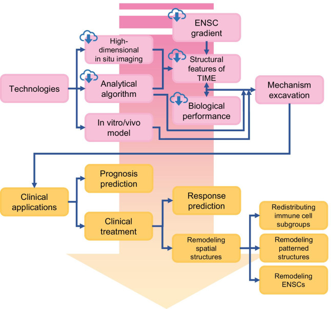
Tumors are not only aggregates of malignant cells but also well-organized complex ecosystems. The immunological components within tumors, termed the tumor immune microenvironment (TIME), have long been shown to be strongly related to tumor development, recurrence and metastasis. However, conventional studies that underestimate the potential value of the spatial architecture of the TIME are unable to completely elucidate its complexity. As innovative high-flux and high-dimensional technologies emerge, researchers can more feasibly and accurately detect and depict the spatial architecture of the TIME. These findings have improved our understanding of the complexity and role of the TIME in tumor biology. In this review, we first epitomized some representative emerging technologies in the study of the spatial architecture of the TIME and categorized the description methods used to characterize these structures. Then, we determined the functions of the spatial architecture of the TIME in tumor biology and the effects of the gradient of extracellular nonspecific chemicals (ENSCs) on the TIME. We also discussed the potential clinical value of our understanding of the spatial architectures of the TIME, as well as current limitations and future prospects in this novel field. This review will bring spatial architectures of the TIME, an emerging dimension of tumor ecosystem research, to the attention of more researchers and promote its application in tumor research and clinical practice.

Keywords: Immunotherapy; Spatial architecture; Tumor immune microenvironment; Tumor immunity.


Conflict of interest statement

The authors declare that they have no competing interests.

Figures

Fig. 1
Fig. 1
Definition and components of the spatial architecture of the tumor immune microenvironment (TIME). The spatial architecture is described according to the location of immune cells (a), distance between cells (b), distribution of immunoregulators (c), and specific spatial patterns (d)
Fig. 2
Fig. 2
Emerging techniques used to identify the spatial architecture of the tumor immune microenvironment. Pink area (a), deep-leaning-based HE techniques. Blue area (bd), probe-based in situ technologies. b, CODEX-FFPE; c, seqFISH+ ; d, IMC and MIBI/MIBI-TOF. Green area (ef) spatial omics. e, microarray-based spatial transcriptomics + sc-RNA-seq; f, MALDI MSI. Consult Table 3 for more detailed information. H&E, hematoxylin and eosin; CNN, convolutional neural network; MALDI MSI, matrix-assisted laser desorption/ionization mass spectrometric imaging; UV, ultraviolet; sc-RNAseq, single-cell RNA sequencing; IMC, imaging mass cytometry; MIBI, multiplexed ion beam imaging; MIBI-TOF, multiplexed ion beam imaging by time of flight; CODEX, codetection by indexing; FFPE, formalin-fixed and paraffin-embedded
Fig. 3
Fig. 3
Representative spatial architecture of immune cells in the tumor microenvironment. a Primary tumors are divided into the tumor core, tumor stroma, and invasion margin based on tumor compartments. b Special immune structures, such as perivascular niches and tertiary lymphoid structures (TLSs), are also involved in the construction of architectures. Moreover, computational technology identified cellular neighborhoods (CNs) as regions with a characteristic local stoichiometry of cellular components. CXCL4, C-X-C chemokine ligand type 4; CCL2, C–C chemokine ligand type 2; CX3CL1, C-X3-C motif ligand 1; TGF-β, transforming growth factor-β; IFN, interferon; IL-2, interleukin 2; ADCC, antibody-dependent cellular cytotoxicity; MDSC, myeloid-derived suppressor cell
Fig. 4
Fig. 4
Spatial evolution of the tumor immune microenvironment (TIME) structure during tumor progression. The process of tumor initiation, expansion, and metastases is accompanied by a gamble between the tumor and the TIME, where antitumor immune and immunosuppressive factors coexist and interact with each other. a In the initiation stage, the immune components around the lesion evolve from immune surveillance to immune escape during the evolution of "normal tissue", precancerous lesions, and carcinoma in situ (CIS). b In the expansion phase, the TIME functions in a contact-dependent or distance-dependent manner. c In the metastatic phase, the specific arrangement of immune cells in the metastatic niche establishes a favorable environment for the formation and growth of metastases. PD-1, programmed cell death 1; PD-L1, programmed cell death ligand 1; Lag-3, lymphocyte-activation gene 3; TLSs, tertiary lymphoid structures; TLR, Toll-like receptor; CXCL12, C-X-C chemokine ligand type 12; CXCR4, C-X-C chemokine receptor type 4; TGF-β, transforming growth factor-β; IL, interleukin; VEGF, vascular endothelial growth factor; ROS, reactive oxygen species; NO, nitric oxide; EGF, endothelial growth factor; Arg1, Arginase-1; CCL5, C–C chemokine ligand type 5; TNF-α, tumor necrosis factor α
Fig. 5
Fig. 5
Oxygen serves as a pivotal extracellular nonspecific chemical (ENSC), and its gradient orchestrates the tumor immune microenvironment (TIME). The aberrant structure of tumor vessels and the abnormal distribution of oxygen in tumors feature a consecutive normoxia-hypoxia-anoxia gradient from feeding vessels toward the tumor center. Chemotactic factors such as CXCL12, CCL5, ET-1, ET-2, VEGF-A and Sema3A released by hypoxic tumor cells, as well as damage-associated molecular patterns (DAMPs) and ATP released by dead/dying tumor cells, attract macrophages to infiltrate into the hypoxic tumor core. Cytokines such as oncostatin, IL-6, IL-10, TGF-β, and HMBG-1 and lactate produced by hypoxic tumor cells further promote the differentiation of macrophages into protumor M2 macrophages, while macrophages remaining next to normoxic feeding vessels display an antitumor phenotype. The oxygen gradient may serve as a marker of the distance from feeding vessels and correlates with other ENSC gradients in the TIME, such as glucose, lactate and hydrion. CXCL12, C-X-C chemokine ligand type 12; CCL5, C–C chemokine ligand type 5; ET, endothelin; VEGF, vascular endothelial growth factor; IL, interleukin; TC, tumor cell; TGF, transforming growth factor; HMBG-1, high mobility group box 1 protein;Sema3A, semaphorin-3A
Fig. 6
Fig. 6
Future development of the tumor immune microenvironment. The development of technologies in high-dimensional in situ imaging, analytical algorithms and in vitro/vivo models will promote the further elucidation of mechanisms underlying the tumor immune microenvironment (TIME) (boxes with cloud marks refer to content that future online databases of TIME might include). Profound advances in the clinical application of TIME rely on deeper insights into the TIME, which will help physicians with determining both a precise diagnosis and therapy

Similar articles

Cited by

References

    1. Hanahan D, Weinberg RA. Hallmarks of cancer: the next generation. Cell. 2011;144(5):646–674. doi: 10.1016/j.cell.2011.02.013. - DOI - PubMed
    1. Anderson NM, Simon MC. The tumor microenvironment. Curr Biol. 2020;30(16):R921–R925. doi: 10.1016/j.cub.2020.06.081. - DOI - PMC - PubMed
    1. Riera-Domingo C, Audige A, Granja S, Cheng WC, Ho PC, Baltazar F, et al. Immunity, hypoxia, and metabolism-the menage a trois of cancer: implications for immunotherapy. Physiol Rev. 2020;100(1):1–102. doi: 10.1152/physrev.00018.2019. - DOI - PubMed
    1. Gajewski TF, Schreiber H, Fu YX. Innate and adaptive immune cells in the tumor microenvironment. Nat Immunol. 2013;14(10):1014–1022. doi: 10.1038/ni.2703. - DOI - PMC - PubMed
    1. Bader JE, Voss K, Rathmell JC. Targeting metabolism to improve the tumor microenvironment for cancer immunotherapy. Mol Cell. 2020;78(6):1019–1033. doi: 10.1016/j.molcel.2020.05.034. - DOI - PMC - PubMed

Publication types