. 2021 Jun 25;7(26):eabg9923.
doi: 10.1126/sciadv.abg9923. Print 2021 Jun.

Structure of the mycobacterial ESX-5 type VII secretion system pore complex

Affiliations

Structure of the mycobacterial ESX-5 type VII secretion system pore complex

Katherine S H Beckham et al. Sci Adv. .

Abstract

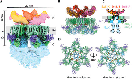
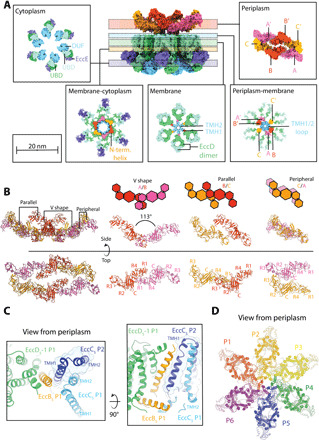
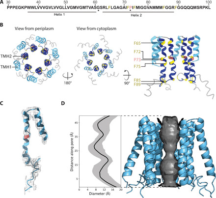
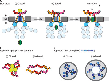
The ESX-5 type VII secretion system is a membrane-spanning protein complex key to the virulence of mycobacterial pathogens. However, the overall architecture of the fully assembled translocation machinery and the composition of the central secretion pore have remained unknown. Here, we present the high-resolution structure of the 2.1-megadalton ESX-5 core complex. Our structure captured a dynamic, secretion-competent conformation of the pore within a well-defined transmembrane section, sandwiched between two flexible protein layers at the cytosolic entrance and the periplasmic exit. We propose that this flexibility endows the ESX-5 machinery with large conformational plasticity required to accommodate targeted protein secretion. Compared to known secretion systems, a highly dynamic state of the pore may represent a fundamental principle of bacterial secretion machineries.


Figures

Fig. 1
Fig. 1. Overall architecture of the ESX-5 complex.
(A) Composite EM map of the ESX-5 complex highlighting the periplasmic (P), inner-membrane (IM) and cytosolic (C) regions of the complex. Periplasmic regions are colored in yellow, orange, and pink. TM and membrane-proximal cytosolic region are colored according to the scheme shown in (C). The distal cytosolic segment tentatively corresponding to the ATPase domains is shown in light blue, and the mycobacterial inner membrane (IM) is indicated in light gray. (B) Side view of the composite model of the ESX-5 complex generated by combining the atomic coordinate model, with an ensemble of models built using integrative approaches. (C) Schematic of the ESX-5 complex showing the organization of the components EccB5 (orange), EccC5 (light blue), EccD5 (green, light green), and EccE5 (purple). The different EccB5 domains are colored to show their location in the segmented EM map in (A). Low-resolution density has been shown as a lighter shade for EccC5 and EccE5. (D) Top and bottom views of the rigid core of the ESX-5 membrane complex.
Fig. 2
Fig. 2. Subunit interactions and assemblies within the ESX-5 complex.
(A) Cross-sections of the ESX-5 complex displayed as a surface representation of the atomic model shown as insets. Each inset highlights the subunit/subunit interactions occurring on the periplasmic, periplasmic-membrane, membrane, membrane-cytoplasmic, and cytoplasmic level. Key features described in the text are highlighted. (B) Overview of the assembly of EccB5 in the periplasm into defined dimers, parallel, V-shape, and peripheral. In the parallel dimer, EccB5-B forms an interface with EccB5-C, where B and C are shifted with respect to each other by about 5 nm and is stabilized by interactions between the central C-domains: (EccB5-B)/R1 + R4 (EccB5-C) and C (EccB5-C)/R2 + R3 (EccB5-B). The trimers along the long axis of the keel-shaped arrangement form the peripheral dimeric interface, mediated by R2 + R3 of EccB5-C from one trimer and by R2 + R3 of EccB5-A′ from the other trimer. These three EccB5 dimers assemble into the characteristic dimer-of-trimer arrangement of the periplasmic face structure of the overall ESX-5 complex (left), which does not follow the sixfold symmetry of other parts of the complex. In the top view, the subdomains in each EccB5 protomer are labeled, indicating specific EccB5 domain/domain interactions for the three distinct EccB5 dimeric interfaces observed. (C) Interprotomer interactions occurring between the EccC5 TMH1 arising from protomer 1 (P1) and EccB5 TMD arising from protomer 2 (P2). The two panels represent the top and side view, respectively. (D) Overview of domain swap interactions occurring between protomeric units when viewed from the periplasm. Each protomer has a different color.
Fig. 3
Fig. 3. Central pore of the ESX-5 secretion system.
(A) M. xenopi EccC5 sequence near the N terminus (residues 30 to 100) highlighting the two TMHs observed in the structure of the overall ESX-5 complex. Invariant residues are indicated by asterisk (*) (for further details see fig. S8). Colors correspond to the model shown in (B). (B) Top-scoring Rosetta (36) model of the central pore formed by EccC5 TMH1 (light blue) and TMH2 (dark blue) viewed from the periplasm, the cytoplasm, and along the membrane. Conserved residues have been colored (proline, red; phenylalanine, yellow). (C) EM density corresponding to the EccC5 TMHs, the top-scoring Rosetta model is shown. (D) Analysis of the pore diameter from 100 Rosetta models with HOLE (42), median pore diameter (black) and 90% confidence interval (gray). Side view of EccC5 pore helices of a top-scoring model showing the pore diameter analysis.
Fig. 4
Fig. 4. Scheme of conformational changes occurring in type VII secretion.
(A) Side views of the ESX-5 overall conformations proceeding from an inactive to active complex. We propose that the closed state (i) based on the M. tuberculosis ESX-5 structure in the presence of the protease MycP5 (18) represents an inactive conformation. The second step toward active secretion (ii), based on the ESX-5 structure reported here, would represent a gated, secretion-competent conformation capable of initiating substrate translocation. In this conformation, the periplasmic EccB5 subunits are reorganized into a dimer-of-trimers arrangement, generating an elongated central cleft (B). The change in orientation of the EccB5 TMH reorients the EccC5 TMHs in the membrane to allow opening the central TM pore (C). In the closed conformation the same EccC5 TMHs are organized into four-helix bundles formed from two protomeric units without a visible pore. In all presently known structures, the cytosolic EccC5 domains adopt multiple conformations, demonstrating a high level of conformational plasticity requirements for ESX-5 substrate recognition and gating into the pore. We speculate that there could be an even more open conformation of the ESX-5 pore during active translocation of folded substrates, resulting from further reorientation of the EccB5 TMHs and the two pore forming EccC5 TMHs (iii). EccB5 is shown in orange, pink, and red; EccC5 is shown in blue and light blue; substrate is shown in brown; and MycP5 is shown in yellow. For reasons of clarity, the remaining structural EccD5 and EccE5 components of the TM section are shown in gray. In (C), the EccC5 TMH1 is in light blue and TMH2 is in dark blue. The TMHs have been numbered according to the protomer they correspond to. The numbering of the closed conformation is based on Bunduc et al. (18).

Similar articles

Cited by

References

    1. I. Kecerdasan, P. Ikep, Global Tuberculosis Report (World Health Organization, 2020).
    1. Orgeur M., Brosch R., Evolution of virulence in the Mycobacterium tuberculosis complex. Curr. Opin. Microbiol. 41, 68–75 (2018). - PubMed
    1. Gey van Pittius N. C., Sampson S. L., Lee H., Kim Y., van Helden P. D., Warren R. M., Evolution and expansion of the Mycobacterium tuberculosis PE and PPE multigene families and their association with the duplication of the ESAT-6 (esx) gene cluster regions. BMC Evol. Biol. 6, 95 (2006). - PMC - PubMed
    1. Ates L. S., van der Woude A. D., Bestebroer J., van Stempvoort G., Musters R. J. P., Garcia-Vallejo J. J., Picavet D. I., van de Weerd R., Maletta M., Kuijl C. P., van der Wel N. N., Bitter W., The ESX-5 system of pathogenic mycobacteria is involved in capsule integrity and virulence through its substrate PPE10. PLOS Pathog. 12, e1005696 (2016). - PMC - PubMed
    1. Ates L. S., Ummels R., Commandeur S., van der Weerd R., Sparrius M., Weerdenburg E., Alber M., Kalscheuer R., Piersma S. R., Abdallah A. M., Abd El Ghany M., Abdel-Haleem A. M., Pain A., Jiménez C. R., Bitter W., Houben E. N. G., Essential role of the ESX-5 secretion system in outer membrane permeability of pathogenic Mycobacteria. PLOS Genet. 11, e1005190 (2015). - PMC - PubMed

LinkOut - more resources