

Antitumor efficacy and reduced toxicity using an anti-CD137 Probody therapeutic
- PMID: 34172583
- PMCID: PMC8255787
- DOI: 10.1073/pnas.2025930118
Antitumor efficacy and reduced toxicity using an anti-CD137 Probody therapeutic
Abstract
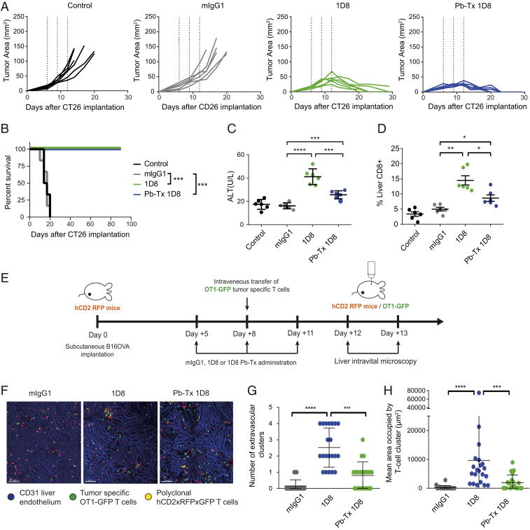
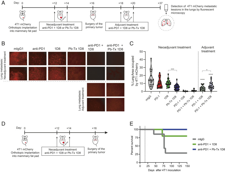
Costimulation via CD137 (4-1BB) enhances antitumor immunity mediated by cytotoxic T lymphocytes. Anti-CD137 agonist antibodies elicit mild liver inflammation in mice, and the maximum tolerated dose of Urelumab, an anti-human CD137 agonist monoclonal antibody, in the clinic was defined by liver inflammation-related side effects. A protease-activated prodrug form of the anti-mouse CD137 agonist antibody 1D8 (1D8 Probody therapeutic, Pb-Tx) was constructed and found to be selectively activated in the tumor microenvironment. This construct, which encompasses a protease-cleavable linker holding in place a peptide that masks the antigen binding site, exerted antitumor effects comparable to the unmodified antibody but did not result in liver inflammation. Moreover, it efficaciously synergized with both PD-1 blockade and adoptive T-cell therapy. Surprisingly, minimal active Pb-Tx reached tumor-draining lymph nodes, and regional lymphadenectomy did not abrogate antitumor efficacy. By contrast, S1P receptor-dependent recirculation of T cells was absolutely required for efficacy. The preferential cleavage of the anti-CD137 Pb-Tx by tumor proteases offers multiple therapeutic opportunities, including neoadjuvant therapy, as shown by experiments in which the Pb-Tx is given prior to surgery to avoid spontaneous metastases.
Keywords: 4-1BB; CD137; Probody; cancer immunotherapy.
Conflict of interest statement
Competing interest statement: W.M.K., O.V., M.B., B.H., B.I., K.T., J.W., and L.M. are full-time employees of CytomX. A.J.K., E.S., and J.J.E. are full-time employees of BMS. I.M. reports receiving commercial research grants from BMS, Bioncotech, Alligator, Pfizer, Leadartis, Genmab, and Roche; has received speakers bureau honoraria from MSD; and is a consultant or advisory board member for BMS, Roche, Genmab, F-Star, Bioncotech, Bayer, Numab, Pieris, Alligator, and Merck Serono.
Figures


Similar articles
-
CD137 Stimulation Enhances Cetuximab-Induced Natural Killer: Dendritic Cell Priming of Antitumor T-Cell Immunity in Patients with Head and Neck Cancer.Clin Cancer Res. 2017 Feb 1;23(3):707-716. doi: 10.1158/1078-0432.CCR-16-0879. Epub 2016 Aug 5. Clin Cancer Res. 2017. PMID: 27496866 Free PMC article.
-
Epitope and Fc-Mediated Cross-linking, but Not High Affinity, Are Critical for Antitumor Activity of CD137 Agonist Antibody with Reduced Liver Toxicity.Mol Cancer Ther. 2020 Apr;19(4):1040-1051. doi: 10.1158/1535-7163.MCT-19-0608. Epub 2020 Jan 23. Mol Cancer Ther. 2020. PMID: 31974274
-
The HIF-1α hypoxia response in tumor-infiltrating T lymphocytes induces functional CD137 (4-1BB) for immunotherapy.Cancer Discov. 2012 Jul;2(7):608-23. doi: 10.1158/2159-8290.CD-11-0314. Epub 2012 Jun 19. Cancer Discov. 2012. PMID: 22719018
-
[The immunotherapy potential of agonistic anti-CD137 (4-1BB) monoclonal antibodies for malignancies and chronic viral diseases].An Sist Sanit Navar. 2006 Jan-Apr;29(1):77-96. doi: 10.4321/s1137-66272006000100007. An Sist Sanit Navar. 2006. PMID: 16670731 Review. Spanish.
-
New emerging targets in cancer immunotherapy: CD137/4-1BB costimulatory axis.ESMO Open. 2020 Jul;4(Suppl 3):e000733. doi: 10.1136/esmoopen-2020-000733. ESMO Open. 2020. PMID: 32611557 Free PMC article. Review.
Cited by
-
CD8+ T cell priming that is required for curative intratumorally anchored anti-4-1BB immunotherapy is constrained by Tregs.Nat Commun. 2024 Mar 1;15(1):1900. doi: 10.1038/s41467-024-45625-0. Nat Commun. 2024. PMID: 38429261 Free PMC article.
-
Prodrug-Activating Chain Exchange (PACE) converts targeted prodrug derivatives to functional bi- or multispecific antibodies.Biol Chem. 2022 Jan 20;403(5-6):495-508. doi: 10.1515/hsz-2021-0401. Print 2022 Apr 26. Biol Chem. 2022. PMID: 35073465 Free PMC article.
-
Agonism of 4-1BB for immune therapy: a perspective on possibilities and complications.Front Immunol. 2023 Aug 17;14:1228486. doi: 10.3389/fimmu.2023.1228486. eCollection 2023. Front Immunol. 2023. PMID: 37662949 Free PMC article. Review.
-
Conditional activation of an anti-IgM antibody-drug conjugate for precise B cell lymphoma targeting.Front Immunol. 2023 Sep 28;14:1258700. doi: 10.3389/fimmu.2023.1258700. eCollection 2023. Front Immunol. 2023. PMID: 37841262 Free PMC article.
-
A Probody T Cell-Engaging Bispecific Antibody Targeting EGFR and CD3 Inhibits Colon Cancer Growth with Limited Toxicity.Cancer Res. 2022 Nov 15;82(22):4288-4298. doi: 10.1158/0008-5472.CAN-21-2483. Cancer Res. 2022. PMID: 36112781 Free PMC article.
References
-
- Mayes P. A., Hance K. W., Hoos A., The promise and challenges of immune agonist antibody development in cancer. Nat. Rev. Drug Discov. 17, 509–527 (2018). - PubMed
-
- Melero I., et al. ., Monoclonal antibodies against the 4-1BB T-cell activation molecule eradicate established tumors. Nat. Med. 3, 682–685 (1997). - PubMed
MeSH terms
Substances
LinkOut - more resources
Full Text Sources
Research Materials
