. 2021 Nov;56(11):2672-2681.
doi: 10.1038/s41409-021-01363-1. Epub 2021 Jun 25.

Itacitinib prevents xenogeneic GVHD in humanized mice

Affiliations

Itacitinib prevents xenogeneic GVHD in humanized mice

Justine Courtois et al. Bone Marrow Transplant. 2021 Nov.

Abstract

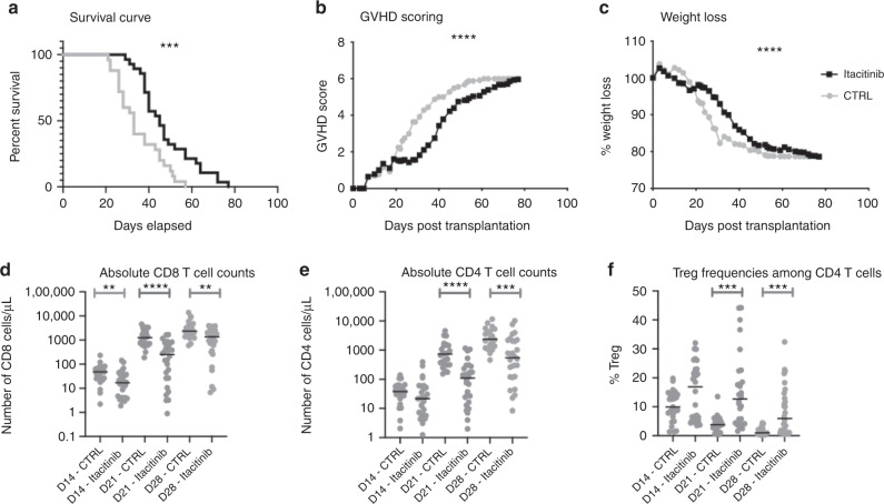
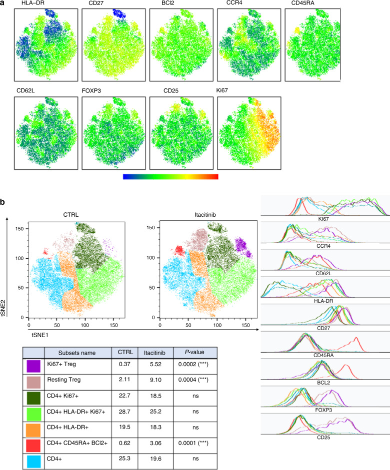
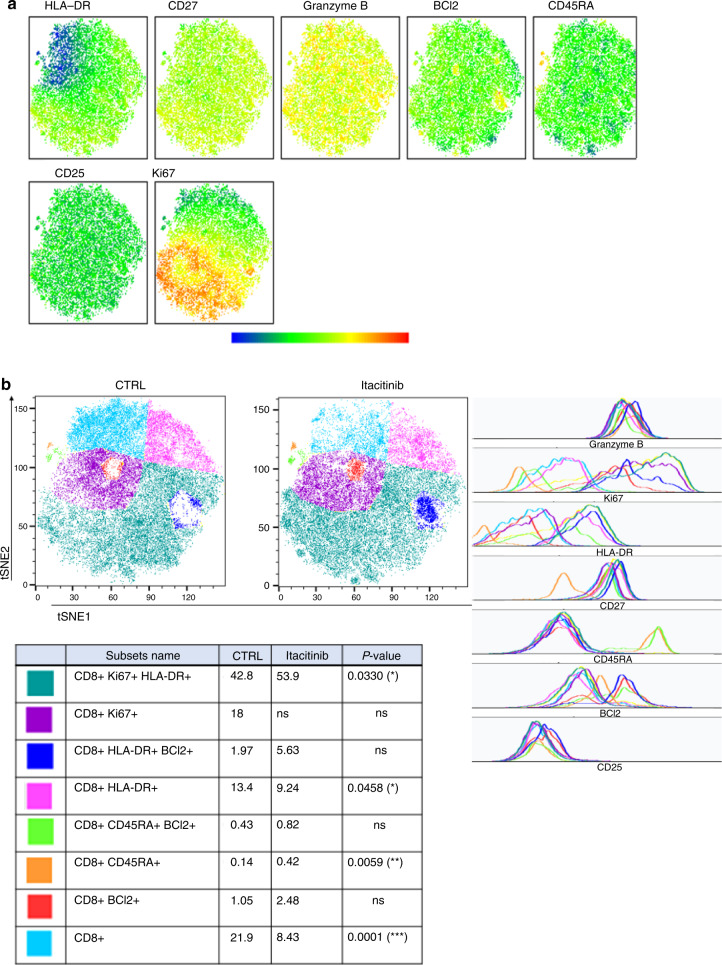
We assessed the impact of the Janus Kinase (JAK) 1 inhibitor itacitinib on xenogeneic graft-versus-host disease (xGVHD). XGVHD was induced by i.v. injection 20 × 106 human peripheral blood mononuclear cells (hPBMC) in NSG mice on day 0. Itacitinib (3 mg, ≈120 mg/kg) or methylcellulose was administered by force-feeding twice a day from day 3 to day 28. Mice were followed for xGVHD score and survival. In addition, human T-cell engraftment and as well as human T-cell subtypes were monitored in blood on days 14, 21, and 28 after transplantation. We observed that itacitinib-treated mice had significantly longer survival than control mice (median 45 versus 33 days; P < 0.001). Further, they also had lower absolute numbers of human CD4+ T cells on days 21 and 28 after transplantation as well as of human CD8+ T cells on days 14, 21, and 28 after transplantation. In addition, itacitinib-treated mice had higher frequencies of human regulatory T cells (Treg) on days 21 and 28 after transplantation. In summary, our data indicate that itacitinib decreases human T-cell engraftment, increases Treg frequencies and attenuates xGVHD in NSG mice transplanted with hPBMC.


Conflict of interest statement

Frédéric Baron has received travel grants from Celgene, Abbvie, Novartis, INCYTE Biosciences, and Sanofi as well as honoraria from Merck and Abbvie. The remaining authors declare that they have no relevant conflict of interest in regard to this study.

Figures

Fig. 1
Fig. 1. Impact of itacitinib on xGVHD.
Survival (a), GVHD scoring (b) and weight loss (c) in the three cohorts (with three different PBMC donors) combined. Gray line with circles shows control mice (n = 25) while black line with squares shows itacitinib mice (n = 28). Absolute CD8 T-cell counts (d), absolute CD4 T-cell counts (e) and Treg frequencies among CD4 T-cells (f) in the three cohorts combined. NSG mice were injected with 2 × 107 human PBMCs. Mice were then treated twice daily with Itacitinib (≈120 mg/kg) or a methylcellulose solution from day 3 to day 28 after transplantation. ac Survival, GVHD scoring and weight loss curves of three groups of mice transplanted with three different healthy donors (n = 15 for donor 1, n = 18 for donor 2 and n = 20 for donor 3, including 25 in the control and 28 in the Itacitinib group). d Comparison of absolute CD8+ T-cell counts for each mouse treated or not with Itacitinib. e Comparison of absolute CD4+ T-cell counts for mice treated or not with Itacitinib. f Comparison of Treg frequencies among CD4+ T-cells for mice treated or not with Itacitinib. Data show median values (*P < 0.05, **P < 0.005, ***P < 0.0005).
Fig. 2
Fig. 2. T-SNE of CD4+ T cells in peripheral blood from mice in cohort 3 on day 28 after transplantation.
T-SNE were created on 20,135 CD4+ T cells and included HLA-DR, CD27, BCl2, CCR4, CD45RA, CD62L, FOXP3, CD25, and Ki67 markers. a Visualization of marker expression (MFI) among CD4+ T cells. b Seven populations were determined based on different marker expressions (geometric mean), i.e., Treg Ki67+ (purple), resting Treg (beige), CD4+ Ki67+ (dark green), CD4+HLA-DR+Ki67+ (light green), CD4+HLA-DR+ (orange), CD4+CD45RA+BCl2+ (red), and other CD4+ (blue) in the control and the itacitinib groups. Ki67+ Treg (purple, P = 0.0002), resting Treg (beige, P = 0.0004) and CD4+CD45RA+BCl2+ T cells (red, P = 0.0001) were increased in the treated group at day 28 in the cohort 3. Histograms show marker expression in subpopulations.
Fig. 3
Fig. 3. T-SNE of CD8+ T cells in peripheral blood from mice in cohort 3 on day 28 after transplantation.
T-SNE were created on 21,000 CD8+ T cells and included HLA-DR, CD27, Granzyme B, BCl2, CD45RA, CD25 and Ki67 markers. a Visualization of marker expression (MFI) among CD8 T cells. b Eight populations were determined based on different marker expressions (geometric mean), i.e. CD8+Ki67+HLA-DR+ (dark green), CD8+Ki67+ (purple), CD8+HLA-DR+BCl2+ (dark blue), CD8+HLA-DR+ (pink), CD8+CD45RA+BCl2+ (light green), CD8+CD45RA+ (orange), CD8+BCl2+ (red), and other CD8+ (light blue) in the control and the treated groups at day 28 in cohort 3. CD8+Ki67+HLA-DR+ (dark green, P = 0.033) and CD8+CD45RA+ (orange, P = 0.0059) were increased in the treated group at day 28 in the cohort 3. CD8+HLA-DR+ (pink, P = 0.0458) and other CD8+ T cells (light blue, P = 0.0001) were decreased in the treated group at day 28 in cohort 3. Histograms show marker expression of subpopulations.
Fig. 4
Fig. 4. Impact of itacitinib on GvL effects.
NSG-A2 mice were infused intravenously with 2 × 107 human PBMCs and 3 × 106 THP-1-luc cells. Mice were then treated twice daily with Itacitinib (≈120 mg/kg) or a methylcellulose solution from day 3 to day 28 after transplantation. a Bioluminescence was monitored. Images acquired at day 31 are shown with the Y axis indicating radiance (photon/sec/cm2/sr) measured from the dorsal view and the ventral view with a region of interest drawn over the entire body of each mouse and compared in subfigures (c, d). b Images acquired at day 40. c Comparison of bioluminescence at day 31 in dorsal view. D Comparison of bioluminescence at day 31 in ventral view. Data show median values (*P < 0.05, **P < 0.005, ***P < 0.0005).

References

    1. Baron F, Efficace F, Cannella L, Muus P, Trisolini S, Halkes CJM, et al. Impact of the type of anthracycline and of stem cell transplantation in younger patients with acute myeloid leukemia: Long-term follow up of a phase III study. Am J Hematol. 2020;95:749–58. doi: 10.1002/ajh.25795. - DOI - PubMed
    1. Baron F, Maris MB, Sandmaier BM, Storer BE, Sorror M, Diaconescu R, et al. Graft-versus-tumor effects after allogeneic hematopoietic cell transplantation with nonmyeloablative conditioning. J Clin Oncol. 2005;23:1993–2003. doi: 10.1200/JCO.2005.08.136. - DOI - PubMed
    1. Hammarén HM, Virtanen AT, Raivola J, Silvennoinen O. The regulation of JAKs in cytokine signaling and its breakdown in disease. Cytokine. 2019;118:48–63. doi: 10.1016/j.cyto.2018.03.041. - DOI - PubMed
    1. Schroeder MA, Choi J, Staser K, DiPersio JF. The role of janus kinase signaling in graft-versus-host disease and graft versus leukemia. Biol Blood Marrow Transpl. 2018;24:1125–34. doi: 10.1016/j.bbmt.2017.12.797. - DOI - PMC - PubMed
    1. Zeiser R, von Bubnoff N, Butler J, Mohty M, Niederwieser D, Or R, et al. Ruxolitinib for glucocorticoid-refractory acute graft-versus-host disease. N. Engl J Med. 2020;382:1800–10. doi: 10.1056/NEJMoa1917635. - DOI - PubMed

Publication types