

Four-Dimensional flow Magnetic Resonance Imaging for Assessment of Pediatric Coarctation of the Aorta
- PMID: 34173693
- PMCID: PMC9084555
- DOI: 10.1002/jmri.27802
Four-Dimensional flow Magnetic Resonance Imaging for Assessment of Pediatric Coarctation of the Aorta
Abstract
Background: Coarctation of the aorta (CoA) typically requires repair, but re-interventions and vascular complications occur, particularly with associated defects like bicuspid aortic valve (BAV). Magnetic resonance imaging (MRI) may identify anatomic and hemodynamic factors contributing to clinical complications.
Purpose: To investigate 4D flow MRI characteristics in pediatric CoA to determine parameters for long-term clinical surveillance.
Study type: Retrospective.
Population: CoA (n = 21), CoA with BAV (n = 24), BAV alone (n = 29), and healthy control (n = 25).
Field strength/sequence: A 1.5 T, 3D CE IR FLASH MRA, 4D flow MRI using 3D time resolved PC-MRI with velocity encoding.
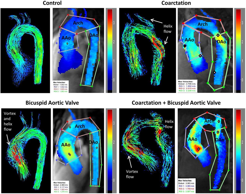
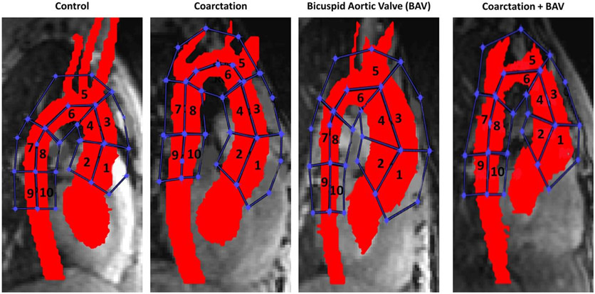
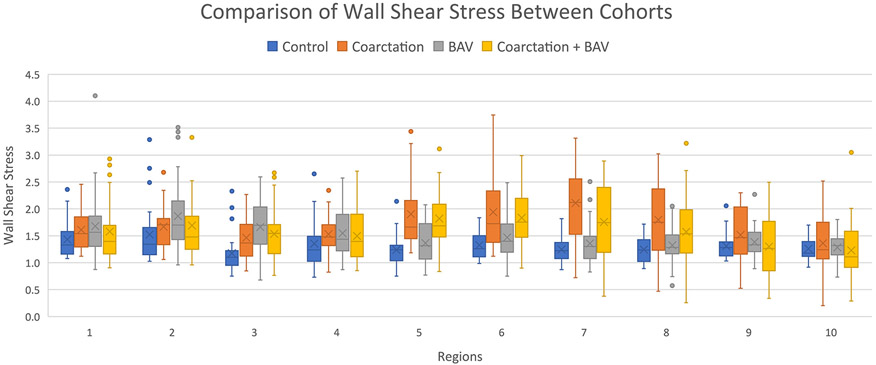
Assessment: Thoracic aorta diameters were measured from 3D CE-MRA. Peak systolic velocities and wall shear stress were calculated and flow patterns were visualized throughout the thoracic aorta using 4D flow. Repair characteristics, re-interventions, and need for anti-hypertensive medications were recorded.
Statistics: Descriptive statistics, ANOVA with post hoc t-testing and Bonferroni correction, Kruskal-Wallis H, intraclass correlation coefficient, Fleiss' kappa.
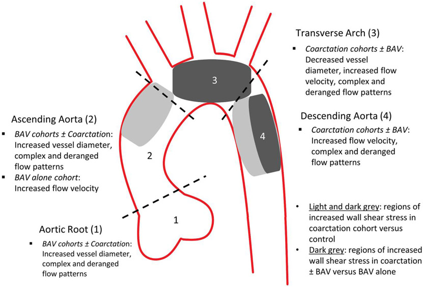
Results: Patients with CoA with or without repair had smaller transverse arch diameters compared to BAV alone and control cohorts (P < 0.05), higher peak systolic flow velocities and wall shear stress compared to controls in the transverse arch and descending aorta (P < 0.05), and flow derangements in the descending aorta. The most common CoA repairs were extended end-to-end anastomosis (n = 22/45, 48.9%, age at repair 1 ± 2 years, seven re-interventions) and stent/interposition graft placement (n = 10/45, 22.2%, age at repair 12 ± 3 years, one re-intervention). Anti-hypertensive medications were prescribed to 33.3% (n = 15/45) of CoA and 34.4% of BAV alone patients (n = 10/29).
Data conclusions: Despite repair, CoA alters hemodynamics and flow patterns in the transverse arch and descending aorta. These findings may contribute to vascular remodeling and secondary complications. 4D flow MRI may be valuable in risk stratification, treatment selection and postintervention assessment. Long-term, prospective studies are warranted to correlate patient and MRI factors with clinical outcomes.
Evidence level: 3 TECHNICAL EFFICACY: Stage 3.
Keywords: 4D flow; cardiac; coarctation of the aorta; congenital heart disease; pediatrics.
© 2021 International Society for Magnetic Resonance in Medicine.
Figures
References
-
- Niaz T, Poterucha JT, Johnson JN, et al. Incidence, morphology, and progression of bicuspid aortic valve in pediatric and young adult subjects with coexisting congenital heart defects. Congenit Heart Dis 2017;129:261–269. - PubMed
-
- Sugimoto A, Ota N, Miyakoshi C, et al. Mid to long-term aortic valve related outcomes after conventional repair for patients with interrupted aortic arch or coarctation of the aorta combined with ventricular septal defect: The impact of bicuspid aortic valve. Eur J Cardiothorac Surg 2014;46:952–960. - PubMed
-
- Connolly HM, Huston J, Brown RD Jr, Warnes CA, Ammash NM, Tajik AJ. Intracranial aneurysms in patients with coarctation of the aorta: A prospective magnetic resonance angiography study of 100 patients. Mayo Clin Proc 2003;78:1491–1499. - PubMed
-
- Brown ML, Burkhart HM, Connolly HM, et al. Coarctation of the aorta: Lifelong surveillance is mandatory following surgical repair. J Am Coll Cardiol 2013;10(62):1020–1025. - PubMed
Publication types
MeSH terms
Grants and funding
LinkOut - more resources
Full Text Sources
