. 2021 Aug 10:336:296-309.
doi: 10.1016/j.jconrel.2021.06.030. Epub 2021 Jun 23.

In vivo targeted delivery of nucleic acids and CRISPR genome editors enabled by GSH-responsive silica nanoparticles

Affiliations

In vivo targeted delivery of nucleic acids and CRISPR genome editors enabled by GSH-responsive silica nanoparticles

Yuyuan Wang et al. J Control Release. .

Abstract

The rapid development of gene therapy and genome editing techniques brings up an urgent need to develop safe and efficient nanoplatforms for nucleic acids and CRISPR genome editors. Herein we report a stimulus-responsive silica nanoparticle (SNP) capable of encapsulating biomacromolecules in their active forms with a high loading content and loading efficiency as well as a well-controlled nanoparticle size (~50 nm). A disulfide crosslinker was integrated into the silica network, endowing SNP with glutathione (GSH)-responsive cargo release capability when internalized by target cells. An imidazole-containing component was incorporated into the SNP to enhance the endosomal escape capability. The SNP can deliver various cargos, including nucleic acids (e.g., DNA and mRNA) and CRISPR genome editors (e.g., Cas9/sgRNA ribonucleoprotein (RNP), and RNP with donor DNA) with excellent efficiency and biocompatibility. The SNP surface can be PEGylated and functionalized with different targeting ligands. In vivo studies showed that subretinally injected SNP conjugated with all-trans-retinoic acid (ATRA) and intravenously injected SNP conjugated with GalNAc can effectively deliver mRNA and RNP to murine retinal pigment epithelium (RPE) cells and liver cells, respectively, leading to efficient genome editing. Overall, the SNP is a promising nanoplatform for various applications including gene therapy and genome editing.

Keywords: CRISPR-Cas9 genome editing; Gene delivery; Silica nanoparticle.


Figures

Figure 1.
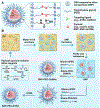
Figure 1.. Design and synthesis of SNPs conjugated with either ATRA or GalNAc.
(A) Illustration of the multifunctional SNP for the delivery of nucleic acids (e.g., DNA and mRNA) and CRISPR genome editor (e.g., RNP, RNP+ssODN). (B) Synthesis scheme for the ATRA- or GalNAc-conjugated SNPs. SNP: silica nanoparticle; TEOS: tetraethyl orthosilicate; TESPIC: N-(3-(triethoxysilyl)propyl)-1H-imidazole-2-carboxamide; BTPD: bis3-[(triethoxysilyl) propyl]-disulfide; PEG: polyethylene glycol; GalNAc: N-acetylgalactosamine; ATRA: all-trans-retinoic acid.
Figure 2.
Figure 2.
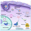
Schematic illustration of the intracellular trafficking pathways of SNP.
Figure 3.
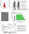
Figure 3.. Characterization of SNP and formulation optimization.
(A) Size distribution of SNPs measured by DLS with an average hydrodynamic diameter of 46 nm. (B) TEM image of SNP. (C) The effect of (1) molar ratio of TESPIC, and (2) surface charge in DNA-delivery by SNP. The transfection efficiency was quantified by the percent of RFP-positive HEK293 cells 48 h post treatment. (D) Effects of GSH concentration in cell culture medium on the DNA transfection efficiency of SNP-PEG. (E) mRNA delivery efficiency of SNP after storage at different conditions. NS: not significant; *: p < 0.05; **: p < 0.01; ****: p<0.0001; n = 3.
Figure 4.
Figure 4.. Intracellular trafficking of SNP.
Colocalization of ATTO-550-tagged RNP and endo/lysosomes was studied at 0.5 h, 2 h, and 6 h post-treatment.
Figure 5.
Figure 5.. Delivery efficiency of nucleic acids and CRISPR-Cas9 genome editors by SNP.
(A) and (B) The transfection efficiency of (A) DNA- and (B) mRNA-loaded SNP-PEG in HEK293 cells. (C) Gene editing efficiency of RNP-loaded SNP-PEG in GFP-expressing HEK 293 cells. (D) Illustration of HDR at a BFP reporter locus induced by the RNP+ssODN. Sequences of unedited (BFP) and edited (GFP) loci are shown. The protospacer adjacent motif sequence of RNP is underlined and the RNP cleavage site is marked by a red arrow. (E) Gene-correction efficiency of RNP+ssODN co-encapsulated SNP-PEG in BFP-expressing HEK 293 cells. NS: not significant *: p < 0.05; **: p < 0.01; n = 3. (F) Viability of HEK 293 cells treated with DNA-loaded SNP-PEG with different concentrations and DNA-complexed Lipo 2000. NS: not significant; ****: p < 0.0001; n = 7.
Figure 6.
Figure 6.. Nucleic acid and RNP delivery efficiency of SNP in Ai14 mice via subretinal injection.
(A) The tdTomato locus in the Ai14 reporter mouse. tdTomato expression can be achieved by Cre-Lox recombination. (B) Illustration of subretinal injection targeting the RPE tissue. (C) The stop cassette containing 3 Ai14 sgRNA target sites prevents downstream tdTomato expression. Excision of two SV40 polyA blocks by Ai14 RNP results in tdTomato expression. (D) Efficient delivery of Cre-mRNA by SNP-PEG-ATRA in mouse RPE. D1, RPE floret of eyes subretinally injected with Cre-mRNA-encapsulated SNP; D2, 20X magnification images of tdTomato+ RPE tissue; D3, RPE floret of PBS controls. (E) Efficient delivery of RNP by SNP-PEG-ATRA in mouse RPE. E1, RPE floret of mouse eyes subretinally injected with Ai14 RNP-encapsulated SNP; E2, 20X magnification images of tdTomato+ RPE tissue; E3, RPE floret of Ai14 mice injected with negative control SNP-PEG-ATRA (SNP-PEG-ATRA encapsulating RNP with negative control sgRNA). The whole RPE layer was outlined with a white dotted line.
Figure 7.
Figure 7.. SNP enabled nucleic acid (A-C) and RNP (D-F) delivery in vivo via systemic administration.
Ai14 mice were injected with Cre-mRNA or RNP encapsulated SNP-PEG or SNP-PEG-GalNAc, and the tdTomato fluorescence of the whole liver (A and D) and homogenized liver tissue (B and E) was detected and analyzed by IVIS imaging. Liver sections were studied by immunofluorescence staining and confocal laser scanning microscopy (C and F). *: p < 0.05; ***: p < 0.001; n = 3.

References

    1. Jin L, Zeng X, Liu M, Deng Y, He N, Current progress in gene delivery technology based on chemical methods and nano-carriers, Theranostics 4 (2014) 240. - PMC - PubMed
    1. Islam MA, Reesor EKG, Xu Y, Zope HR, Zetter BR, Shi J, Biomaterials for mRNA delivery, Biomater. Sci 3 (2015) 1519–1533. - PMC - PubMed
    1. Zhang H, Lee M-Y, Hogg MG, Dordick JS, Sharfstein ST, Gene delivery in three-dimensional cell cultures by superparamagnetic nanoparticles, ACS Nano 4 (2010) 4733–4743. - PubMed
    1. Glass Z, Li Y, Xu Q, Nanoparticles for CRISPR–Cas9 delivery, Nat. Biomed. Eng 1 (2017) 854. - PMC - PubMed
    1. Hsu PD, Lander ES, Zhang F, Development and applications of CRISPR-Cas9 for genome engineering, Cell 157 (2014) 1262–1278. - PMC - PubMed

Publication types