. 2021 Sep-Oct:54:107361.
doi: 10.1016/j.carpath.2021.107361. Epub 2021 Jun 24.

COVID-19 myocarditis: quantitative analysis of the inflammatory infiltrate and a proposed mechanism

Affiliations

COVID-19 myocarditis: quantitative analysis of the inflammatory infiltrate and a proposed mechanism

Sharon E Fox et al. Cardiovasc Pathol. 2021 Sep-Oct.

Abstract

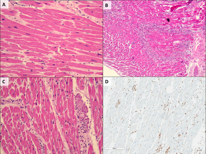
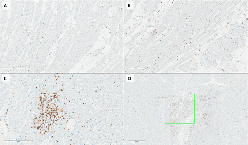
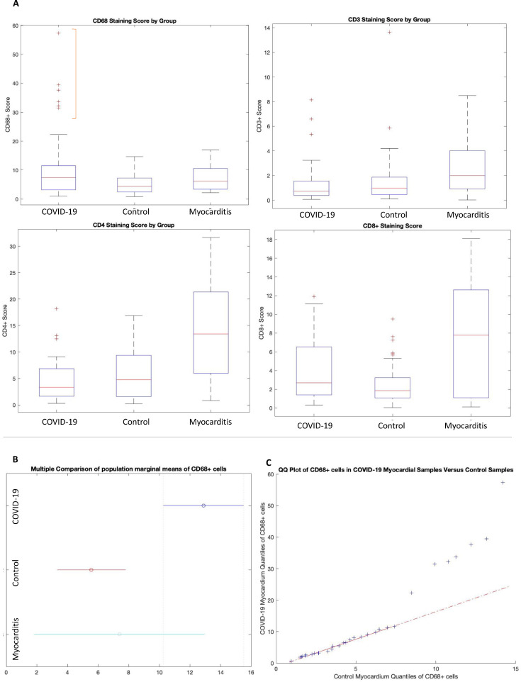
COVID-19 has a significant effect upon the cardiovascular system. While a number of different cardiovascular histopathologies have been described at post-mortem examination, the incidence of typical viral myocarditis in COVID-19 positive patients appears very low [1-3]. In this study, we further characterize and quantify the inflammatory cell infiltrate in a COVID-19 study cohort and compare the findings to both an age and disease matched control cohort and a cohort of patients diagnosed with typical inflammatory myocarditis. All study and control cohorts had 1 or more of the comorbidities most commonly associated with severe disease (hypertension, type II diabetes, obesity, or known cardiovascular disease). The results demonstrate a skewed distribution of the number of CD68+ cells in COVID-19 hearts, with upper quantiles showing a significant increase as compared to both matched control hearts, and those with myocarditis. In contrast, hearts from typical inflammatory myocarditis contained increased numbers of CD4+, and CD8+ cells compared to both COVID-19 and control cohorts. In conclusion, the presence of an increased number of CD68+ cells suggests that COVID-19 may incite a form of myocarditis different from typical viral myocarditis, and associated with diffusely infiltrative cells of monocytes/macrophage lineage.

Keywords: CD68 cells; COVID-19; SARS-CoV-2; autopsy, myocarditis, heart, inflammation, macrophages.


Figures

Fig 1:
Fig. 1
(A) Cardiac myocytes from control patient (H&E). (B) Myocarditis, characterized by patchy, dense inflammation within the myocardium (H&E). (C) Endotheliitis and diffuse, perivascular distribution of inflammation in COVID-19. (D) CD68 immunostaining highlighting the presence of CD68+ cells in a mild, diffuse intravascular and perivascular distribution in a case of COVID-19.
Fig 2:
Fig. 2
(A) CD3 immunostaining, demonstrating a paucity of CD3+ lymphocytes in COVID-19. (B) CD68 immunostaining in COVID-19, highlighting a mild, diffuse distribution of CD68+ cells. (C) CD3 immunostaining in a case of myocarditis, demonstrating intense staining for CD3+ lymphocytes in a patchy distribution. (D) CD68 immunostaining in myocarditis, in a similar patchy distribution to that of lymphocytes, with example of region selection box for analysis.
Fig 3:
Fig. 3
(A) Boxplots showing median and range of CD68, CD3, CD4, and CD8 staining cells for each Patient Group. A red bracket highlights the numerous outliers of CD68+ cells in the upper quantile of the COVID-19 group, which may represent a subset of COVID-19 patients with greater myocardial inflammation. (B) Population marginal means after correction for multiple comparisons within the nested ANOVA performed on CD68+ cells. The population marginal mean of CD68+ cells is significantly higher for the COVID-19 group as compared to controls. C) Q-Q plot of CD68+ cell quantiles in the COVID-19 myocardium samples as compared to control quantiles. The relationship is non-linear at higher quantiles, indicating a difference in distribution of CD68 positivity among the COVID-19 group, with higher values at the upper quantiles.

References

    1. Fox SE, Li G, Akmatbekov A, Harbert JL, Lameira FS, Brown JQ, et al. Unexpected features of cardiac pathology in COVID-19 infection. Circulation. 2020;142:1123–1125. - PubMed
    1. Halushka MK, Vander Heide RS. Myocarditis is rare in COVID-19 autopsies: cardiovascular findings across 277 postmortem examinations. Cardiovasc Pathol. 2021;50 - PMC - PubMed
    1. Hooper JE, Padera RF, Dolhnikoff M, da Silva LFF, Duarte-Neto AN, Kapp ME, et al. A postmortem portrait of the coronavirus disease 2019 (COVID-19) pandemic: a large multiinstitutional autopsy survey study. Arch Pathol Lab Med. 2021 doi: 10.5858/arpa.2020-0786-SA. - DOI - PubMed
    1. Wölfel R, Corman VM, Guggemos W, Seilmaier M, Zange S, Müller MA, et al. Virological assessment of hospitalized patients with COVID-2019. Nature. 2020;581:465–469. - PubMed
    1. Klok FA, Kruip MJHA, van der Meer NJM, Arbous MS, Gommers DAMPJ, Kant KM, et al. Incidence of thrombotic complications in critically ill ICU patients with COVID-19. Thromb Res. 2020;191:145–147. - PMC - PubMed

MeSH terms