

Gene editing in a Myo6 semi-dominant mouse model rescues auditory function
- PMID: 34174443
- PMCID: PMC8753286
- DOI: 10.1016/j.ymthe.2021.06.015
Gene editing in a Myo6 semi-dominant mouse model rescues auditory function
Abstract
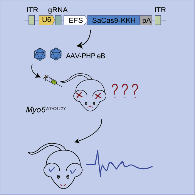
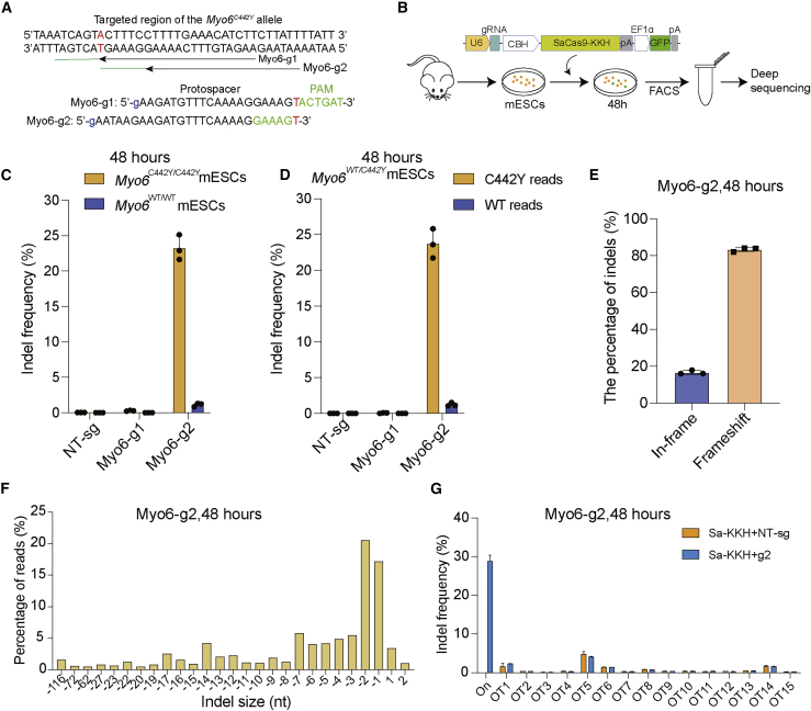
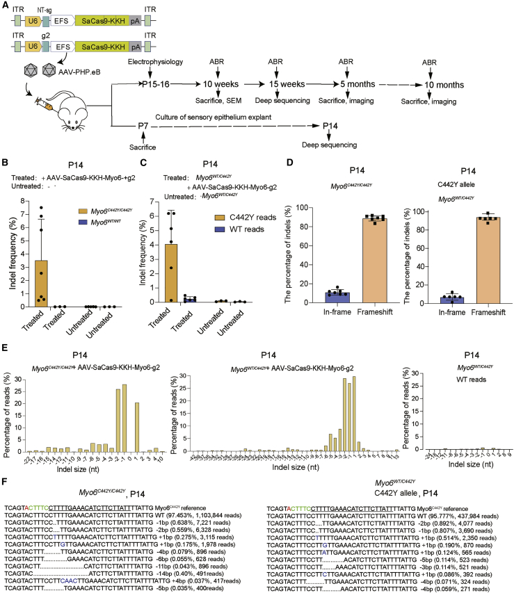
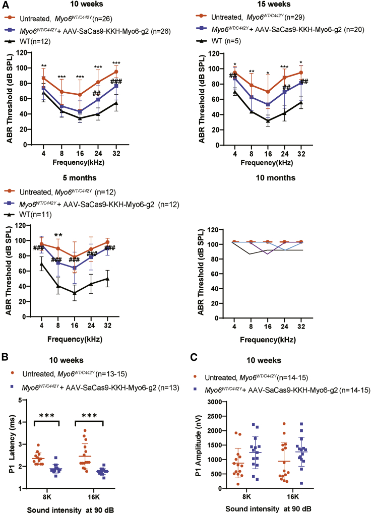
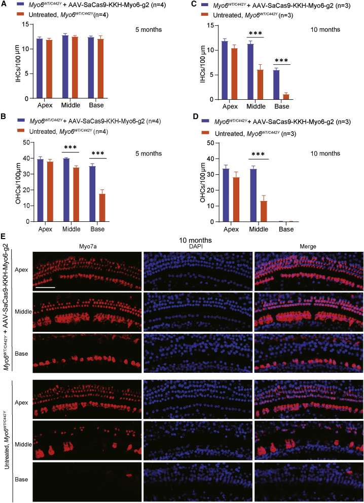
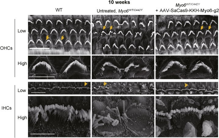
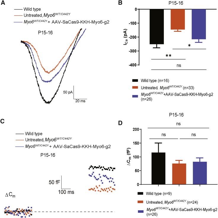
Myosin VI(MYO6) is an unconventional myosin that is vital for auditory and vestibular function. Pathogenic variants in the human MYO6 gene cause autosomal-dominant or -recessive forms of hearing loss. Effective treatments for Myo6 mutation causing hearing loss are limited. We studied whether adeno-associated virus (AAV)-PHP.eB vector-mediated in vivo delivery of Staphylococcus aureus Cas9 (SaCas9-KKH)-single-guide RNA (sgRNA) complexes could ameliorate hearing loss in a Myo6WT/C442Y mouse model that recapitulated the phenotypes of human patients. The in vivo editing efficiency of the AAV-SaCas9-KKH-Myo6-g2 system on Myo6C442Y is 4.05% on average in Myo6WT/C442Y mice, which was ∼17-fold greater than editing efficiency of Myo6WT alleles. Rescue of auditory function was observed up to 5 months post AAV-SaCas9-KKH-Myo6-g2 injection in Myo6WT/C442Y mice. Meanwhile, shorter latencies of auditory brainstem response (ABR) wave I, lower distortion product otoacoustic emission (DPOAE) thresholds, increased cell survival rates, more regular hair bundle morphology, and recovery of inward calcium levels were also observed in the AAV-SaCas9-KKH-Myo6-g2-treated ears compared to untreated ears. These findings provide further reference for in vivo genome editing as a therapeutic treatment for various semi-dominant forms of hearing loss and other semi-dominant diseases.
Keywords: CRISPR-Cas9; gene therapy; hearing loss; myosin VI; semi-dominant hearing loss.
Copyright © 2021 The American Society of Gene and Cell Therapy. Published by Elsevier Inc. All rights reserved.
Conflict of interest statement
Declaration of interests The authors declare no competing interests.
Figures
Similar articles
-
Rescue of autosomal dominant hearing loss by in vivo delivery of mini dCas13X-derived RNA base editor.Sci Transl Med. 2022 Jul 20;14(654):eabn0449. doi: 10.1126/scitranslmed.abn0449. Epub 2022 Jul 20. Sci Transl Med. 2022. PMID: 35857824
-
A humanized mouse model, demonstrating progressive hearing loss caused by MYO6 p.C442Y, is inherited in a semi-dominant pattern.Hear Res. 2019 Aug;379:79-88. doi: 10.1016/j.heares.2019.04.014. Epub 2019 Apr 26. Hear Res. 2019. PMID: 31103816
-
Targeted genome editing restores auditory function in adult mice with progressive hearing loss caused by a human microRNA mutation.Sci Transl Med. 2024 Jul 10;16(755):eadn0689. doi: 10.1126/scitranslmed.adn0689. Epub 2024 Jul 10. Sci Transl Med. 2024. PMID: 38985856 Free PMC article.
-
CRISPR/Cas9: targeted genome editing for the treatment of hereditary hearing loss.J Appl Genet. 2020 Feb;61(1):51-65. doi: 10.1007/s13353-019-00535-6. Epub 2020 Jan 7. J Appl Genet. 2020. PMID: 31912450 Review.
-
Association of Audiometric Measures with plasma long chain polyunsaturated fatty acids in a high-fish eating population: The Seychelles Child Development Study.Neurotoxicology. 2020 Mar;77:137-144. doi: 10.1016/j.neuro.2020.01.005. Epub 2020 Jan 23. Neurotoxicology. 2020. PMID: 31982419 Free PMC article. Review.
Cited by
-
Gene Therapy for Inherited Hearing Loss: Updates and Remaining Challenges.Audiol Res. 2023 Dec 4;13(6):952-966. doi: 10.3390/audiolres13060083. Audiol Res. 2023. PMID: 38131808 Free PMC article. Review.
-
Recent Therapeutic Progress and Future Perspectives for the Treatment of Hearing Loss.Biomedicines. 2023 Dec 18;11(12):3347. doi: 10.3390/biomedicines11123347. Biomedicines. 2023. PMID: 38137568 Free PMC article. Review.
-
Hearing loss: a global view for gene therapy approaches and challenges.Eur J Pediatr. 2025 Aug 27;184(9):578. doi: 10.1007/s00431-025-06426-9. Eur J Pediatr. 2025. PMID: 40858759 Free PMC article. Review.
-
Gene therapy restores auditory function and rescues damaged inner hair cells in an aged Vglut3 knockout mouse model.Gene Ther. 2025 Aug 21. doi: 10.1038/s41434-025-00558-1. Online ahead of print. Gene Ther. 2025. PMID: 40841774
-
Exploring AAV-Mediated Gene Therapy for Inner Ear Diseases: from Preclinical Success to Clinical Potential.Adv Sci (Weinh). 2025 Sep;12(33):e08397. doi: 10.1002/advs.202408397. Epub 2025 Jun 20. Adv Sci (Weinh). 2025. PMID: 40538303 Free PMC article. Review.
References
-
- Avraham K.B., Hasson T., Steel K.P., Kingsley D.M., Russell L.B., Mooseker M.S., Copeland N.G., Jenkins N.A. The mouse Snell’s waltzer deafness gene encodes an unconventional myosin required for structural integrity of inner ear hair cells. Nat. Genet. 1995;11:369–375. - PubMed
-
- Melchionda S., Ahituv N., Bisceglia L., Sobe T., Glaser F., Rabionet R., Arbones M.L., Notarangelo A., Di Iorio E., Carella M., et al. MYO6, the human homologue of the gene responsible for deafness in Snell’s waltzer mice, is mutated in autosomal dominant nonsyndromic hearing loss. Am. J. Hum. Genet. 2001;69:635–640. - PMC - PubMed
Publication types
MeSH terms
Substances
Grants and funding
LinkOut - more resources
Full Text Sources
