

Interaction of porcine circovirus-like virus P1 capsid protein with host proteins
- PMID: 34174877
- PMCID: PMC8235626
- DOI: 10.1186/s12917-021-02926-6
Interaction of porcine circovirus-like virus P1 capsid protein with host proteins
Abstract
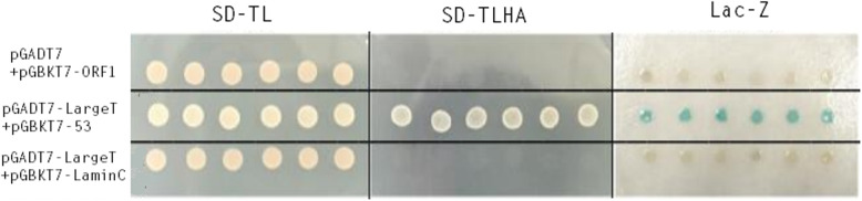
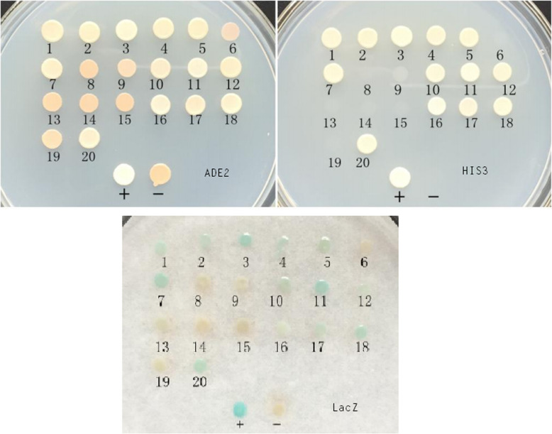
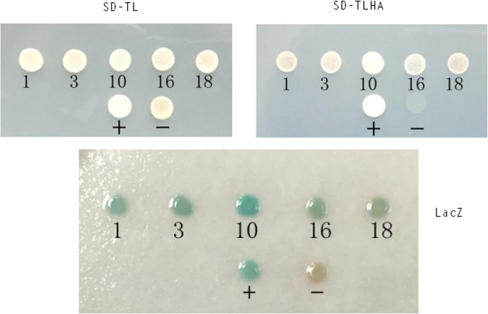
Background: Porcine circovirus-like virus P1 is a relatively new kind of virus that is closely related to the post-weaning multisystemic wasting syndrome, congenital tremors, and abortions in swine. The molecular mechanisms of P1 virus infection and pathogenesis are fully unknown. To analyze P1 and its host interactions, we used a yeast two-hybrid (Y2H) assay to identify cellular proteins interacting with the Cap of the P1 virus. In this study, the Cap of the P1 virus exhibited no self-activation and toxicity to yeast cells and was used as bait to screen the Y2H library prepared from the pancreas tissue.
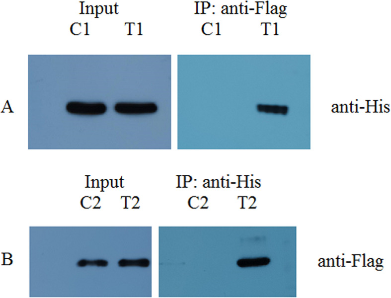
Results: Five cellular proteins (EEP, Ral GDS, Bcl-2-L-12, CPS1, and one not identified) were found to interact with P1 Cap. The interaction between Cap and Ral GDS was confirmed by co-immunoprecipitation.
Conclusions: Our data are likely to support the future investigation of the underlying mechanism of P1 infection and pathogenesis.
Keywords: Cap; Porcine circovirus-like virus P1; cellular protein; co-immunoprecipitation; yeast two-hybrid assay.
Conflict of interest statement
The authors declare that they have no competing interests.
Figures
Similar articles
-
Detection of porcine circovirus-like virus P1 in Hebei, China.Transbound Emerg Dis. 2018 Oct;65(5):1133-1136. doi: 10.1111/tbed.12896. Epub 2018 May 14. Transbound Emerg Dis. 2018. PMID: 29761653
-
Porcine MKRN1 Modulates the Replication and Pathogenesis of Porcine Circovirus Type 2 by Inducing Capsid Protein Ubiquitination and Degradation.J Virol. 2018 May 14;92(11):e00100-18. doi: 10.1128/JVI.00100-18. Print 2018 Jun 1. J Virol. 2018. PMID: 29514908 Free PMC article.
-
Ubiquitination-dependent degradation of DHX36 mediated by porcine circovirus type 3 capsid protein.Virology. 2025 Mar;604:110419. doi: 10.1016/j.virol.2025.110419. Epub 2025 Jan 21. Virology. 2025. PMID: 39862752
-
Porcine circovirus type 2 (PCV2): pathogenesis and interaction with the immune system.Annu Rev Anim Biosci. 2013 Jan;1:43-64. doi: 10.1146/annurev-animal-031412-103720. Epub 2013 Jan 3. Annu Rev Anim Biosci. 2013. PMID: 25387012 Review.
-
Interactions of porcine circovirus 2 with its hosts.Virus Genes. 2016 Aug;52(4):437-44. doi: 10.1007/s11262-016-1326-x. Epub 2016 Mar 25. Virus Genes. 2016. PMID: 27016220 Review.
Cited by
-
The Isoforms of Ral Guanine Nucleotide Dissociation Stimulator (RalGDS) in LLC-PK1 Cells.Curr Issues Mol Biol. 2025 Jul 18;47(7):566. doi: 10.3390/cimb47070566. Curr Issues Mol Biol. 2025. PMID: 40729035 Free PMC article.
References
-
- Harding JCS, Clark EG. Recognizing and diagnosing postweaning multisystemic wasting syndrome (PMWS) Swine Health Prod. 1997;5:201–203.
MeSH terms
Substances
Grants and funding
LinkOut - more resources
Full Text Sources
Miscellaneous
