. 2021 Jun 26;18(1):144.
doi: 10.1186/s12974-021-02123-0.

Beyond the lesion site: minocycline augments inflammation and anxiety-like behavior following SCI in rats through action on the gut microbiota

Affiliations

Beyond the lesion site: minocycline augments inflammation and anxiety-like behavior following SCI in rats through action on the gut microbiota

Emma K A Schmidt et al. J Neuroinflammation. .

Abstract

Background: Minocycline is a clinically available synthetic tetracycline derivative with anti-inflammatory and antibiotic properties. The majority of studies show that minocycline can reduce tissue damage and improve functional recovery following central nervous system injuries, mainly attributed to the drug's direct anti-inflammatory, anti-oxidative, and neuroprotective properties. Surprisingly the consequences of minocycline's antibiotic (i.e., antibacterial) effects on the gut microbiota and systemic immune response after spinal cord injury have largely been ignored despite their links to changes in mental health and immune suppression.

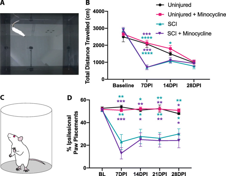
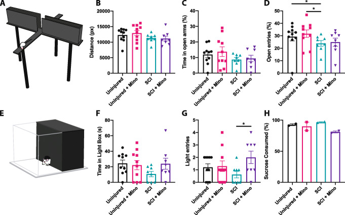
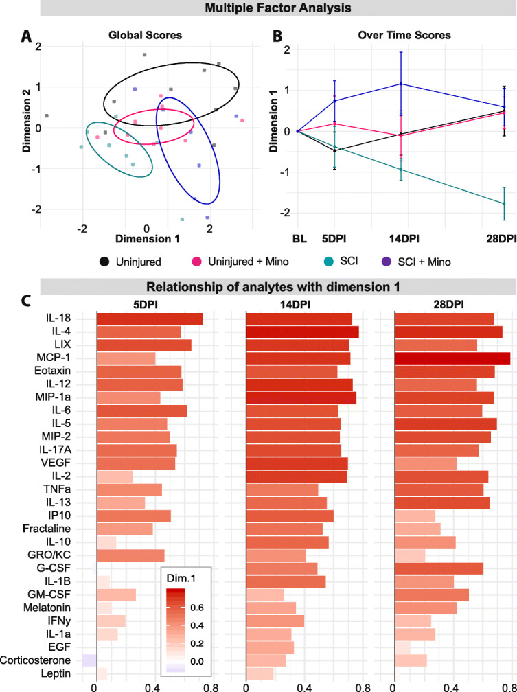
Methods: Here, we sought to determine minocycline's effect on spinal cord injury-induced changes in the microbiota-immune axis using a cervical contusion injury in female Lewis rats. We investigated a group that received minocycline following spinal cord injury (immediately after injury for 7 days), an untreated spinal cord injury group, an untreated uninjured group, and an uninjured group that received minocycline. Plasma levels of cytokines/chemokines and fecal microbiota composition (using 16s rRNA sequencing) were monitored for 4 weeks following spinal cord injury as measures of the microbiota-immune axis. Additionally, motor recovery and anxiety-like behavior were assessed throughout the study, and microglial activation was analyzed immediately rostral to, caudal to, and at the lesion epicenter.

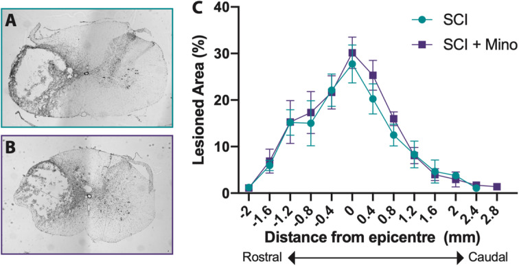
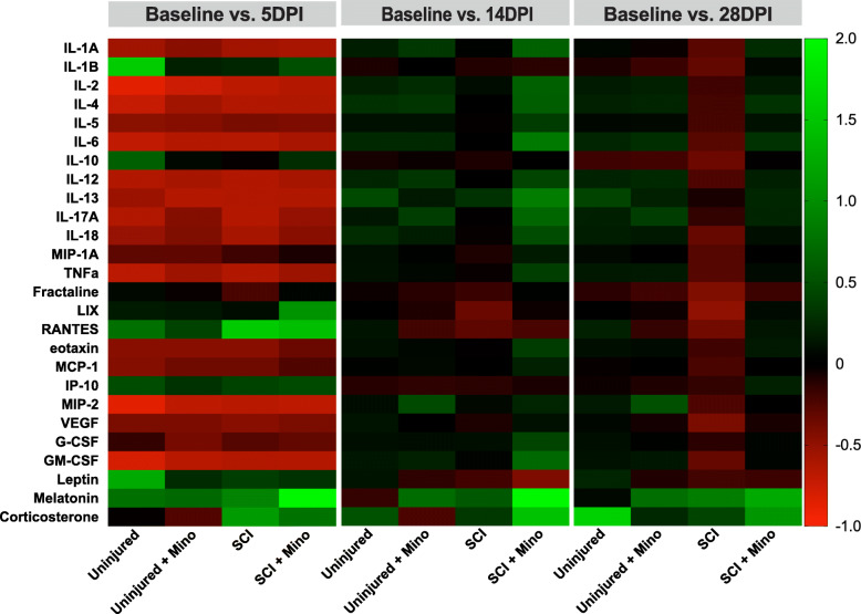
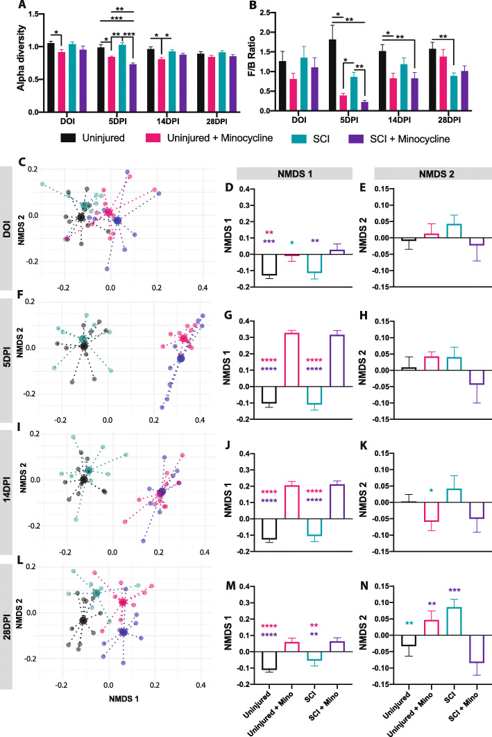
Results: We found that minocycline had a profound acute effect on the microbiota diversity and composition, which was paralleled by the subsequent normalization of spinal cord injury-induced suppression of cytokines/chemokines. Importantly, gut dysbiosis following spinal cord injury has been linked to the development of anxiety-like behavior, which was also decreased by minocycline. Furthermore, although minocycline attenuated spinal cord injury-induced microglial activation, it did not affect the lesion size or promote measurable motor recovery.

Conclusion: We show that minocycline's microbiota effects precede its long-term effects on systemic cytokines and chemokines following spinal cord injury. These results provide an exciting new target of minocycline as a therapeutic for central nervous system diseases and injuries.

Keywords: Inflammation; Microbiota; Minocycline; Spinal cord injury.


Conflict of interest statement

The authors declare that they have no competing interests.

Figures

Fig. 1
Fig. 1
Minocycline treatment had no effect on lesion size. Representative images of the maximum lesioned area for the SCI group (a) and SCI + minocycline group (b). c The rostral (negative numbers) to caudal (positive numbers) extension of the lesion was quantified and expressed as the percentage of the lesioned area for each coronal section. Error bars represent the standard error of the mean
Fig. 2
Fig. 2
Minocycline treatment altered the microglial density and morphology around the lesion site. a IBA1 immunohistochemical marker was used to visualize the microglia in the cervical spinal cord at C4, at the maximum lesion site (C5), and caudal to the lesion site at C6. Representative spinal cord images are shown from each group at C4 (b), C5 (c), and C6 (d). Quantification of the area of IBA+ staining is shown at C4 (e), C5 (f), and C6 (g) on the contralesional (left graphs) and ipsilesional (right graphs) spinal cord. Microglial morphology in the ventral gray matter was assessed by quantifying the length and number of endpoints per cell. h Image of a ramified microglia and i the automated analysis shows the soma in blue, processes in red, and the endpoints in green. j Image of an activated microglia and k the corresponding output of the analysis. Quantification of the average process length per cell is shown at C4 (l), C5 (m), and C6 (n) on the contralesional (left graphs) and ipsilesional (right graphs) spinal cord. Quantification of the average number of process endpoints per cell is shown at C4 (o), C5 (p), and C6 (q) on the contralesional (left graphs) and ipsilesional (right graphs) spinal cord. Error bars represent the standard error of the mean. Colored asterisks represent which group is significantly different. Scale bar represents 1 mm. *p < 0.05, **p < 0.01. ***p < 0.001, ****p < 0.0001
Fig. 3
Fig. 3
Minocycline treatment did not affect motor recovery in the open field or cylinder task. a Image showing a rat in the center of the open field apparatus. b Both SCI and SCI + minocycline groups traveled significantly less distance than uninjured rats in the open field at 7 days post-SCI. c The cylinder test was used to assess forepaw use asymmetry. d SCI resulted in decreased use of the ipsilesional paw compared to uninjured rats. Error bars represent the standard error of the mean. *p < 0.05, **p < 0.01, ***p < 0.001, ****p < 0.0001. Top asterisks represent the uninjured + minocycline groups vs. SCI (green) and SCI + minocycline (purple). Bottom asterisks represent uninjured vs. SCI (green) and SCI + minocycline (purple)
Fig. 4
Fig. 4
Minocycline treatment attenuated spinal cord injury-induced anxiety-like behavior. a Rat in the open arm of the elevated plus maze. b The total distance traveled in the maze. c The percent time in the open arms was calculated as a percentage of the time spent in the open arms divided by the total time spent in the maze. d The percent of open arm entries was calculated as the percentage of the number of open arm entries divided by the total open and closed arm entries. e Schematic showing a rat entering the light component of the light-dark box. f The amount of time spent in the light component and g the number of entries made into the light component of the light-dark box. h The percent of sucrose water consumed where each data point represents a cage. Error bars represent the standard error of the mean. *p < 0.05
Fig. 5
Fig. 5
Minocycline treatment prevented spinal cord injury-induced long-term changes in plasma cytokines. a Multiple factor analysis of plasma analytes (measured with respect to the baseline values) shows the relationship of each subject with the multidimensional factors 1 and 2 across all time points. b Scores in multidimensional factor 1 are shown for each group over time. c The importance of each plasma analyte to multidimensional factor 1 is shown over time using their loadings. Ellipses in a represent the 50% bivariate distribution. Error bars represent the standard error of the mean
Fig. 6
Fig. 6
Minocycline prevents spinal cord injury-induced suppression of plasma cytokines and chemokines. Heatmap showing the relative change of plasma analytes at 5, 14, and 28 days post-injury. Positive numbers reflect an increase and negative numbers reflect a decrease from baseline values
Fig. 7
Fig. 7
Minocycline treatment altered the gut microbiota composition. a The Shannon index of alpha diversity and b the Firmicutes/Bacteroidetes ratio are shown over time for each treatment group. cn Non-metric multidimensional scaling (NMDS) was used to visualize the overall microbiota composition at the phylum level. c 2D plot of the NMDS first 2 components shows the centroid of each group (large points) and each individual rat (small points) on the day of injury. Individual NMDS 1 scores (d) and NMDS 2 scores (e) are shown for each group on the day of injury. Similar plots are shown for 5 days post-injury (fh), 14 days post-injury (ik), and 28 days post-injury (ln). All data is normalized to baseline values. Error bars represent the standard error of the mean. Colored asterisks represent which group is significantly different. *p < 0.05, **p < 0.01, ***p < 0.001

Similar articles

Cited by

References

    1. Kennedy P, Rogers BA. Anxiety and depression after spinal cord injury: a longitudinal analysis. Arch Phys Med Rehabil. 2000;81:932–937. doi: 10.1053/apmr.2000.5580. - DOI - PubMed
    1. Weaver LC, Marsh DR, Gris D, Brown A, Dekaban GA. Autonomic dysreflexia after spinal cord injury: central mechanisms and strategies for prevention. Prog Brain Res. 2006:245–63 Elsevier. [cited 2020 Oct 20]. Available from: https://linkinghub.elsevier.com/retrieve/pii/S0079612305520168. - PubMed
    1. Riegger T, Conrad S, Liu K, Schluesener HJ, Adibzahdeh M, Schwab JM. Spinal cord injury-induced immune depression syndrome (SCI-IDS): SCI-IDS. Eur J Neurosci. 2007;25:1743–1747. doi: 10.1111/j.1460-9568.2007.05447.x. - DOI - PubMed
    1. Riegger T, Conrad S, Schluesener HJ, Kaps H-P, Badke A, Baron C, et al. Immune depression syndrome following human spinal cord injury (SCI): a pilot study. Neuroscience. 2009;158:1194–1199. doi: 10.1016/j.neuroscience.2008.08.021. - DOI - PubMed
    1. Krassioukov A, Warburton DE, Teasell R, Eng JJ. A systematic review of the management of autonomic dysreflexia after spinal cord injury. Arch Phys Med Rehabil. 2009;90:682–695. doi: 10.1016/j.apmr.2008.10.017. - DOI - PMC - PubMed

MeSH terms