

Reductions in midbrain GABAergic and dopamine neuron markers are linked in schizophrenia
- PMID: 34174930
- PMCID: PMC8235806
- DOI: 10.1186/s13041-021-00805-7
Reductions in midbrain GABAergic and dopamine neuron markers are linked in schizophrenia
Abstract
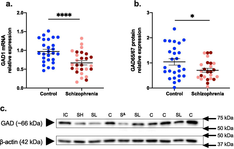
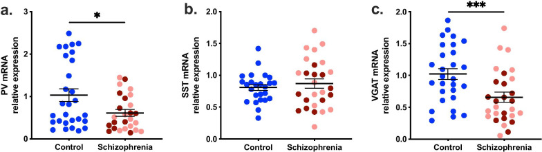
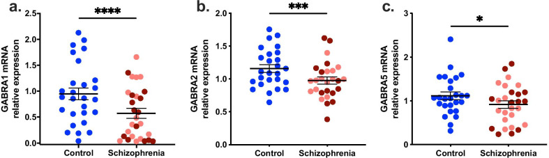
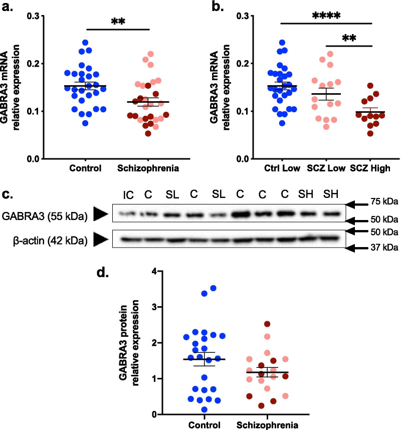
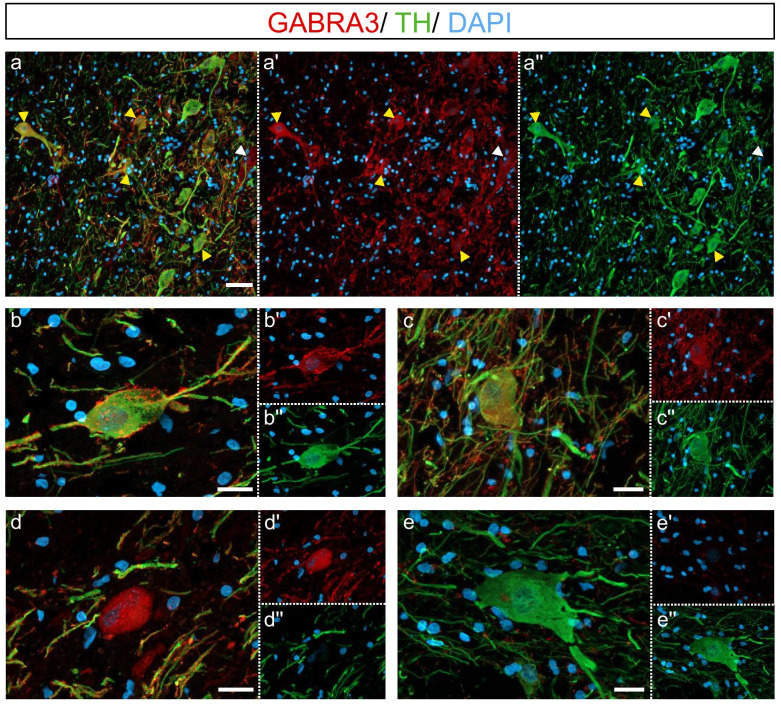
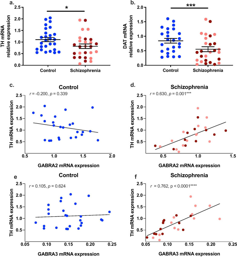
Reductions in the GABAergic neurotransmitter system exist across multiple brain regions in schizophrenia and encompass both pre- and postsynaptic components. While reduced midbrain GABAergic inhibitory neurotransmission may contribute to the hyperdopaminergia thought to underpin psychosis in schizophrenia, molecular changes consistent with this have not been reported. We hypothesised that reduced GABA-related molecular markers would be found in the midbrain of people with schizophrenia and that these would correlate with dopaminergic molecular changes. We hypothesised that downregulation of inhibitory neuron markers would be exacerbated in schizophrenia cases with high levels of neuroinflammation. Eight GABAergic-related transcripts were measured with quantitative PCR, and glutamate decarboxylase (GAD) 65/67 and GABAA alpha 3 (α3) (GABRA3) protein were measured with immunoblotting, in post-mortem midbrain (28/28 and 28/26 control/schizophrenia cases for mRNA and protein, respectively), and analysed by both diagnosis and inflammatory subgroups (as previously defined by higher levels of four pro-inflammatory cytokine transcripts). We found reductions (21 - 44%) in mRNA encoding both presynaptic and postsynaptic proteins, vesicular GABA transporter (VGAT), GAD1, and parvalbumin (PV) mRNAs and four alpha subunits (α1, α2, α3, α5) of the GABAA receptor in people with schizophrenia compared to controls (p < 0.05). Gene expression of somatostatin (SST) was unchanged (p = 0.485). We confirmed the reduction in GAD at the protein level (34%, p < 0.05). When stratifying by inflammation, only GABRA3 mRNA exhibited more pronounced changes in high compared to low inflammatory subgroups in schizophrenia. GABRA3 protein was expressed by 98% of tyrosine hydroxylase-positive neurons and was 23% lower in schizophrenia, though this did not reach statistical significance (p > 0.05). Expression of transcripts for GABAA receptor alpha subunits 2 and 3 (GABRA2, GABRA3) were positively correlated with tyrosine hydroxylase (TH) and dopamine transporter (DAT) transcripts in schizophrenia cases (GABRA2; r > 0.630, GABRA3; r > 0.762, all p < 0.001) but not controls (GABRA2; r < - 0.200, GABRA3; r < 0.310, all p > 0.05). Taken together, our results support a profound disruption to inhibitory neurotransmission in the substantia nigra regardless of inflammatory status, which provides a potential mechanism for disinhibition of nigrostriatal dopamine neurotransmission.
Keywords: GABA; GABRA; GAD1; GAD65/67; Neuroinflammation; Parvalbumin; Schizophrenia; Somatostatin; Substantia nigra; Tyrosine hydroxylase.
Conflict of interest statement
CSW is on an advisory board for Lundbeck, Australia Pty Ltd and in collaboration with Astellas Pharma Inc., Japan. The other authors all declare that they have no competing interests.
Figures
Similar articles
-
Putative presynaptic dopamine dysregulation in schizophrenia is supported by molecular evidence from post-mortem human midbrain.Transl Psychiatry. 2017 Jan 17;7(1):e1003. doi: 10.1038/tp.2016.257. Transl Psychiatry. 2017. PMID: 28094812 Free PMC article.
-
Altered cortical expression of GABA-related genes in schizophrenia: illness progression vs developmental disturbance.Schizophr Bull. 2015 Jan;41(1):180-91. doi: 10.1093/schbul/sbt178. Epub 2013 Dec 22. Schizophr Bull. 2015. PMID: 24361861 Free PMC article.
-
The neonatal ventral hippocampal lesion model of schizophrenia: effects on dopamine and GABA mRNA markers in the rat midbrain.Eur J Neurosci. 2003 Dec;18(11):3097-104. doi: 10.1111/j.1460-9568.2003.03047.x. Eur J Neurosci. 2003. PMID: 14656305
-
GABAergic dysfunction in schizophrenia: new treatment strategies on the horizon.Psychopharmacology (Berl). 2005 Jul;180(2):191-205. doi: 10.1007/s00213-005-2212-8. Epub 2005 Apr 28. Psychopharmacology (Berl). 2005. PMID: 15864560 Review.
-
[Schizophrenia and cortical GABA neurotransmission].Seishin Shinkeigaku Zasshi. 2010;112(5):439-52. Seishin Shinkeigaku Zasshi. 2010. PMID: 20560363 Review. Japanese.
Cited by
-
Using the excitation/inhibition ratio to optimize the classification of autism and schizophrenia.Transl Psychiatry. 2025 Jul 9;15(1):234. doi: 10.1038/s41398-025-03455-8. Transl Psychiatry. 2025. PMID: 40634290 Free PMC article.
-
Transcriptional evidence of reduced BDNF trophic capacity in the post-mortem human midbrain of schizophrenia cases with high inflammation.Transl Psychiatry. 2025 May 7;15(1):162. doi: 10.1038/s41398-025-03359-7. Transl Psychiatry. 2025. PMID: 40335479 Free PMC article.
-
Age-associated changes in lineage composition of the enteric nervous system regulate gut health and disease.Elife. 2023 Dec 18;12:RP88051. doi: 10.7554/eLife.88051. Elife. 2023. PMID: 38108810 Free PMC article.
-
Molecular diversity and migration of GABAergic neurons in the developing ventral midbrain.iScience. 2024 Oct 23;27(11):111239. doi: 10.1016/j.isci.2024.111239. eCollection 2024 Nov 15. iScience. 2024. PMID: 39569362 Free PMC article.
-
Inflammation in Schizophrenia: The Role of Disordered Oscillatory Mechanisms.Cells. 2025 Apr 29;14(9):650. doi: 10.3390/cells14090650. Cells. 2025. PMID: 40358174 Free PMC article. Review.
References
-
- Waszczak BL, Bergstrom DA, Walters JR. Single unit responses of substantia nigra and globus pallidus neurons to GABA agonist and antagonist drugs. Adv Biochem Psychopharmacol. 1981;30:79–94. - PubMed
Publication types
MeSH terms
Substances
LinkOut - more resources
Full Text Sources
Medical
