. 2021 Jun 26;52(1):95.
doi: 10.1186/s13567-021-00964-4.

CRISPR/Cas9-based generation of a recombinant double-reporter pseudorabies virus and its characterization in vitro and in vivo

Affiliations

CRISPR/Cas9-based generation of a recombinant double-reporter pseudorabies virus and its characterization in vitro and in vivo

Peng-Fei Fu et al. Vet Res. .

Abstract

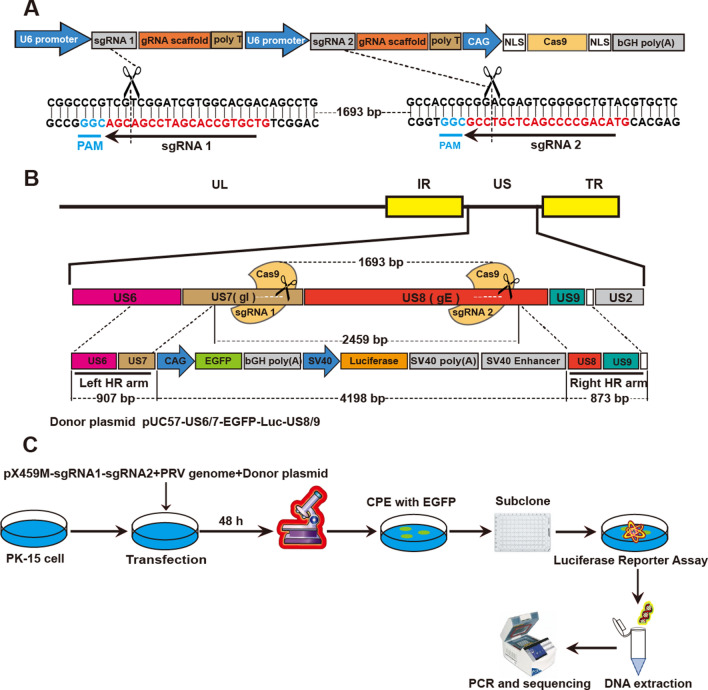
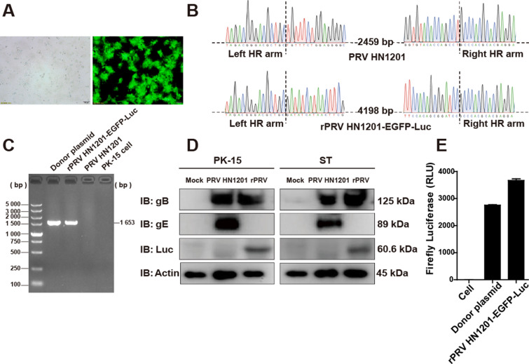
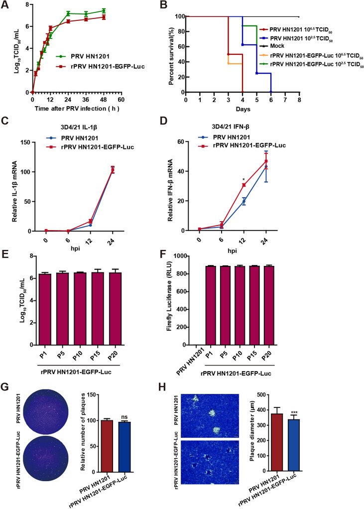
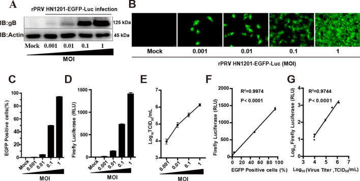
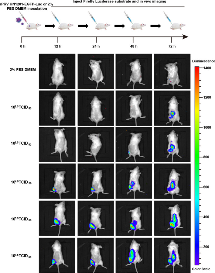
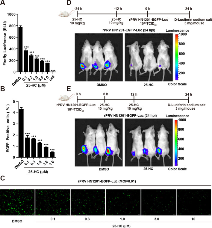
Pseudorabies, caused by pseudorabies virus (PRV) variants, has broken out among commercial PRV vaccine-immunized swine herds and resulted in major economic losses to the pig industry in China since late 2011. However, the mechanism of virulence enhancement of variant PRV is currently unclear. Here, a recombinant PRV (rPRV HN1201-EGFP-Luc) with stable expression of enhanced green fluorescent protein (EGFP) and firefly luciferase as a double reporter virus was constructed on the basis of the PRV variant HN1201 through CRISPR/Cas9 gene-editing technology coupled with two sgRNAs. The biological characteristics of the recombinant virus and its lethality to mice were similar to those of the parental strain and displayed a stable viral titre and luciferase activity through 20 passages. Moreover, bioluminescence signals were detected in mice at 12 h after rPRV HN1201-EGFP-Luc infection. Using the double reporter PRV, we also found that 25-hydroxycholesterol had a significant inhibitory effect on PRV both in vivo and in vitro. These results suggested that the double reporter PRV based on PRV variant HN1201 should be an excellent tool for basic virology studies and evaluating antiviral agents.

Keywords: 25-hydroxycholesterol; CRISPR/Cas9; EGFP; Firefly luciferase; Imaging in vivo; Pseudorabies virus.


Conflict of interest statement

The authors declare that they have no competing interests.

Figures

Figure 1
Figure 1
The double-sgRNA CRISPR/Cas9 system and the protocol used to generate the recombinant virus rPRV HN1201-EGFP-Luc. A Schematic diagrams of the double -sgRNA CRISPR/Cas9 system and the CRISPR/Cas9 cleavage positions. B Diagrams showing the PRV HN1201 genome and the donor plasmid. In the donor plasmid, the left homologous recombination arm (left HR arm) and the right HR arm are located upstream and downstream of the sgRNA1 and sgRNA2 target sites, respectively. There were 1693 base pairs (bp) between the two sgRNA target sites. After the PRV genome is cut at the two sgRNA target sites and homologous recombination occurs, the PRV genome loses a 2459 bp DNA fragment, and the cassette containing the EGFP and luciferase genes is knocked in at the DNA deletion sites. C Diagram depicting the protocol used to obtain and purify recombinant PRV for the expression of EGFP and luciferase genes. A mixture of PRV HN1201 genomes, Cas9/sgRNAs and donor plasmid was cotransfected into PK-15 cells. CPEs with EGFP were observed 2–4 days after transfection. Cells and media were collected after three freeze–thaw cycles and then inoculated into cells in 96-well plates after serial dilutions to obtain single viral clones. Subcloned viruses were subjected to luciferase assays and sequence analysis.
Figure 2
Figure 2
Identification of the recombinant virus rPRV HN1201-EGFP-Luc. A PK-15 cells were infected with purified recombinant rPRV HN1201-EGFP-Luc virus and visualized by fluorescence and bright field microscopy. B Sequence chromatogram showing the DNA sequences of PRV HN1201 (top) and rPRV HN1201-EGFP-Luc (bottom) at the genetic homologous recombination sites. C Cells were infected with rPRV HN1201-EGFP-Luc (MOI = 0.1) for 12 h. The mRNA of firefly luciferase in the cells was detected with RT-PCR. D PK-15 cells (left) and ST cells (right) were either mock infected or infected with PRV HN1201 or rPRV HN1201-EGFP-Luc (MOI = 0.1) for 24 h and then lysed for immunoblotting analysis with antibodies against firefly luciferase and the viral proteins gB and gE. β-Actin was used as the loading control. E PK-15 cells were infected with rPRV HN1201-EGFP-Luc (MOI = 0.1) for 24 h and then lysed for luciferase assays. PK-15 cell lysate was used as a negative control, and PK-15 cells transfected with donor plasmid lysate were used as a positive control. This experiment was performed three times, and the results are shown as the mean ± SD.
Figure 3
Figure 3
Biological characteristics of the recombinant virus rPRV HN1201-EGFP-Luc. A One-step growth curves of PRV HN1201 (MOI = 0.1) and rPRV HN1201-EGFP-Luc (MOI = 0.1) in PK-15 cells. This experiment was performed three times, and the results are shown as the mean ± SD. B Percentage survival of mice (eight mice per group) infected I.M. with 106.5 TCID50 and 102.5 TCID50 of PRV HN1201 and rPRV HN1201-EGFP-Luc. C, D Equal numbers of 3D4/21 cells were infected with PRV HN1201 or rPRV HN1201-EGFP-Luc at an MOI of 1. Total RNA was collected at 6, 12 and 24 hpi, and then, IL-1β (C) and IFN-β (D) mRNA expression was quantified by RT-qPCR. This experiment was performed three times, and the results are shown as the mean ± SD. E The rPRV HN1201-EGFP-Luc passage experiments were performed in PK-15 cells (passages 1–20), and then, viral titre was determined with TCID50 assays at specific passages (1, 5, 10, 15 and 20) of rPRV HN1201-EGFP-Luc. Data are shown as the mean ± SD from three independent experiments. F PK-15 cells were infected with specific passages (1, 5, 10, 15 or 20) of rPRV HN1201-EGFP-Luc (MOI = 0.1). Luciferase assays were performed in triplicate at the indicated times after infection. Data are shown as the mean ± SD from three independent experiments. G The plaque assay standardized on genome copy numbers. The number of rPRV HN1201-EGFP-Luc plaques was normalized to the number of PRV HN1201 plaques to detect the effect of gI/gE gene knockout on PRV packaging. Representative data from triplicate experiments are shown. Mean + SD, ns, no significant difference. H Plaque sizes of PK-15 cells infected with PRV HN1201 or rPRV HN1201-EGFP-Luc at 36 hpi. The mean plaque diameter of 50 plaques from one representative experiment out of three independent experiments is shown. Mean + SD, *** P < 0.001.
Figure 4
Figure 4
Firefly luciferase activity, GFP expression and viral titre of recombinant virus. A PK-15 cells were infected with rPRV HN1201-EGFP-Luc (MOI = 0.001, 0.01, 0.1 or 1) for 18 h and then lysed for immunoblotting analysis with antibodies against viral protein gB. β-Actin was used as the loading control. Fluorescence microscopy (B), fluorescence-activated cell sorting (FACS) analysis (C), luciferase assays (D) and TCID50 assays (E) of rPRV HN1201-GFP-Luc (MOI = 0.001, 0.01, 0.1 or 1) proliferation in PK-15 cells for 18 h. Data are shown as the mean ± SD from three independent experiments. Scale bar, 100 μm. F Correlation between luminescence intensity and GFP expression (R2 = 0.9974, p < 0.0001; GraphPad Prism 5, La Jolla, CA, USA). G Correlation between luminescence intensity and infectious viral titres (TCID50) (R2 = 0.9744, p < 0.0001; GraphPad Prism 5, La Jolla, CA, USA).
Figure 5
Figure 5
The replication and distribution of rPRV HN1201-EGFP-Luc in a mouse model. 6-week-old BALB/c mice were infected I.M. with 2% FBS DMEM or recombinant pseudorabies virus rPRV HN1201-EGFP-Luc (106.5, 105.5, 104.5, 103.5 or 102.5 TCID50 per mouse) in the left leg. Viral replication and distribution were quantified at the indicated times after infection by whole-animal bioluminescence imaging.
Figure 6
Figure 6
The 25-HC antiviral tests in vivo and in vitro. Treatment with 25-HC inhibited the proliferation of rPRV HN1201-EGFP-Luc in vitro. PK-15 cells were pretreated with the indicated concentrations of 25-HC for 4 h and then infected with rPRV HN1201-EGFP-Luc (MOI = 0.01) along with the same concentrations of 25-HC for 18 h. Luciferase assays (A), FACS analysis (B) and fluorescence microscopy (C) were performed. Data are shown as the mean ± SD from three independent experiments. Scale bar, 200 μm. ***P < 0.001. D Bioluminescent images of mice pretreated with 25-HC or DMSO at 24 h after rPRV HN1201-EGFP-Luc infection. E Bioluminescent images of mice in which 25-HC or DMSO was injected later upon rPRV HN1201-EGFP-Luc infection. Whole animal bioluminescence imaging was performed 24 h after rPRV HN1201-EGFP-Luc infection.

Similar articles

Cited by

References

    1. Mettenleiter TC. Immunobiology of pseudorabies (Aujeszky’s disease) Vet Immunol Immunopathol. 1996;54:221–229. doi: 10.1016/S0165-2427(96)05695-4. - DOI - PubMed
    1. Klupp BG, Hengartner CJ, Mettenleiter TC, Enquist LW. Complete, annotated sequence of the pseudorabies virus genome. J Virol. 2004;78:424–440. doi: 10.1128/JVI.78.1.424-440.2004. - DOI - PMC - PubMed
    1. Mettenleiter TC. Aujeszky’s disease (pseudorabies) virus: the virus and molecular pathogenesis—state of the art, June 1999. Vet Res. 2000;31:99–115. - PubMed
    1. Koyuncu OO, Hogue IB, Enquist LW. Virus infections in the nervous system. Cell Host Microbe. 2013;13:379–393. doi: 10.1016/j.chom.2013.03.010. - DOI - PMC - PubMed
    1. Qi H, Wu H, Abid M, Qiu HJ, Sun Y. Establishment of a fosmid library for pseudorabies virus SC strain and application in viral neuronal tracing. Front Microbiol. 2020;11:1168. doi: 10.3389/fmicb.2020.01168. - DOI - PMC - PubMed

LinkOut - more resources