Review
. 2021 Oct:70:24-33.
doi: 10.1016/j.conb.2021.05.005. Epub 2021 Jun 24.

From synapse to network: models of information storage and retrieval in neural circuits

Affiliations
Review

From synapse to network: models of information storage and retrieval in neural circuits

Johnatan Aljadeff et al. Curr Opin Neurobiol. 2021 Oct.

Abstract

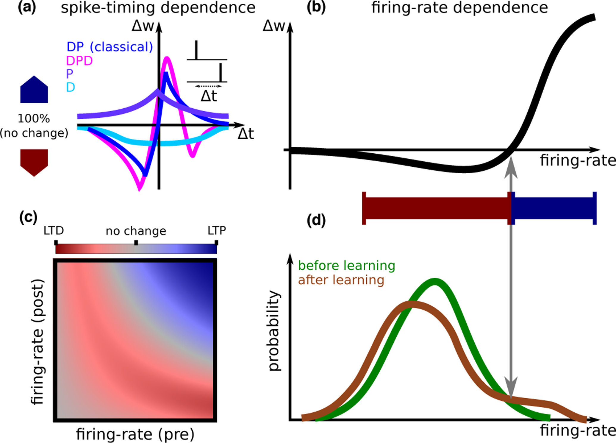
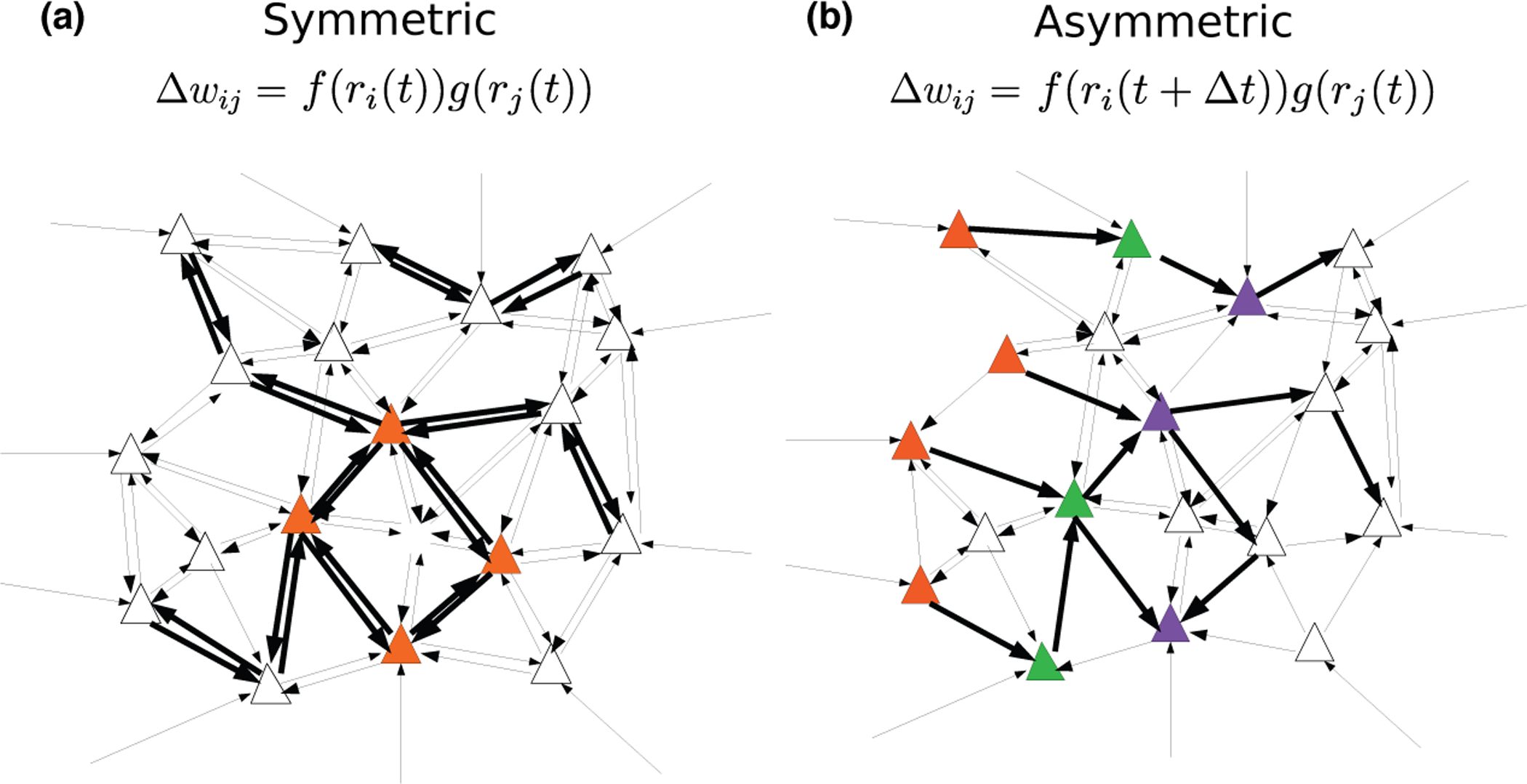
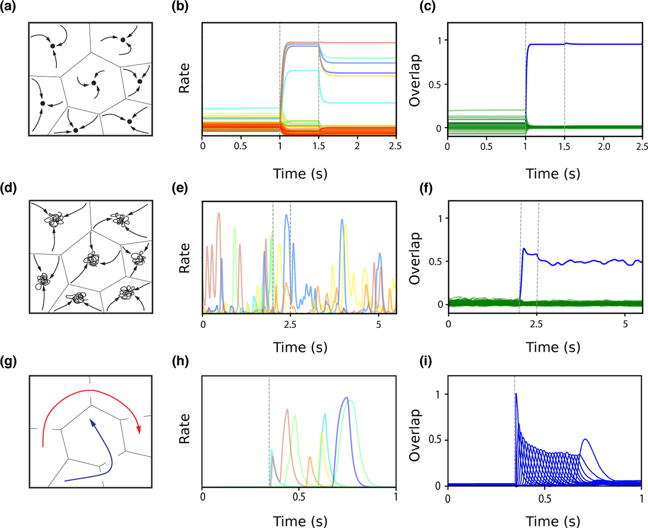
The mechanisms of information storage and retrieval in brain circuits are still the subject of debate. It is widely believed that information is stored at least in part through changes in synaptic connectivity in networks that encode this information and that these changes lead in turn to modifications of network dynamics, such that the stored information can be retrieved at a later time. Here, we review recent progress in deriving synaptic plasticity rules from experimental data and in understanding how plasticity rules affect the dynamics of recurrent networks. We show that the dynamics generated by such networks exhibit a large degree of diversity, depending on parameters, similar to experimental observations in vivo during delayed response tasks.


Conflict of interest statement

Conflict of interest statement Nothing declared.

Figures

Figure 1:
Figure 1:. Synaptic plasticity.
A. STDP (spike timing dependent plasticity) curves (i.e., dependence of synaptic plasticity on the timing difference between pre- and postsynaptic spikes Δt) observed experimentally. A number of qualitatively distinct STDP curve shapes have been reported experimentally: The classical curve (dark blue, seen e.g., in hippocampal cultures [2] and cortical slices [3]) exhibits a depression window (D) followed by a potentiation window (P); A curve with a second depression window (DPD, magenta, seen in CA3 to CA1 connections at high calcium concentration[4]); A curve with only potentiation (P, purple, seen in area CA3 [5], in hippocampal cultures in the presence of dopamine[6]); and finally a curve with only depression (D, light blue, in CA3 to CA1 connections at low extra-cellular calcium concentrations[7, 4]).A calcium based model can account for all curve shapes with different choices of parameters [8], and for the transition between different curves as a function of the extracellular calcium concentration [4]. B. Models from each of the classes discussed in the text are typically fit to in vitro data based on plasticity protocols where the same pre-post pattern of activity is repeated. These models all give qualitatively similar firing rate dependence, which is similar to the BCM rule [9]. The specific shape of the non-linearity, and in particular the threshold separating LTD and LTP, depends on the model and its parameters, and on whether pre/postsynaptic (or both) firing-rate(s) are varied. A learning rule inferred from in vivo data [10] gives a similar dependence on firing rate. C. Models can also be used to predict synaptic plasticity for independent pre- and postsynaptic firing-rates. Shown here qualitatively is the magnitude of LTD (red) and LTP (blue), in a scenario where synaptic modifications are well approximated by the form Δw = g(rpre)f(rpost) [10]. The dependence of plasticity on both pre- and postsynaptic firing rate, varying as two independent variables, has not yet been characterized experimentally. D. Inferring plasticity rules from in vivo data. The firing-rate distribution of a single neuron in response to sensory stimuli exhibits significant differences between novel and familiar stimuli. These differences can be used to reverse-engineer a learning rule that causes such differences. This learning rule has a dependence on the postsynaptic firing rate that is qualitatively similar to the curve in panel B. Such a learning rule sparsens the representation of sensory stimuli, decreasing the response of most neurons (those that respond with a firing rate that is smaller than the threshold between LTD and LTP), but leading to an increased response for a small subset of neurons that have initially the strongest response [10].
Figure 2:
Figure 2:
Network connectivity after learning with temporally symmetric (A) and asymmetric (B) plasticity rules. A. An external input leads to increased activity of the set of orange neurons. With a temporally symmetric plasticity rule, this activity leads in turn to strengthening of synapses connecting these neurons in a bidirectional fashion, leading to strongly symmetric synaptic connectivity. B. Temporally varying external inputs lead to successive activation of the orange, green and purple sets of neurons. With a temporally asymmetric plasticity rule, connectivity is strengthened in a unidirectional fashion, leading to an asymmetric, effectivety feedforward connectivity between these sets of neurons.
Figure 3:
Figure 3:
Network dynamics following learning. A. Schematic of fixed point attractor retrieval states occupying discrete subspaces. The space of network states is divided into basins of attraction, within which network activity will converge to a fixed point (black circle), representing a learned item. B. Single unit firing rates during background (0–1s), presentation of a pattern stored in memory (between dashed lines, 1–1.5s), and ‘delay’ period following stimulus presentation (1.5–2.5s), in a network in which distributions of stored patterns and learning rule have been inferred from data [28]. In the background state, single neurons fire with a wide (~ log-normal) distribution of rates. During the stimulus presentation, a small fraction of neurons are driven to high rates, while most neurons are suppressed. After the stimulus is removed, the network converges to a selective attractor states, in which a small fraction of neurons exhibits persistent activity. C. Overlap between network state and the presented pattern (blue) increases during stimulus presentation, and remains high following removal of external input, while overlaps between network state and other stored patterns (green) remains small. D. Schematic of chaotic attractor retrieval states. Network activity is confined to a subspace, but does not converge to a stable fixed point. E–F. Similar as in panel B–C, except the network has stronger recurrent connections and stimulus presentation is between 2 and 2.5s [28]. The network is in a chaotic state before stimulus presentation, with all single neuron firing rates fluctuating widely. After stimulus presentation, the network switches to a different chaotic state that is strongly correlated with the stored pattern (see blue overlap in F). G. Schematic of sequential pattern retrieval for two separate stored sequences (red and blue). Activity transitions from one subspace to the next. H–I. Similar as in panels B–C, except the network stores sequences, and is initialized close to the first stored pattern in a sequence of 24 patterns at 0.35s [29]. Single neurons are transiently activated at different times, and the network successively visits the neighborhoods of the different patterns composing the sequence as shown by transient overlap activations in I.

Similar articles

Cited by

References

    1. Kamiński J and Rutishauser U (2020). Between persistently active and activity-silent frameworks: novel vistas on the cellular basis of working memory. Ann N Y Acad Sci 1464, 64–75. - PMC - PubMed
    1. Bi GQ and Poo MM (1998). Synaptic modifications in cultured hippocampal neurons: dependence on spike timing, synaptic strength, and postsynaptic cell type. J. Neurosci 18, 10464–10472. - PMC - PubMed
    1. Markram H, Lubke J, Frotscher M, and Sakmann B (1997). Regulation of synaptic efficacy by coincidence of postsynaptic APs and EPSPs. Science 275, 213–215. - PubMed
    1. Inglebert Y, Aljade J, Brunel N, and Debanne D (2020). Synaptic plasticity rules with physiological calcium levels. Proc Natl Acad Sci U S A 117, 33639–33648. - PMC - PubMed
    2. STDP experiments at physiological calcium concentrations show no significant synaptic changes, while high firing rates and/or bursts restore plasticity. Experimental data is consistent with a calcium-based rule with non-linear summation between calcium transients due to pre and post-synaptic spikes.

    1. Mishra RK, Kim S, Guzman SJ, and Jonas P (2016). Symmetric spike timing-dependent plasticity at CA3-CA3 synapses optimizes storage and recall in autoassociative networks. Nat Commun 7, 11552. - PMC - PubMed

Publication types