Case Reports
. 2022 Feb;38(2):441-445.
doi: 10.1007/s00381-021-05273-x. Epub 2021 Jun 26.

Malignant cerebral infarction associated with COVID-19 in a child

Affiliations
Case Reports

Malignant cerebral infarction associated with COVID-19 in a child

Maria Rosaria Scala et al. Childs Nerv Syst. 2022 Feb.

Abstract

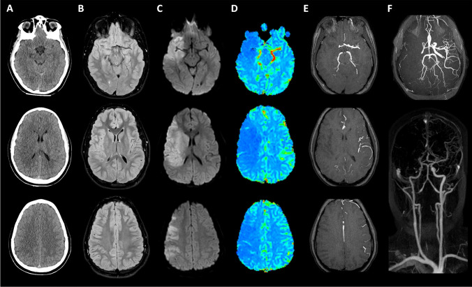
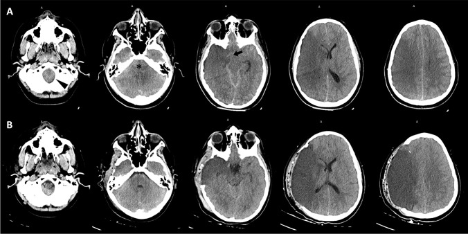
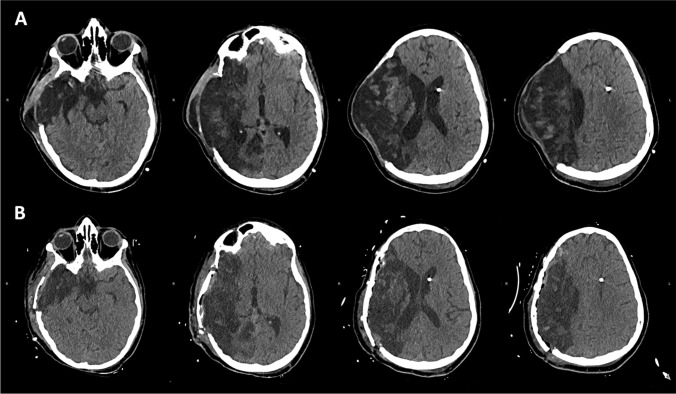
Neurological manifestations, such as encephalitis, meningitis, ischemic, and hemorrhagic strokes, are reported with increasing frequency in patients affected by Coronavirus disease 2019 (COVID-19). In children, acute ischemic stroke is usually multifactorial: viral infection is an important precipitating factor for stroke. We present a case of a child with serological evidence of SARS-CoV-2 infection whose onset was a massive right cerebral artery ischemia that led to a malignant cerebral infarction. The patient underwent a life-saving decompressive hemicraniectomy, with good functional recovery, except for residual hemiplegia. During rehabilitation, the patient also developed a lower extremity peripheral nerve neuropathy, likely related to a long-Covid syndrome.

Keywords: Acute ischemic stroke; Decompressive craniectomy; Long-Covid syndrome; Peripheral nerve neuropathy.


Conflict of interest statement

The authors declare that they have no conflict of interest.

Figures

Fig. 1
Fig. 1
Early CT scan and fast MR study at admission. First CT scan (A) showed only slight hypoattenuation and obscuration of the head of the caudate nucleus as well as a loss of gray/white matter definition in the lateral margins of the right insula, findings suggestive of “early signs” of ischemic stroke; slight hyperattenuating band on the right middle cerebral artery (MCA) was also observed, due to intravascular occluding thrombus. Similarly, MR FLAIR images (B) were nearly normal, with only subtle hyperintensity along with the cortical insular ribbon. Instead, diffusion-weighted [b 1000 s/mm2] images (C) clearly showed acute stroke–induced cytotoxic edema in the right MCA territory; the discrepancy between DWI and FLAIR images was suggestive of an early stage of stroke. The MR perfusion CBF map (D) demonstrated a large wedge-shaped area of significantly reduced perfusion in the same region, also corresponding to a lack of vascular representation on MRA TOF3D MIP reconstructions (E). MR-angiography (F) performed with TOF3D, and contrast-enhanced techniques confirmed the occlusion of M1 segment of the right MCA
Fig. 2
Fig. 2
Brain CT monitoring. Post-endovascular treatment CT scan (A), performed after the onset of anisocoria, demonstrated extensive swelling in the right hemisphere, right transtentorial uncal herniation (black arrow), effacement of the CSF spaces in the posterior fossa (asterisk), incipient tonsillar herniation (arrowhead) and leftward midline shift due to malignant cerebral edema. These compressive findings were improved on CT images (B) obtained after DHC
Fig. 3
Fig. 3
Late brain CT exams. Surveillance CT scan (A) showed progressive malacic evolution of MCA infarction, with brain expanded through the skull defect. Last CT exam at discharge (B) demonstrated a good result of bone cranioplasty

References

    1. Koralnik IJ, Tyler KL. COVID-19: A global threat to the nervous system. Ann Neurol. 2020;88:1–11. doi: 10.1002/ana.25807. - DOI - PMC - PubMed
    1. Liu JM, Tan BH, Wu S, Gui Y, Suo JL, Li YC. Evidence of central nervous system infection and neuroinvasive routes, as well as neurological involvement, in the lethality of SARS-CoV-2 infection. J Med Virol. 2021;93:1304–1313. doi: 10.1002/jmv.26570. - DOI - PMC - PubMed
    1. Zhou Z, Kang H, Li S, Zhao X. Understanding the neurotropic characteristics of SARS-CoV-2: from neurological manifestations of COVID-19 to potential neurotropic mechanisms. J Neurol. 2020;267:2179–2184. doi: 10.1007/s00415-020-09929-7. - DOI - PMC - PubMed
    1. Zubair AS, McAlpine LS, Gardin T, Farhadian S, Kuruvilla DE, Spudich S. Neuropathogenesis and neurologic manifestations of the coronaviruses in the age of coronavirus disease 2019: a review. JAMA Neurol. 2020;77:1018–1027. doi: 10.1001/jamaneurol.2020.2065. - DOI - PMC - PubMed
    1. Ranucci M, Ballotta A, Di Dedda U, Bayshnikova E, Dei Poli M, Resta M, Falco M, Albano G, Menicanti L. The procoagulant pattern of patients with COVID-19 acute respiratory distress syndrome. J Thromb Haemost. 2020;18:1747–1751. doi: 10.1111/jth.14854. - DOI - PMC - PubMed

Publication types