Review
. 2021 Aug;58(8):4188-4215.
doi: 10.1007/s12035-021-02457-z. Epub 2021 Jun 26.

COVID-19 Infection and Circulating Microparticles-Reviewing Evidence as Microthrombogenic Risk Factor for Cerebral Small Vessel Disease

Affiliations
Review

COVID-19 Infection and Circulating Microparticles-Reviewing Evidence as Microthrombogenic Risk Factor for Cerebral Small Vessel Disease

Che Mohd Nasril Che Mohd Nassir et al. Mol Neurobiol. 2021 Aug.

Abstract

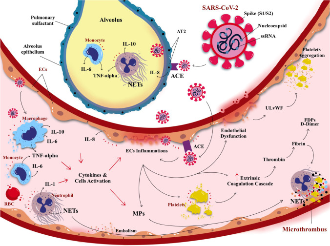
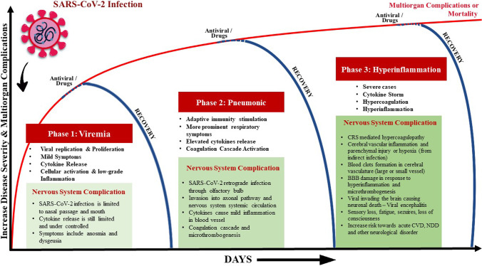
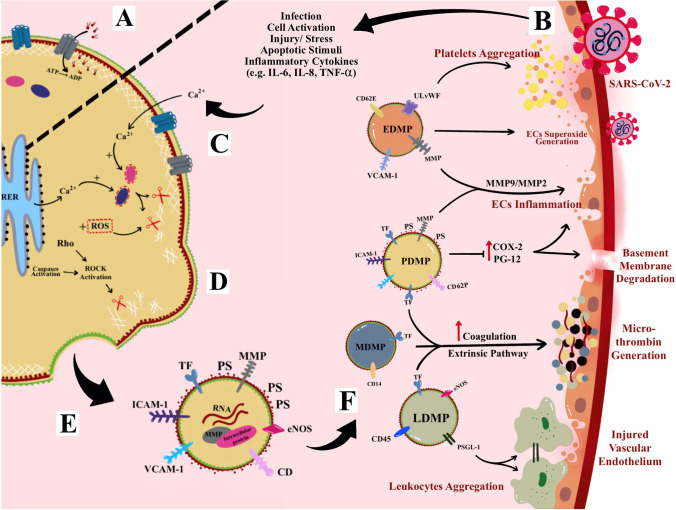
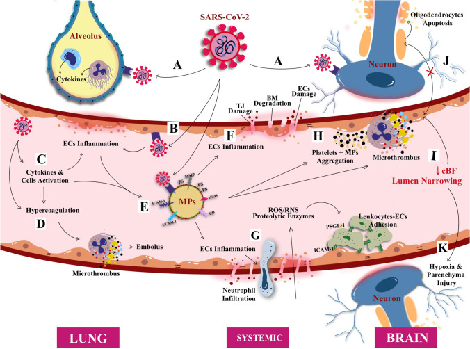
Severe acute respiratory syndrome corona virus-2 (SARS-CoV-2) due to novel coronavirus disease 2019 (COVID-19) has affected the global society in numerous unprecedented ways, with considerable morbidity and mortality. Both direct and indirect consequences from COVID-19 infection are recognized to give rise to cardio- and cerebrovascular complications. Despite current limited knowledge on COVID-19 pathogenesis, inflammation, endothelial dysfunction, and coagulopathy appear to play critical roles in COVID-19-associated cerebrovascular disease (CVD). One of the major subtypes of CVD is cerebral small vessel disease (CSVD) which represents a spectrum of pathological processes of various etiologies affecting the brain microcirculation that can trigger subsequent neuroinflammation and neurodegeneration. Prevalent with aging, CSVD is a recognized risk factor for stroke, vascular dementia, and Alzheimer's disease. In the background of COVID-19 infection, the heightened cellular activations from inflammations and oxidative stress may result in elevated levels of microthrombogenic extracellular-derived circulating microparticles (MPs). Consequently, MPs could act as pro-coagulant risk factor that may serve as microthrombi for the vulnerable microcirculation in the brain leading to CSVD manifestations. This review aims to appraise the accumulating body of evidence on the plausible impact of COVID-19 infection on the formation of microthrombogenic MPs that could lead to microthrombosis in CSVD manifestations, including occult CSVD which may last well beyond the pandemic era.

Keywords: COVID-19; Cerebral small vessel disease; Coagulopathy; Microparticles; Stroke.


Conflict of interest statement

The authors declare no competing interests.

Figures

Fig. 1
Fig. 1
A schematic illustration of direct SARS-CoV-2 infection from the lung alveolus and blood circulation. The virus SARS-CoV-2 (and its main structure) acquired through respiratory droplets attacking angiotensin converting enzymes (ACE) type 2 receptors that are present on the surface of alveolus epithelium, namely, the alveolar epithelial type (AT2). The attachment of SARS-CoV-2 with ACE elicited the inflammatory reaction of the AT2 cells, releasing pro-inflammatory cytokines; i.e., interleukin-8 (IL-8) alongside the activation of monocytes and neutrophil elevate the inflammation causing lung parenchymal injury. In addition, SARS-CoV-2 also can enter blood circulation and raise activation of circulating cells (i.e., macrophage, monocytes, platelets, and neutrophil) to release pro-inflammatory cytokines causing endothelial inflammation (endotheliitis). Endotheliitis then activates the coagulation cascade and production of thrombin followed by fibrinolysis and fibrin. If left untreated, the infection will progress to cause hypercoagulation state leading to coagulopathy. Collectively, cytokines, cellular activation, and endothelial inflammation drive the production of microparticles (MPs) which further instigate the production of microthrombus, cell-endothelium adhesion, and aggregation0
Fig. 2
Fig. 2
The known phases of COVID-19 infection: from viremia, pulmonary to multi-system manifestations; emphasis made on the impacts on the nervous system. Also shown are the simplified underlying COVID-19 likely pathomechanisms as the infection progresses, and the current corresponding therapeutic targets in the clinical management of each phase. It is probable that even after the recovery at any phase of the disease, the involvement of cellular activation by-product (such as microparticles) may persist and result in an undesirable health sequel
Fig. 3
Fig. 3
Neuroimaging correlates of CSVD based on STRIVE method. A Recent small subcortical infarct (RSBI) on diffusion weighted imaging (DWI) (red arrow). Usual diameter is around 3–15 mm, with hyperintense rim surrounding ovoid cavity. RSBI seen as increased T2-weighted, fluid attenuated inverse recovery (FLAIR), and DWI signal intensities and decreased T1-weighted signal and iso-intense in T2*-weighted gradient recoiled echo (GRE) signal and susceptibility weighted imaging (SWI). RSBI is best identified through DWI with usual infarct diameter of ≤ 20 mm. B Lacunar infracts on FLAIR (red arrow). Lacunar infarcts appeared as increase hyperintensity in T2-weighted signal, decrease T1-weighted, and FLAIR signal and iso-intense in DWI. Usual diameter is around 3–15 mm, with hyperintense rim surrounding ovoid cavity. C White matter hyperintensities (WMHs) of presumed vascular origin on FLAIR (arrow). WMHS seen as increase intensity or hyperintensity on T2-weighted imaging, T2*-weighted GRE and FLAIR (best identified); iso-intense on DWI; and hypointense (decrease intensity) on T1-weighted imaging. D FLAIR WMHs at left superior frontal gyrus and left anterior cingulate cortex, from a 60-year-old COVID-19 patient without history of seizures. E Enlarged perivascular spaces (PVS) on T1-weighted imaging (red arrow) with usual diameter of ≤ 2 mm. PVS is seen as decrease FLAIR and T1-weighted signal intensity, with increase T2-weighted signal. Meanwhile, T2*-weighted GRE and DWI appeared iso-intense, and they also appeared in similar signal intensity with cerebrospinal fluid (CSF). F Cerebral microbleeds (CMBs) on T2*-GRE (red arrow). CMBs are small, rounded areas of signal void with blooming, whereby they were visualized as iso-intense T1- and T2-weighted signal, FLAIR, and DWI. They are best identified under T2*-weighted GRE or SWI as reduced signal intensities. Usual diameter is around ≤ 10 mm (mostly 2–5 mm). G 3 Tesla-MRI representation of cortical microinfarcts (red arrow) on T1-weighted (hypointense). H FLAIR WMHs in multiple foci, including the deep white matter, periventricular, and subcortical regions in COVID-19 patient with CADASIL. Notes: (A), (B), (C), (E), and (F) were adapted from Mustapha et al. [70]; (D) was adapted from Muhammedi et al. [73]; (G) was adapted from Takasugi et al. [74], and (H) was adapted from Williams et al. [75]
Fig. 4
Fig. 4
Microparticles (MPs) formation and mechanism of action. A Active translocase transporting phosphatidylserine (PS) from outside to inside layer through adenosine triphosphate (ATP)-dependent manner. B Cellular activation due to infection or other cellular stressor such as increase cytokines and apoptotic stimuli. C The activation causes an increase in intracellular cytosolic calcium release by stress endoplasmic reticulum (ER) and acquired from extracellular space and hence activates enzymes calpain and gelsolin that cleave cell membrane cytoskeleton. D The cleaved cytoskeleton causes inactivation of translocase and, hence, induces phospholipid “flip-flopping.” E Externalization of PS produces MPs that bring their parent surface molecules and protein antigens. F MPs production can trigger series of micro-thrombotic cascades. For example, leukocytes-derived MPs (PDMPs) contain P-selectin glycoprotein ligand-1 (PSGL-1) on its surface that enables leukocytes-endothelial cell (ECs) adhesion. Most MPs contain tissue factor (TF) associated with an increase in the extrinsic coagulation cascade and production of microthrombus. In fact, PDMPs and endothelial cell-derived MPs (EDMPs) may bring pro-inflammatory antigens such as matrix metalloproteinase (MMP) that can cause endotheliitis. EDMPs also possess ultra-large von Willebrand factor (ULVWF) that further assists in the recruitment and aggregation of platelets on endothelium
Fig. 5
Fig. 5
Proposed interaction between COVID-19 infection and the formation of circulating microparticles (MPs) as plausible microthrombogenic risk factor for cerebral small vessel disease (CSVD) in addition to existing co-morbidity with conventional CVD risk factors—for overt symptomatic stroke events to occult (asymptomatic) manifestations. A SARS-CoV-2 infection in lung alveolus and central nervous system through angiotensin converting enzymes type 2 (ACE2) receptor present on the surface of lung alveolus and nerves cells. B SARS-CoV-2 also enters vascular microcirculation causing endothelial cells (ECs) activation and inflammation, C cytokines releases causing further inflammation and cellular activation and D hypercoagulation causing elevated clots/microthrombus formation and embolus to other organ/s. E cytokines release and cellular activation induced the formation of circulating microparticles (MPs). F MPs bring surface matrix metalloproteinase that can cause ECs inflammation and induce blood brain barrier (BBB) disruption, through (1) tight junction (TJ) damage, (2) basement membrane (BM) degradation, and (3) the EC damage and dysfunction. G BBB damage and endothelial dysfunction elevate the cellular (i.e., neutrophil) infiltration and hence increase cellular oxidative stress through increment of reactive oxygen species (ROS), reactive nitrogen species (RNS), and proteolytic enzymes, followed by leukocytes-ECs adhesion on the endothelium lining hence causing arterial wall blockage. H MPs also cause aggregation and platelet aggregations at the endothelium wall causing lumen narrowing; besides, the thrombo-emboli from microcirculation also can settle at the wall and cause blockage and narrowing of lumen and reduce cerebral blood flow (CBF). I reduced CBF and lumen narrowing can cause J no crosstalk between ECs and neuronal oligodendrocytes and hence cause oligodendrocytes apoptosis, i.e., demyelination disease and K neuronal/glial hypoxia and cerebral parenchymal injury. Thus, this emerges as a potential pathogenesis of occult CSVD

Similar articles

Cited by

References

    1. Dhama K, Khan S, Tiwari R, Sircar S, Bhat S, Malik YS, et al. Coronavirus disease 2019–COVID-19. Clin Microbiol Rev. 2020;33:e00028–e120. - PMC - PubMed
    1. Wu JT, Leung K, Bushman M, Kishore N, et al. Estimating clinical severity of COVID-19 from the transmission dynamics in Wuhan, China. Nat Med. 2020;26:506–510. - PMC - PubMed
    1. COVID-19 Map (2020). Coronavirus Resource Centre, John Hopkins University (JHU). https://coronavirus.jhu.edu/map.html. Accessed Jan 15, 2021
    1. Lai CC, Liu HY, Wang CY, Wang YH, Hsueh SC, Yen MY, et al. Asymptomatic carrier state, acute respiratory disease, and pneumonia due to severe acute respiratory syndrome coronavirus 2 (SARS-CoV-2): facts and myths. J Microbiol Immunol Infect. 2020;53(3):404–412. - PMC - PubMed
    1. Kronbichler A, Kresse D, Yoon S, Lee KH, Effenberger M, Shin JI. Asymptomatic patients as a source of COVID-19 infections: a systematic review and meta-analysis. Int J Infect Dis. 2020;98:180–186. - PMC - PubMed