

Hyperoxia-induced S1P1 signaling reduced angiogenesis by suppression of TIE-2 leading to experimental bronchopulmonary dysplasia
- PMID: 34176100
- PMCID: PMC8551021
- DOI: 10.1007/s12013-021-01014-8
Hyperoxia-induced S1P1 signaling reduced angiogenesis by suppression of TIE-2 leading to experimental bronchopulmonary dysplasia
Abstract
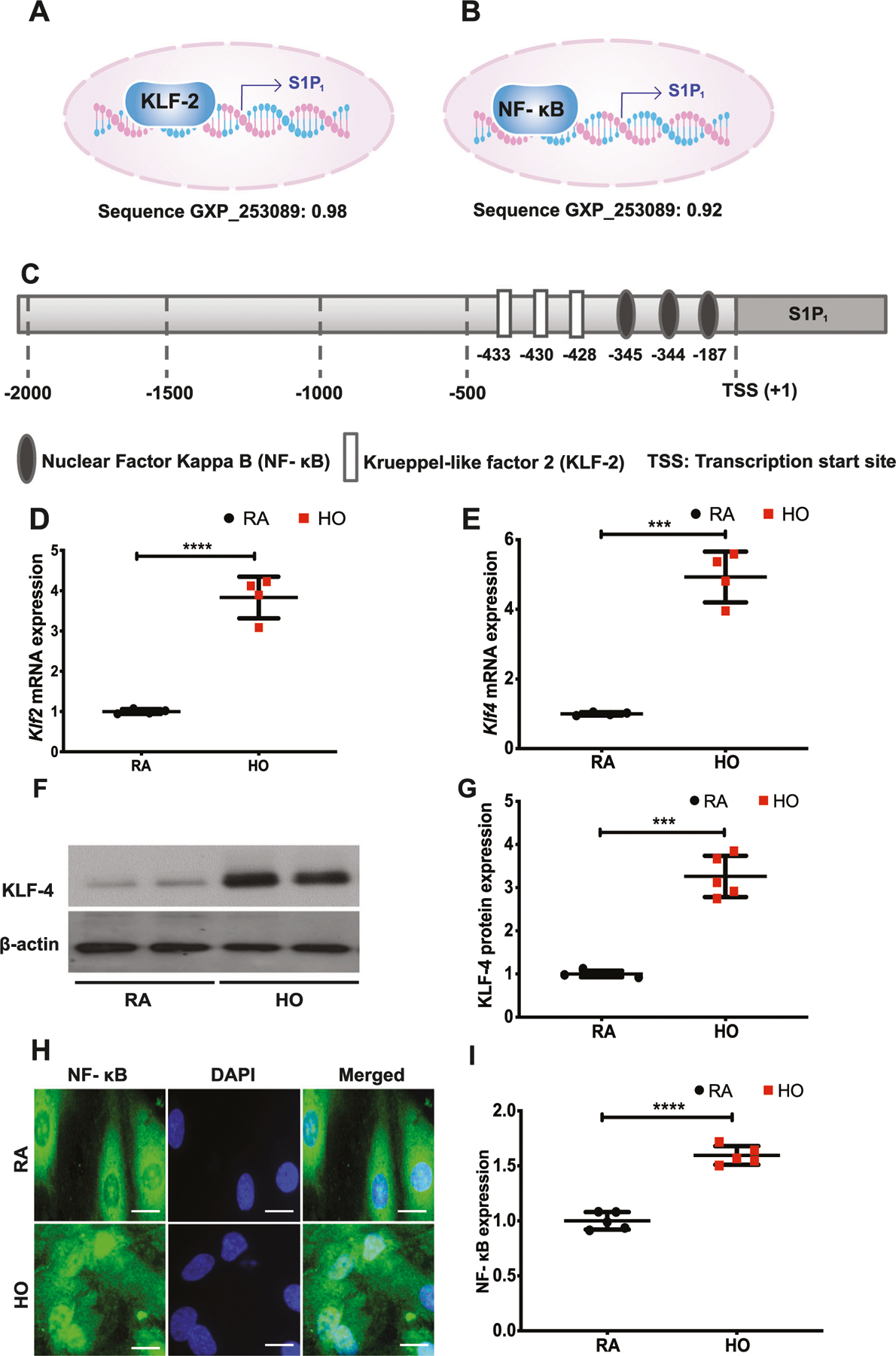
Introduction: We have earlier shown that hyperoxia (HO)-induced sphingosine kinase 1 (SPHK1)/sphingosine-1-phosphate (S1P) signaling contribute to bronchopulmonary dysplasia (BPD). S1P acts through G protein-coupled receptors, S1P1 through S1P5. Further, we noted that heterozygous deletion of S1pr1 ameliorated the HO-induced BPD in the murine model. The mechanism by which S1P1 signaling contributes to HO-induced BPD was explored.
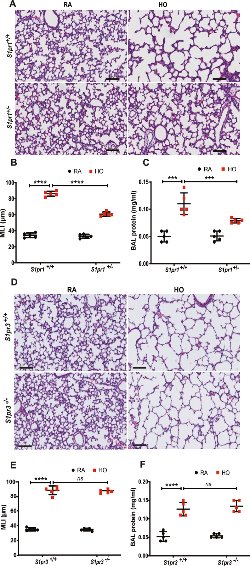
Methods: S1pr1+/+ and S1pr1+/- mice pups were exposed to either room air (RA) or HO (75% oxygen) for 7 days from PN 1-7. Lung injury and alveolar simplification was evaluated. Lung protein expression was determined by Western blotting and immunohistochemistry (IHC). In vitro experiments were performed using human lung microvascular endothelial cells (HLMVECs) with S1P1 inhibitor, NIBR0213 to interrogate the S1P1 signaling pathway.
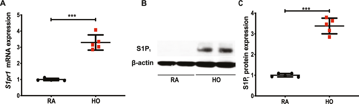
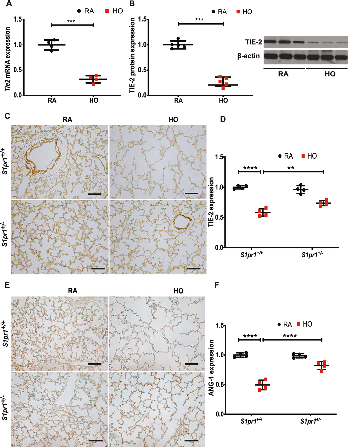
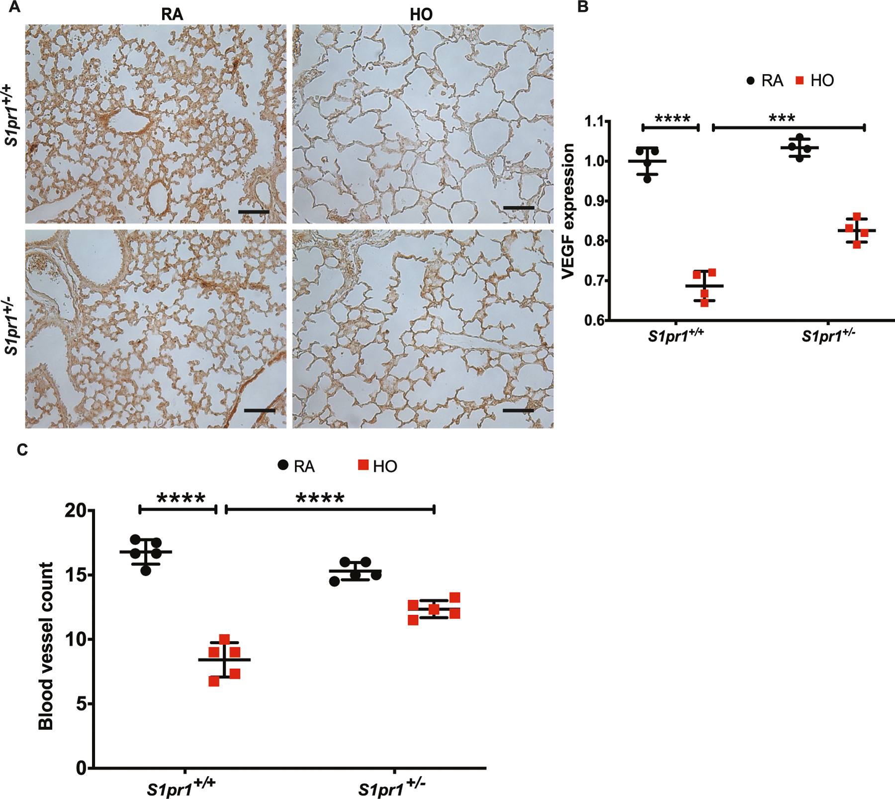
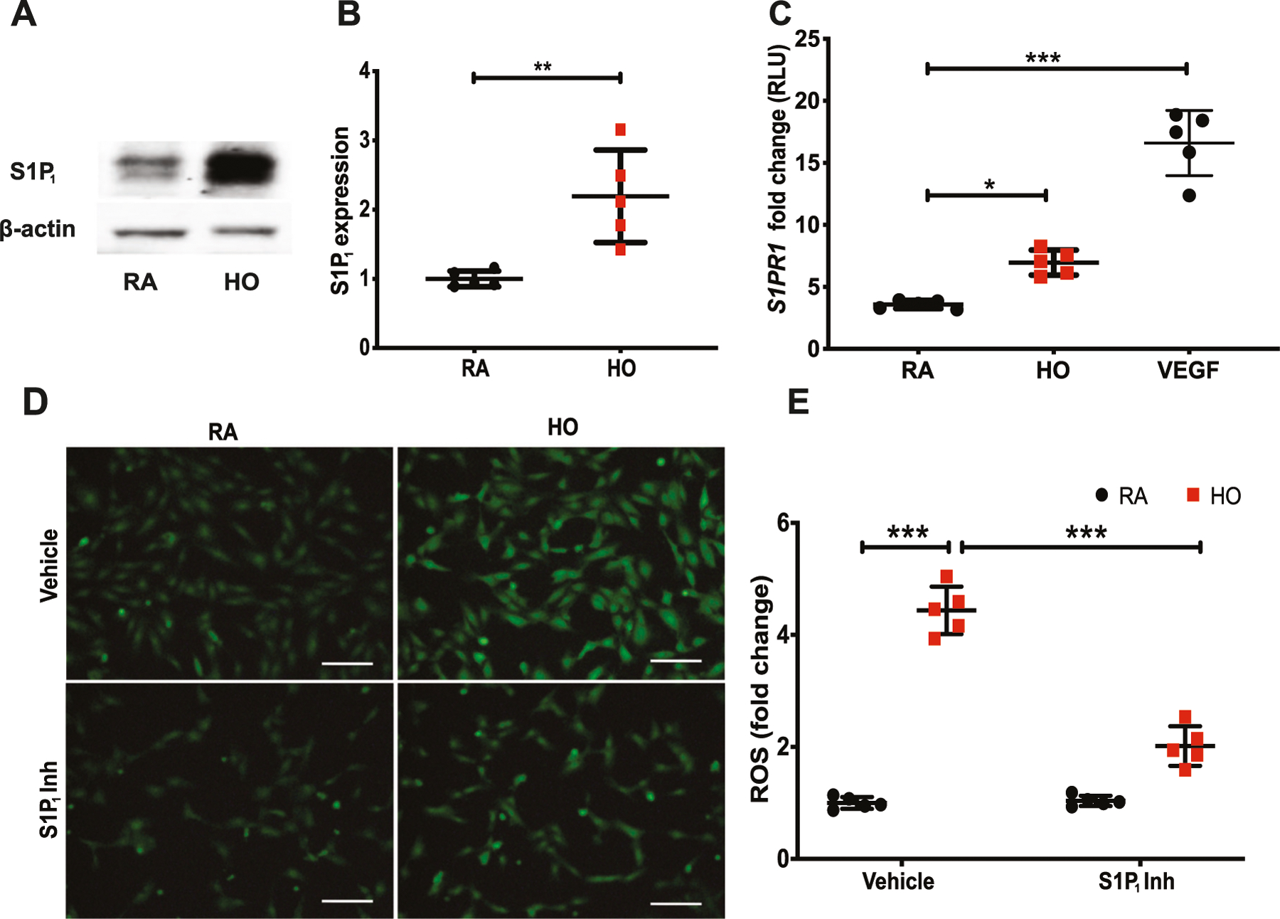
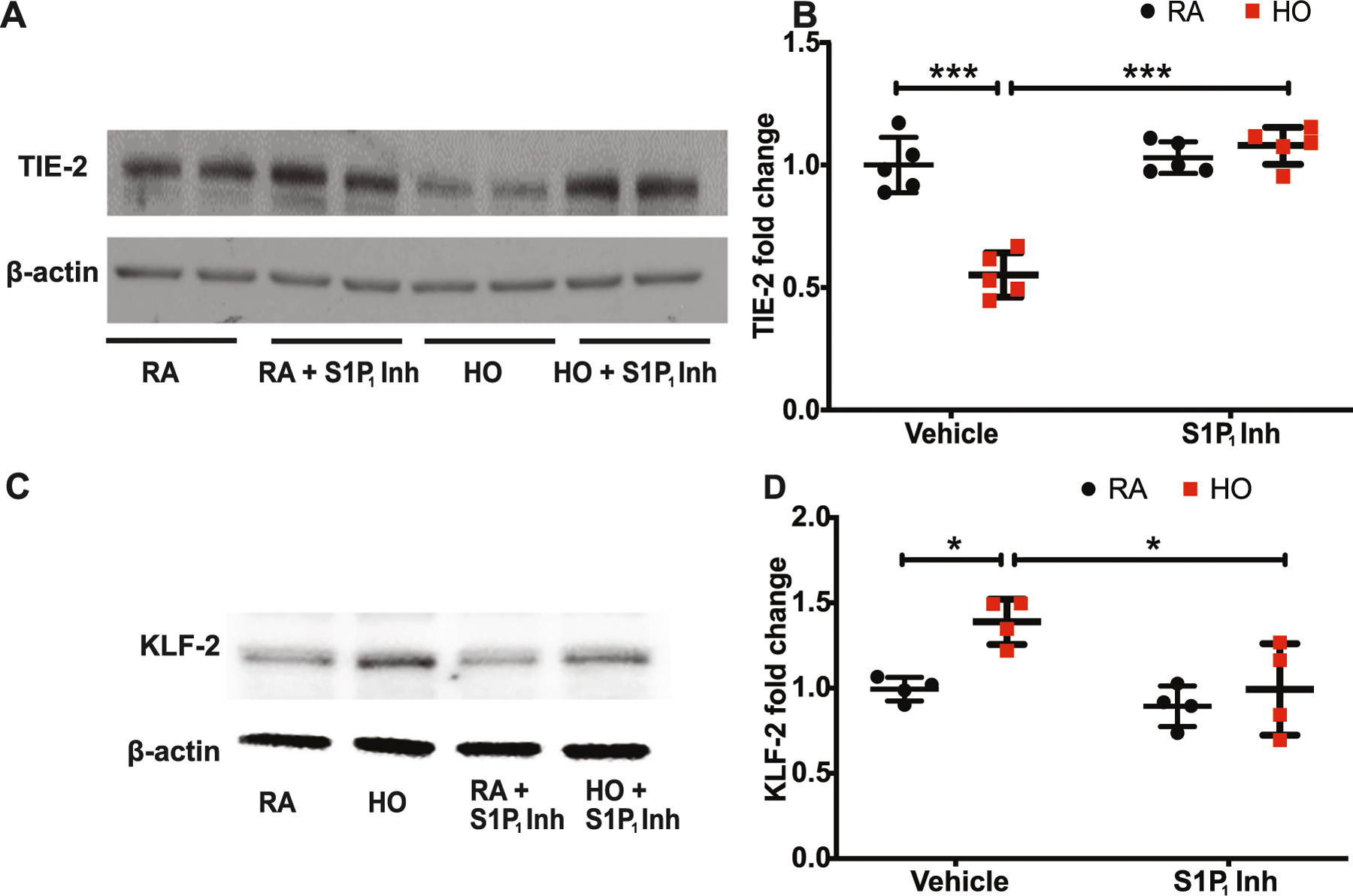
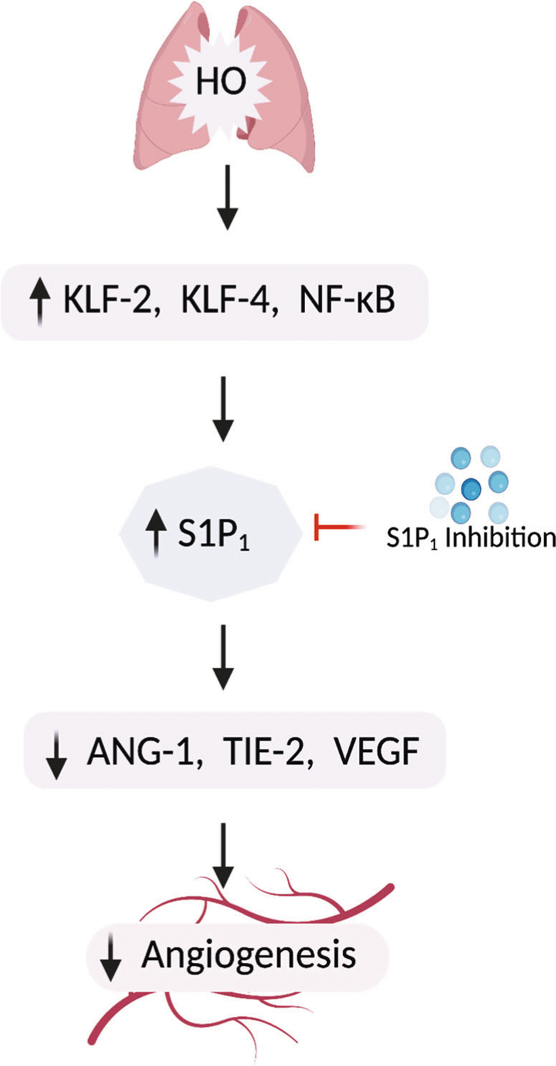
Results: HO increased the expression of S1pr1 gene as well as S1P1 protein in both neonatal lungs and HLMVECs. The S1pr1+/- neonatal mice showed significant protection against HO-induced BPD which was accompanied by reduced inflammation markers in the bronchoalveolar lavage fluid. HO-induced reduction in ANG-1, TIE-2, and VEGF was rescued in S1pr1+/- mouse, accompanied by an improvement in the number of arterioles in the lung. HLMVECs exposed to HO increased the expression of KLF-2 accompanied by reduced expression of TIE-2, which was reversed with S1P1 inhibition.
Conclusion: HO induces S1P1 followed by reduced expression of angiogenic factors. Reduction of S1P1 signaling restores ANG-1/ TIE-2 signaling leading to improved angiogenesis and alveolarization thus protecting against HO-induced neonatal lung injury.
Keywords: Angiogenesis.; Neonatal lung disease; Oxidative stress; Sphingosine 1 phosphate receptor.
© 2021. The Author(s), under exclusive licence to Springer Science+Business Media, LLC, part of Springer Nature.
Conflict of interest statement
Figures
Similar articles
-
Fingolimod, a sphingosine-1-phosphate receptor modulator, prevents neonatal bronchopulmonary dysplasia and subsequent airway remodeling in a murine model.J Appl Physiol (1985). 2024 Nov 1;137(5):1231-1242. doi: 10.1152/japplphysiol.00311.2024. Epub 2024 Sep 12. J Appl Physiol (1985). 2024. PMID: 39262336
-
Sphingosine kinase 1 deficiency confers protection against hyperoxia-induced bronchopulmonary dysplasia in a murine model: role of S1P signaling and Nox proteins.Am J Pathol. 2013 Oct;183(4):1169-1182. doi: 10.1016/j.ajpath.2013.06.018. Epub 2013 Aug 8. Am J Pathol. 2013. PMID: 23933064 Free PMC article.
-
Expression profiling of genes regulated by sphingosine kinase1 signaling in a murine model of hyperoxia induced neonatal bronchopulmonary dysplasia.BMC Genomics. 2017 Aug 29;18(1):664. doi: 10.1186/s12864-017-4048-0. BMC Genomics. 2017. PMID: 28851267 Free PMC article.
-
The Role of Sphingolipid Signaling in Oxidative Lung Injury and Pathogenesis of Bronchopulmonary Dysplasia.Int J Mol Sci. 2022 Jan 23;23(3):1254. doi: 10.3390/ijms23031254. Int J Mol Sci. 2022. PMID: 35163176 Free PMC article. Review.
-
Advancements in understanding the role of lysophospholipids and their receptors in lung disorders including bronchopulmonary dysplasia.Biochim Biophys Acta Mol Cell Biol Lipids. 2020 Jul;1865(7):158685. doi: 10.1016/j.bbalip.2020.158685. Epub 2020 Mar 10. Biochim Biophys Acta Mol Cell Biol Lipids. 2020. PMID: 32169655 Free PMC article. Review.
Cited by
-
Sphingosine kinase 1-specific inhibitor PF543 reduces goblet cell metaplasia of bronchial epithelium in an acute asthma model.Am J Physiol Lung Cell Mol Physiol. 2024 Mar 1;326(3):L377-L392. doi: 10.1152/ajplung.00269.2023. Epub 2024 Jan 30. Am J Physiol Lung Cell Mol Physiol. 2024. PMID: 38290992 Free PMC article.
-
NOX4 Mediates Epithelial Cell Death in Hyperoxic Acute Lung Injury Through Mitochondrial Reactive Oxygen Species.Front Pharmacol. 2022 May 19;13:880878. doi: 10.3389/fphar.2022.880878. eCollection 2022. Front Pharmacol. 2022. PMID: 35662702 Free PMC article.
-
Research progress of microvascular development in bronchopulmonary dysplasia.Pediatr Investig. 2024 Jul 12;8(4):299-312. doi: 10.1002/ped4.12441. eCollection 2024 Dec. Pediatr Investig. 2024. PMID: 39720284 Free PMC article. Review.
-
[Expression and regulatory mechanism of miR-34a in neonatal rat model of bron-chopulmonary dysplasia induced by hyperoxia].Beijing Da Xue Xue Bao Yi Xue Ban. 2025 Apr 18;57(2):237-244. doi: 10.19723/j.issn.1671-167X.2025.02.003. Beijing Da Xue Xue Bao Yi Xue Ban. 2025. PMID: 40219551 Free PMC article. Chinese.
-
IRF4 affects the protective effect of regulatory T cells on the pulmonary vasculature of a bronchopulmonary dysplasia mouse model by regulating FOXP3.Mol Med. 2024 Jan 9;30(1):6. doi: 10.1186/s10020-023-00770-y. Mol Med. 2024. PMID: 38195465 Free PMC article.
References
-
- Thébaud B, Ladha F, Michelakis ED, Sawicka M, Thurston G, Eaton F, & Archer SL (2005). Vascular endothelial growth factor gene therapy increases survival, promotes lung angiogenesis, and prevents alveolar damage in hyperoxia-induced lung injury: Evidence that angiogenesis participates in alveolarization. Circulation, 112(16), 2477–2486. 10.1161/CIRCULATIONAHA.105.541524. - DOI - PubMed
-
- Ha AW, Sudhadevi T, Ebenezer DL, Fu P, Berdyshev EV, Ackerman SJ, & Harijith A (2020). Neonatal therapy with PF543, a sphingosine kinase 1 inhibitor, ameliorates hyperoxia-induced airway remodeling in a murine model of bronchopulmonary dysplasia. American Journal of Physiology Lung Cellular and Molecular Physiology, 319(3), L497–L512. 10.1152/ajplung.00169.2020. - DOI - PMC - PubMed
MeSH terms
Substances
Grants and funding
LinkOut - more resources
Full Text Sources
Miscellaneous
